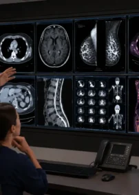
A recent study published in European Radiology highlights a significant increase in the use of cardiac imaging techniques such as MRIs and CT scans between 2011 and 2022 across 32 countries. The data, gathered from the European Society of Cardiovascular Radiology’s MR-CT registry, showed a 3.8-fold increase in MRIs and a 4.5-fold increase in CT scans for cardiac concerns during this period.
Radiologists, either independently or in collaboration with non-radiologists, primarily reported these examinations. The study emphasized the importance of radiologists in providing cardiac imaging services, attributing their expertise to the expanding availability of these modalities in both academic and non-academic centers.
Challenges with Interpretations
Interpreting cardiac imaging presents a range of challenges due to the complexity of the heart’s structure, function, and the dynamic nature of cardiac activity. Here are some specific examples of these challenges:
- Complex Anatomy and Physiology
Detailed Anatomy: The heart’s intricate structures, such as the coronary arteries, valves, myocardium, and chambers, require careful analysis. Identifying subtle anomalies like small congenital defects or early signs of disease can be difficult.
Example: Diagnosing a small atrial septal defect (ASD) in a transthoracic echocardiogram (TTE) can be challenging due to its subtle presentation and the need to differentiate it from normal anatomical variations.
- Motion Artifacts
Heart Motion: The constant movement of the heart can create artifacts, making it difficult to obtain clear and accurate images.
Example: In cardiac MRI, the rapid motion of the heart can blur images, especially if the patient cannot hold their breath adequately during the scan.
- Image Quality and Resolution
Image Clarity: Achieving high-resolution images is crucial for accurate diagnosis, but various factors can degrade image quality.
Example: In echocardiography, poor acoustic windows due to obesity, lung disease, or previous surgeries can obscure critical details, making it hard to assess valve function or wall motion abnormalities.
- Differentiating Normal Variants from Pathology
Physiological Variants: Distinguishing between normal anatomical variants and pathological findings requires expertise.
Example: Differentiating between a benign variant like a prominent trabeculae in the left ventricle and early signs of cardiomyopathy in a cardiac MRI requires careful interpretation.
- Dynamic Functional Assessment
Real-Time Functionality: Assessing the dynamic function of the heart, including systolic and diastolic function, valve movement, and blood flow, can be complex.
Example: Evaluating diastolic dysfunction on an echocardiogram involves interpreting multiple parameters such as mitral inflow patterns, tissue Doppler imaging, and left atrial volume, which can be nuanced and interdependent.
- Contrast Agents and Artifacts
Use of Contrast: While contrast agents can enhance visualization of cardiac structures and perfusion, they can also introduce artifacts and complications.
Example: In cardiac CT angiography (CTA), contrast-induced artifacts, such as streak artifacts from dense iodinated contrast, can obscure coronary artery details, complicating the assessment of stenosis.
- Interpreting Complex Cases
Multifactorial Disease: Patients with multiple coexisting cardiac conditions present a challenge for comprehensive interpretation.
Example: A patient with ischemic heart disease, heart failure, and arrhythmias may have overlapping imaging findings on a cardiac MRI, requiring a detailed and integrated interpretation to delineate the contribution of each condition.
- Stress Imaging
Inducing and Interpreting Stress Conditions: Stress echocardiography or cardiac MRI stress tests involve interpreting the heart’s response to induced stress (exercise or pharmacological agents).
Example: Identifying stress-induced wall motion abnormalities in a stress echocardiogram requires comparing pre- and post-stress images, which can be subtle and influenced by technical factors and patient effort.
- Integration of Multimodal Imaging
Combining Data from Multiple Modalities: Integrating information from various imaging techniques like echocardiography, MRI, and CT to provide a comprehensive diagnosis.
Example: Correlating findings from a cardiac MRI showing myocardial fibrosis with a CT angiogram revealing coronary artery stenosis requires synthesizing data from both modalities to understand the patient’s overall cardiac condition.
These challenges underscore the need for advanced training, experience, and often subspecialty expertise in cardiac imaging to ensure accurate and reliable interpretations.
Vesta Teleradiologists: Specialists in Cardiac Imaging
In conclusion, the surge in cardiac imaging underscores the critical role radiologists play in providing accurate and timely diagnoses for heart patients. With subspecialties in cardiac imaging, Vesta’s board-certified radiologists are well-equipped to meet the growing demand for accurate cardiac imaging interpretation for outpatient centers, mobile radiology units, and hospitals alike, whether on-site or remotely. As the field of cardiac imaging continues to evolve, radiologists remain at the forefront, leveraging their specialized knowledge to support healthcare providers and deliver high-quality imaging services across diverse clinical settings.
Sources:
radiologybusiness.com
ncbi.nlm.nih.gov
acc.org
openai.com