Mammography is one of the necessary tests physicians use to detect the early stages of breast cancer and other breast diseases. Fortunately, mammogram technology has advanced rapidly within the last few years and has positively impacted women’s health and wellness.
Radiological mammography has been in use through most of the 1900s, but the FDA didn’t approve digital mammography until 2000. The digital technology advancement opened up a whole new world for physicians to diagnose breast cancer earlier. Digital mammography accesses computer technology to enhance the X-ray images of the breast.
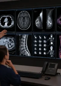
After digital mammography came into use, 3D breast imaging technology emerged in 2011. The 3D digital mammography (also known as 3D tomosynthesis) is where a technician takes multiple breast images from different angles. The technician then processes these images using computer software to create a three-dimensional reproduction of the breast.
With a three-dimensional reproduction of the breast, a radiologist can analyze the imaging slice-by-slice in great detail. This process has reduced many of the physician’s false-positive diagnoses given to women and reduced the stress of call-back appointments.
Since the 3D technology, companies have developed more advanced mammography equipment, tests, and computer-aided diagnosis systems (CAD). Researchers also have advanced imaging tools like whole breast ultrasound (WBUS) and magnetic resonance imaging (MRI) to aid the mammography process.
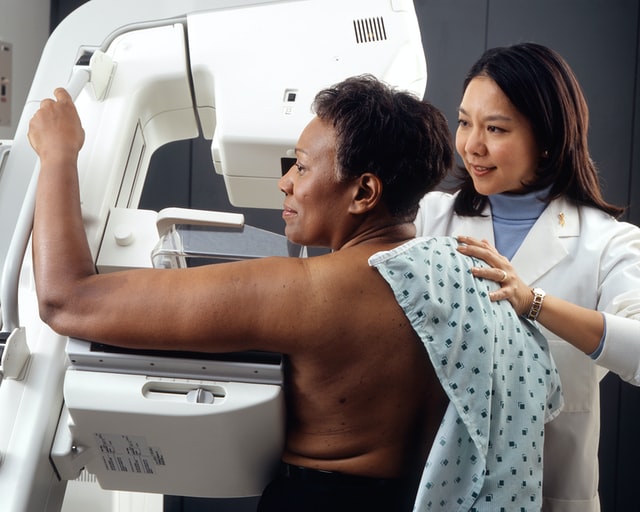
Physicians may recommend patients perform regular year-to-year screening mammograms so any changes in the patient’s breast that may cause concern can be detected. A physician orders a diagnostic mammogram when the screening mammogram shows an abnormality or if the patient notes other extraordinary symptoms.
A diagnostic mammogram is similar to a screening mammogram, except the technician will take more images using more positions to get more explicit photos of the area. A diagnostic mammogram can define if a biopsy is needed.
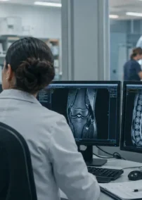
Throughout mammogram use, the human eye has been depended on to detect abnormalities in a patient’s breast X-rays, leading to false positives and false negative exams. With the advancements in equipment, technology, and software, radiologists can detect any abnormality in breast tissue with more certainty.
Increased research and equipment advancements in mammograms have also decreased patients’ exposure to radiation. Studies have concluded that the benefits of mammograms nearly always outweigh the potential harm from radiation exposure. However, patients should always disclose to the X-ray technicians if they are pregnant or have other health issues at risk by using any level of radiation.
Newer mammography imaging tests help physicians diagnose the smallest of tumors and most minimal cell defects. These tests include positron emission mammography (PEM), optical imaging, electrical impedance tomography (EIT), and molecular breast imaging (MBI).
Positron emission mammography (PEM) is a scan that uses sugar attached to a radioactive particle to look for cancer cells. This test is sometimes a replacement for an MRI.

Optical imaging is a test where technicians monitor the light passed into the woman’s breast and compare it to the measurement of light passing through the breast tissue. An altered reading of light will detect an area of the breast that warrants further exploration. Researchers are using this test with MRIs or 3D mammograms.
Since breast cancer cells conduct electricity differently than normal cells, physicians sometimes use electrical impedance tomography (EIT) as a diagnostic tool. During the test, a technician passes a bit of current through the patient’s breast and looks for changes with small electrodes applied to the skin.
Another test that researchers have developed is molecular breast imaging (MBI). This test is used with mammograms for women who have dense breasts. Doctors inject a radioactive drug into a patient’s vein, and the drug attaches to cancer cells, and a special camera can locate those cancer cells through the imaging process.
Researchers are continuing their efforts to improve mammogram results. Safe and effective screening and diagnostic mammograms will continue to improve survival statistics for women no matter what their genetic makeup, family history, or any other risk factor may indicate.
Vesta Teleradiology
At Vesta, our US Board Certified Radiologists are trained to read mammography scans as well as an entire host of other types of diagnostic imaging results. Look to us to support your team. Learn more about our teleradiology services here.