Why Work with Vesta Teleradiology? Explore a Rewarding Career in Radiology

Are you a radiologist seeking a fulfilling career with flexibility, competitive pay, and an unmatched work-life balance? Look no further than Vesta Teleradiology! Whether you’re interested in onsite or remote opportunities, Vesta is growing and is hiring for multiple positions to fit your unique needs and career goals, both full and even part-time!

Who We Are

Vesta Teleradiology is a leader in radiology services, trusted by clients in over 40 states. With accreditation from The Joint Commission, we pride ourselves on delivering high-quality interpretations 24x7x365. Our radiologists are at the forefront of patient care, supported by cutting-edge technology and a dynamic, collaborative team.

What Makes Vesta Unique?

At Vesta, we understand that every radiologist has different career aspirations. That’s why we offer a range of positions—part-time, locums, and full-time roles—with opportunities for both onsite and remote work. Whether you’re looking to make a big impact by covering critical daytime shifts or prefer overnight flexibility, there’s a spot for you at Vesta.

Flexible Opportunities Tailored to You

  • Remote Freedom: Work from the comfort of your home while delivering life-changing diagnoses to patients across the country.
  • Onsite Engagement: Be part of a team in a hospital or healthcare facility, making a direct difference in patient care.
  • Part-Time Roles: Just a few hours a week can make a big impact while giving you time for your other passions.

Why Radiologists Love Working with Vesta

  1. Competitive Compensation

We believe in rewarding your expertise. With industry-leading pay, your hard work and dedication won’t go unnoticed.

  1. Flexible Work Schedules

Looking for balance? Vesta offers flexible shifts, whether you prefer daytime hours or overnight coverage. Choose when and where you work to match your lifestyle.

  1. Work from Anywhere

Say goodbye to long commutes! Remote radiologists can provide interpretations from anywhere in the country. Vesta’s seamless technology ensures that you’re always connected, no matter your location.

  1. No Startup Costs

Joining Vesta is hassle-free. We cover the essentials so you can focus on what matters most—providing exceptional patient care.

  1. Comprehensive Malpractice Coverage

We’ve got your back. Our malpractice insurance includes tail coverage, giving you peace of mind every step of the way.

Your Ideal Candidate Profile

We’re seeking dedicated professionals who:

  • Hold active state licenses, especially in CA, TX, FL, GA, AZ, or NM.
  • Are looking for meaningful opportunities to grow, contribute, and thrive in a supportive environment.
Why Now Is the Perfect Time to Join Vesta

Radiology is at the cutting edge of medical innovation, and Vesta Teleradiology offers you the chance to be part of that evolution. With daytime, overnight, and weekend opportunities, your options are limitless. Plus, our growing client base means your expertise is in high demand.

Apply Today and Transform Your Career

Take the next step in your radiology career with Vesta Teleradiology. Discover how flexibility, competitive pay, and a supportive team can create the ideal working environment for you.

Click here to apply now: bit.ly/3XKhTH7

Don’t wait—join a team that values your skills, supports your ambitions, and empowers you to excel. Vesta Teleradiology: Where Excellence Meets Opportunity.


Advancements in 3D and 4D Imaging: Revolutionizing Diagnostic Radiology

As diagnostic imaging technology continues to evolve, 3D and 4D imaging are paving the way for more accurate and dynamic visualizations of the human body. These innovations offer healthcare providers critical tools for enhanced diagnosis and treatment planning, especially in areas like orthopedics, oncology, and cardiology. The ability to capture real-time motion in 4D imaging adds an extra layer of depth to how medical professionals view and understand various conditions.

The Power of 3D and 4D Imaging in Medical Diagnostics

3D imaging transforms traditional scans, like MRIs or CTs, into fully realized three-dimensional representations. This allows healthcare providers to explore anatomy from different angles, improving diagnostic precision. For example, in oncology, 3D imaging helps in detecting and mapping tumors, providing a more comprehensive understanding of the disease.

Meanwhile, 4D imaging—essentially adding the dimension of time to 3D scans—offers dynamic views, capturing motion in real time. This is particularly impactful in fields like cardiology and orthopedics, where movement plays a significant role in disease progression. A recent study highlights how 4D CT imaging is helping radiologists detect biomechanical abnormalities in joints, providing earlier interventions for conditions like osteoarthritis​.

By visualizing joint motion during specific movements, radiologists gain a clearer understanding of how diseases develop and progress over time.

The Role of AI in 3D and 4D Imaging

As these technologies advance, the integration of artificial intelligence (AI) is playing a critical role in enhancing the accuracy and speed of diagnostic interpretations. AI tools, for instance, are now capable of analyzing complex 3D imaging data to detect patterns or abnormalities that might otherwise be missed by the human eye. In 2024, AI continues to push the boundaries of medical imaging by assisting radiologists in reading complex scans faster and more accurately, particularly in complex cases like oncology and neurology​


Furthermore, AI-driven predictive analytics can analyze 3D and 4D imaging data to forecast disease progression, offering earlier diagnoses and personalized treatment plans. This combination of AI with cutting-edge imaging tools not only improves patient outcomes but also optimizes workflows in radiology departments.

Future Applications of 3D and 4D Imaging

The future of diagnostic imaging lies in the continued refinement of these technologies. For example, 4D ultrasound is growing in popularity, particularly in obstetrics, where real-time imaging is crucial for monitoring fetal development. Additionally, the trend of using portable imaging devices for 3D and 4D scans is making diagnostic tools more accessible, especially in remote or underserved areas​

In musculoskeletal imaging, 4D CT offers detailed insight into joint motion, allowing radiologists to better understand biomechanical derangements and intervene earlier to prevent further deterioration, such as the development of osteoarthritis​

How Vesta’s Teleradiology Services Can Support Your Imaging Needs

As the field of diagnostic imaging continues to evolve, we’re here to help healthcare providers interpret these advanced 3D and 4D scans. Our team of board-certified radiologists offers subspecialty expertise in areas like orthopedics, cardiology, and oncology, ensuring accurate and timely interpretations of complex imaging studies. With the integration of AI and the latest imaging technologies, our teleradiology services are equipped to support your practice as you navigate the future of diagnostic imaging.

By partnering with Vesta, you gain access to the latest innovations in imaging and expert interpretations that can enhance patient outcomes—one scan at a time.


Sources:


Rsna.org
insights.omnia-health.com
spectrumxray.com
openai.com


Why Second Opinions Matter: How Our Teleradiology Services Support Healthcare Facilities with Subspecialty Expertise

When it comes to accurate diagnoses and effective patient care, getting a second opinion on imaging results can make all the difference. Our teleradiology company is here to support healthcare facilities that need a reliable and expert review of radiology reports. Whether you’re looking for a second set of eyes for complex cases or want to ensure the highest level of diagnostic accuracy, our team of board-certified radiologists—with subspecialties in areas such as neuroradiology, musculoskeletal imaging, and oncology—are ready to assist.

Why Choose a Teleradiology Partner for Second Opinions?

Second opinions are often requested in cases where findings are ambiguous, complex, or where the stakes are high. By partnering with a teleradiology company like ours, healthcare providers can access subspecialized expertise quickly and cost-effectively, regardless of their location. This can be a game-changer for facilities with limited in-house radiology staff or those handling a high volume of specialized imaging studies.

In fact, a multidisciplinary tumor board at a National Comprehensive Cancer Network (NCCN) center found that 43% of patients referred for second opinions on breast cancer imaging had their initial diagnosis changed. This statistic highlights the significant impact that expert second opinions can have on patient outcomes​.


Types of Imaging Studies That Often Need a Second Opinion

Not all imaging studies are straightforward, and some require deeper insight to reach a definitive diagnosis. Here’s a look at the imaging tests that most frequently benefit from a second opinion:

MRI (Magnetic Resonance Imaging)

    • Neuroradiology (Brain and Spine Imaging): Brain and spinal MRIs are complex studies that may reveal subtle findings like micro-bleeds, white matter lesions, or small tumors. Our neuroradiologists can provide expert insight into these challenging cases.
    • Musculoskeletal MRI: Joint injuries, ligament tears, and early-stage bone lesions often require subspecialty evaluation to avoid misdiagnosis.

CT (Computed Tomography) Scans

      • Abdominal and Pelvic CT: Complex abdominal findings, such as small tumors or liver masses, can be challenging to interpret. Our abdominal imaging specialists ensure accurate diagnoses for conditions like pancreatic cancer or complex GI issues.
      • Thoracic CT: Pulmonary nodules or lung masses require careful analysis to differentiate benign from malignant findings, especially in patients with a history of smoking or cancer.

Mammography and Breast Imaging

        • Given the sensitivity and potential impact of findings on patient care, a second opinion can confirm initial readings and prevent over- or under-treatment. Our radiologists with breast imaging expertise can re-evaluate mammograms, breast MRIs, and ultrasounds.

Providing second opinions on breast imaging cases not only ensures diagnostic accuracy but also places significant demands on radiologists’ workloads. A study found in the Journal of the American College of Radiology revealed that second opinions for breast cancer cases required an estimated 3,135 to 3,804 work relative value units (wRVUs) over a three-year period, but the reimbursement received did not match the effort involved​.

Ultrasound

    • Obstetric and Gynecologic Ultrasounds: Subtle findings such as ovarian cysts or fetal abnormalities often require expert review to confirm a diagnosis.
    • Vascular Ultrasound: Evaluating blood flow and vessel abnormalities can benefit from a second opinion to ensure treatment decisions are accurate.

Oncologic Imaging

    • PET-CT and Whole Body MRIs: Cancer staging is critical for treatment planning, and our subspecialized oncologic radiologists can help refine staging or detect subtle metastases that may have been missed.

Who Benefits from Our Teleradiology Second Opinion Services?

Our second opinion teleradiology services are a valuable resource for a wide variety of healthcare facilities, each with unique needs:

Hospitals and Health Systems

    • From large urban hospitals to smaller rural facilities, hospitals often have to handle complex imaging cases where a second opinion is invaluable. This is especially true for trauma cases, oncology patients, and complex neurological conditions.

Outpatient Imaging Centers

    • Standalone imaging centers that offer diagnostic imaging services such as MRI, CT, and X-ray can enhance the quality of their reports by providing second opinions from subspecialized radiologists. This builds trust with referring physicians and patients.

Urgent Care Centers

    • Urgent care facilities frequently deal with musculoskeletal injuries and abdominal pain that require precise interpretations. A teleradiology partner with musculoskeletal and abdominal imaging expertise can help confirm initial findings and ensure appropriate follow-up care.

Primary Care and Family Practice Clinics

    • When general practitioners encounter unexpected or unusual imaging findings, a second opinion from a subspecialist can guide the next steps in patient management.

Specialty Clinics (Neurology, Orthopedics, Oncology)

    • Specialty clinics can use our second opinion services to validate complex imaging studies, such as intricate spinal MRIs or oncology staging scans, ensuring the highest standard of care for their patients.

Our Subspecialized Expertise Sets Us Apart

Our team includes radiologists who are not only board-certified but also have subspecialty training in fields such as:

  • Neuroradiology
  • Musculoskeletal Imaging
  • Breast Imaging
  • Abdominal Imaging
  • Cardiothoracic Imaging
  • Pediatric Radiology

This means that when you request a second opinion, your cases are reviewed by experts who focus on these specific areas every day, providing a level of precision that general radiology might not offer.

How Our Teleradiology Process Works

  1. Submit the Case Securely: Our HIPAA-compliant platform allows you to upload imaging studies quickly and securely.
  2. Subspecialized Review: We assign your case to a radiologist with the relevant subspecialty training to review the images and provide an expert second opinion.
  3. Receive a Detailed Report: You receive a comprehensive report with detailed findings, recommendations, and any necessary follow-up steps, ensuring that your patients get the best possible care.choosing a teleradiologist
Ready to Partner for Second Opinions? Choose Vesta Teleradiology

If your healthcare facility needs expert support for complex or high-stakes imaging cases, contact us today. Our team is dedicated to helping you provide accurate, timely, and comprehensive care for your patients through our teleradiology second opinion services.

Optimize patient outcomes and build trust in your imaging results by partnering with us for your second opinion needs!

Contact Vesta now to learn more about our subspecialty teleradiology services.


Sources:

jacr.org
pubmed.ncbi.nlm.nih.gov
openai.com


Understanding the New Mammography Quality Standards Act (MQSA) Amendment: What It Means for Healthcare Facilities and Patients

In a significant move to enhance breast cancer detection and patient care, the U.S. Food and Drug Administration (FDA) recently enacted a final rule under the Mammography Quality Standards Act (MQSA). Effective as of September 2024, this new amendment mandates that all mammography facilities include information about breast density in their mammography reports and results letters to patients.

This change brings critical updates to mammography practices, promising greater transparency and more personalized healthcare. Let’s explore what this new rule entails and what it means for healthcare facilities and their patients.

What is the Mammography Quality Standards Act ?

The Mammography Quality Standards Act (MQSA) was originally enacted in 1992 to ensure high standards in mammography for the detection of breast cancer. The act requires mammography facilities to meet quality standards, which are regulated by the FDA, to be certified and able to legally operate in the United.  Over the years, the MQSA has played a vital role in improving the accuracy and reliability of mammography, ultimately enhancing early detection and treatment outcomes for breast cancer.

mammogram

What’s New with the Recent MQSA Amendment?

As of September 2024, the FDA has implemented an important amendment to the MQSA. The new rule requires all mammography facilities to include detailed information about breast density in both the patient’s mammography report and the results letter provided to the patient. This update is a step forward in making breast density a standard part of mammogram interpretations.

Breast density refers to the amount of fibroglandular tissue compared to fatty tissue in the breast, visible on a mammogram. Dense breast tissue, which appears white on a mammogram, can obscure cancerous masses, also white, making it harder to detect breast cancer. Furthermore, women with dense breasts have a higher risk of developing breast cancer.

By including breast density information in mammography reports, healthcare providers and patients can better understand an individual’s specific risk profile and decide on the most appropriate next steps for screening and prevention.

What Does This Mean for Healthcare Facilities?

For healthcare facilities offering mammography services, this new amendment brings several critical changes:

  1. Updated Reporting Requirements: Facilities must now ensure that mammography reports include standardized information about breast density. This includes classifying breast density into one of four categories:
    • Almost entirely fatty
    • Scattered areas of fibroglandular density
    • Heterogeneously dense, which may obscure small masses
    • Extremely dense, which lowers the sensitivity of mammography

These classifications help provide a more precise picture of a patient’s breast tissue composition, allowing for better decision-making in follow-up care.

  1. Enhanced Communication with Patients: The new rule requires that breast density information be clearly communicated in the results letter sent to patients. This step increases transparency, empowers patients to be proactive about their health, and ensures they are aware of potential challenges in detecting cancer due to dense breast tissue.
  2. Compliance and Training: Healthcare facilities must update their protocols to comply with the new requirements. This may involve additional staff training to accurately assess and report breast density, update reporting systems, and educate patients on what breast density means for their health.
  3. Increased Demand for Supplemental Screening: With more awareness of breast density, facilities may see an increased demand for additional screening methods, such as 3D mammograms, breast MRIs, ultrasounds, or contrast-enhanced mammography. Facilities should be prepared to offer these services or provide referrals to facilities that do.

What Does This Mean for Patients?

For patients, particularly women undergoing routine mammography, this amendment provides several benefits:

  1. Greater Awareness of Breast Density: Many women are unaware of their breast density and its impact on cancer detection. By receiving information directly in their mammography results, patients can better understand their personal risk factors and discuss potential concerns with their healthcare providers.
  2. Informed Decision-Making: Knowing their breast density allows patients to make informed decisions about their screening options. For instance, those with dense breasts may opt for additional imaging tests that could provide a more comprehensive evaluation than a standard mammogram alone (American Cancer Society).
  3. Empowerment Through Education: The requirement for breast density information fosters greater patient education. Patients are empowered to ask questions, seek second opinions, or request further testing if they have concerns about the accuracy of their mammogram results.
  4. Improved Early Detection: By understanding their breast density, patients and healthcare providers can work together to develop a tailored screening plan, potentially catching breast cancer earlier when it is most treatable. This could lead to better outcomes and a higher chance of survival.

What Are the Challenges?

While the new MQSA amendment offers many advantages, there are some challenges to consider:

  • Increased Anxiety: Some patients may feel anxious upon learning they have dense breast tissue and are at higher risk for breast cancer. It is essential for healthcare providers to provide clear, compassionate communication to help patients understand their situation without causing unnecessary worry.
  • Potential Costs: Additional screening methods, like breast MRIs or 3D mammography, may involve out-of-pocket costs for some patients, depending on their insurance coverage. Patients and providers must discuss these costs and determine the most appropriate and affordable screening options.
  • Adjustment Period for Facilities: Healthcare facilities will need time to fully implement the new requirements, including training staff, updating reporting procedures, and ensuring compliance with the new rule.

Moving Forward

The recent MQSA amendment represents a significant step toward improving breast cancer detection and patient care in the United States. By mandating the inclusion of breast density information in mammography reports, the FDA aims to enhance early detection and empower patients with critical knowledge about their breast health.

For healthcare facilities, this means updating practices and procedures to meet the new standards, while for patients, it offers a clearer understanding of their risk factors and screening options. While challenges remain, the new rule is ultimately a move toward better, more personalized healthcare and could play a crucial role in the fight against breast cancer.


How Vesta Helps with Mammogram Readings and the Amendment


At Vesta, our experienced radiologists are fully equipped to handle the new MQSA amendments for mammogram interpretations, ensuring that you receive the most accurate and comprehensive information about your breast health. We are here to provide you with the highest standard of care and support every step of the way. Reach out to us to schedule your mammogram or to learn more about what these new standards mean for you.


Sources:

Fda.gov
Acr.org
ncbi.nlm.nih.gov
Healthline.com


Update on ChatGPT and Radiology Readings

ChatGPT-4 Vision and Radiology Exam Questions

ChatGPT-4 Vision is an enhanced version of OpenAI’s GPT-4 that can interpret both text and images. This multimodal capability allows it to analyze visual content, such as photos, diagrams, and medical images, in addition to understanding text.

Applications of ChatGPT-4 Vision include assisting with medical imaging analysis, enhancing accessibility by describing images, extracting data from visual documents, and supporting creative tasks. However, it has limitations, such as occasional inaccuracies when interpreting complex images, especially in specialized fields like radiology.

A study published in Radiology evaluated the performance of ChatGPT-4 Vision on radiology exam questions, revealing that while the model performed well on text-based questions, it struggled with image-related questions. ChatGPT-4 Vision is the first version of the language model capable of interpreting both text and images.

The study, led by Dr. Chad Klochko, used 377 retired questions from the American College of Radiology’s Diagnostic Radiology In-Training Examinations. The model answered 65.3% of all questions correctly, achieving 81.5% accuracy on text-only questions but only 47.8% on questions with images. The model performed best on image-based questions in chest and genitourinary radiology, and worst in nuclear medicine.


The study explored different prompting techniques and found that the model declined to answer 120 questions, primarily image-based, and showed hallucinatory responses when interpreting images, suggesting incorrect interpretations leading to correct diagnoses. Dr. Klochko emphasized the need for more specialized evaluation methods, as the model’s current limitations in accurately interpreting radiologic images restrict its applicability in clinical settings.


ChatGPT-4 For Summarizing Radiology Reports for Pancreatic Ductal Adenocarinoma

A study published in Radiology found that ChatGPT-4 outperforms GPT-3.5 in creating structured, summarized radiology reports for pancreatic ductal adenocarcinoma (PDAC), potentially improving surgical decision-making. Led by Dr. Rajesh Bhayana from the University of Toronto, the study demonstrated that GPT-4 generated near-perfect PDAC synoptic reports and achieved high accuracy in categorizing resectability using a chain-of-thought prompting strategy, resulting in more accurate and efficient surgical decision-making.


The study included 180 PDAC staging CT reports from Princess Margaret Cancer Centre in 2018. Two radiologists set a reference standard for 14 key features and the National Comprehensive Cancer Network (NCCN) resectability category. ChatGPT-4 was found to have equal or higher F1 scores than GPT-3.5 for all features and outperformed GPT-3.5 in categorizing resectability. Surgeons using AI-generated reports reduced their review time by 58%.


The findings suggest that ChatGPT-4 can improve standardization, communication, and efficiency in pancreatic cancer care. However, Paul Chang, MD, from the University of Chicago, emphasized the need to integrate these AI capabilities into scalable and comprehensive workflows, acknowledging the gap between feasibility and operational solutions.


Sources:

Auntminnie.com
medicalexpress.com
openai.com


MQSA Regulations: Are You Ready?

Effective September 10, 2024, the FDA has mandated updates to the Mammography Quality Standards Act (MQSA) regulations. Facilities must comply with new requirements, including breast density notifications in mammography reports and patient summaries.

What are the Key Updates?

Mammography Reports: Must include the facility’s name and location, a final assessment of findings in specific categories, and an overall assessment of breast density.

Patient Lay Summaries: Must include the patient’s name, facility information, and a breast density notification statement.

Communication of Results: For findings categorized as “Suspicious” or “Highly Suggestive of Malignancy,” reports must be provided to healthcare providers and patients within seven days. For incomplete assessments, follow-up reports must be issued within 30 days.

Medical Outcomes Audit: Annual audits must include metrics such as positive predictive value, cancer detection rate, and recall rate for each interpreting physician and the facility.

Additional requirements include maintaining personnel records for a specified duration, stringent recordkeeping of original mammograms and reports, and protocols for transferring or releasing mammography records within 15 days upon request.

Facilities failing accreditation three times cannot reapply for one year, and all mammography devices must meet FDA premarket authorization requirements.

These updates aim to improve the quality and accuracy of mammography services and ensure better patient communication and record management​.


Facilities that must comply with the Mammography Quality Standards Act (MQSA) include:

  1. Mammography Facilities: Any facility that provides mammography services, which includes hospitals, outpatient imaging centers, and private radiology practices.
  2. Mobile Mammography Units: These are mobile facilities that travel to various locations to provide mammography services and must meet the same MQSA standards as stationary facilities.
  3. Diagnostic Clinics: Clinics that perform diagnostic mammography to further investigate abnormalities found during screening mammograms.
  4. Screening Centers: Facilities that focus on providing routine mammograms to screen for breast cancer in asymptomatic women.


These facilities are required to comply with MQSA regulations to ensure high standards of care, including the quality of mammography equipment, the qualifications of personnel, and the quality of mammogram images​. If you partner with a teleradiology company like Vesta, we ensure reports adhere to these updates. Vesta is always ahead of the curve when it comes to regulations and working with their clients not only to educate them on what is coming but also work closely with them to put in place and roll out any new requirements. 


Sources:

fda.gov/radiation-emitting-products/mammography-quality-standards-act-and-program/important-information-final-rule-amend-mammography-quality-standards-act-mqsa
openai.com


Radiologists Say They’d Accept Lower Pay for Better Work-Life Balance

A recent Medscape survey, published on Friday, reveals that around 60% of radiologists would accept lower pay for a better work-life balance. Many radiologists face challenges balancing family and professional demands, with 39% feeling conflicted as parents and 33% struggling to achieve balance. Michael Ziffra, MD, from Northwestern, noted the persistent time issue for doctors, who wish to spend more time with family while also feeling compelled to prioritize patient care.

The survey found that 65% of radiologists consider prioritizing personal and family time very important, while 30% view it as somewhat important. Regarding exercise, 30% of radiologists work out 2-3 times a week, 24% do so 4-5 times, and only 13% do not exercise at all.

Most radiologists do not prioritize reducing screen time, with 63% not attempting to cut back on personal online hours. For personal internet use, 55% spend 2-4 hours daily, while 46% spend fewer than 2 hours on professional use.

Vacation time also varies, with 45% taking more than six weeks annually, 25% taking 3-4 weeks, and 13% taking 5-6 weeks. Only 5% take less than a week off each year.


Can Yoga Help Radiologists with Burnout?

A study published on January 31 in JAMA Network Open suggests that Yoga, specifically Sudarshan Kriya Yoga (SKY), could help alleviate burnout symptoms in radiologists. Led by Duygu Sag, PhD, and Fahri Saatcioglu, PhD, the research found that SKY reduced psychological distress and burnout while increasing wellness in physicians, including radiologists and radiation oncologists. The study emphasizes SKY as a safe and practical method to manage stress and burnout, which are significant issues in the medical profession.

yoga practice
Can yoga help with burnout?


Burnout negatively impacts physicians’ well-being, personal lives, and patient care, and has been a factor in their decision to leave the workforce. The study, conducted online from November 2021 to March 2022, involved 129 physicians from Turkey, Germany, and Dubai, comparing SKY with a stress management education program. Participants in the SKY group practiced daily and attended weekly follow-up sessions.

The researchers used various scales to measure the programs’ effectiveness and found that the SKY group experienced lower stress, decreased depression, reduced insomnia, increased professional fulfillment, and reduced work exhaustion and burnout compared to the control group. However, there was no significant effect on self-reported medical errors. The authors conclude that SKY could be a practical tool to mitigate stress and burnout in various professions facing similar challenges.


Is this Affecting Radiologist Staffing?

High workloads and mental demands contribute to burnout among radiologists, leading many to retire early or leave the field. The COVID-19 pandemic has exacerbated these issues, accelerating the rate at which radiologists are exiting the profession​


How Teleradiology Helps

Teleradiology offers a vital solution to the current radiologist shortage by enabling remote reading of imaging studies, thus bridging the gap between demand and available workforce. By leveraging advanced technology, teleradiology ensures timely and accurate interpretations, especially for underserved and rural areas that struggle to attract full-time radiologists. As a top teleradiology company, Vesta is committed to providing high-quality remote radiology services, enhancing patient care, and supporting healthcare facilities in managing their workloads efficiently. We support small, medium and large volumes for ERs, private facilities, outpatient imaging and mobile radiology.


Sources:

Radiologybusiness.com
diagnosticimaging.com
openai.com

An Update to the Physician Shortage Problem

The AAMC (Association of American Medical Colleges) has released new projections indicating a physician shortage of up to 86,000 physicians in the United States by 2036. This underscores the critical need for sustained and increased investments in training new physicians to address the country’s healthcare needs. The report, conducted by GlobalData Plc, includes various scenarios based on trends in healthcare delivery and the workforce. While the projected shortfall is smaller than previous estimates, it still highlights the necessity for additional investments in graduate medical education (GME). Demographics, particularly population growth and aging, are driving the increasing demand for physicians. The report also notes a significant portion of the physician workforce nearing retirement age, which will further decrease the physician supply. Addressing underserved communities could require approximately 202,800 more physicians than current estimates. Lifting the federal cap on Medicare support for GME and bipartisan legislation like the Resident Physician Shortage Reduction Act aim to alleviate the shortage, but further efforts are needed to meet future healthcare demands.


Rural Americans’ Healthcare Challenges

Rural Americans face significant healthcare challenges, with fewer available doctors compared to urban areas, exacerbating existing health issues. Dr. Bruce A. Scott, President of the American Medical Association, emphasizes the urgent need for policymakers to address these disparities. Rural communities experience higher rates of various illnesses, exacerbated by economic pressures and limited access to healthy living conditions. The shortage of specialists and the closure of rural hospitals further compound the problem. Insufficient access to primary care physicians is a pressing issue, with inadequate residency spots and decreasing applications from rural areas. The AMA advocates for changes to the Medicare physician payment system, which has seen a decline in rates over the years. Administrative burdens, such as prior authorizations, are also contributing to physician burnout and compromising patient care. To combat the doctor shortage and rural health challenges, the AMA advocates for healthcare reforms, including overhauling the Medicare payment system, expanding telehealth, increasing residency positions, incentivizing rural practice, and addressing workforce stresses.

hospitals in rural America

Radiology Is Being Hit, Too

Radiology departments are grappling with worsening staffing shortages alongside declining reimbursements. During the RSNA 2023 meeting, Ashish Sant from Merge by Merative discussed key trends and challenges. Staffing and cost management remain top concerns due to burnout and insufficient replacements for retiring radiologists. To address these issues, there’s a push towards cloud-based solutions, with a modular approach easing concerns about data security and patient information management. The pandemic has accelerated the shift towards cloud adoption, highlighting benefits such as accessibility and cost reduction. Integrating AI into radiology workflows is another focus, though challenges persist in seamlessly embedding AI solutions. Merge’s partnership with Microsoft Azure aims to provide customers with cloud solutions tailored to their needs.


Radiology Support for the US

Addressing radiology staffing shortages is crucial for ensuring efficient and effective healthcare delivery. Whether you’re a hospital, outpatient center, or part of the Indian Health Service (IHS), Vesta is here to help. Our team can provide on-site radiologists or teleradiologists to meet the specific needs of your facility. By partnering with us, you can ensure timely and accurate radiology services, ultimately improving patient care and outcomes. Don’t let staffing shortages hinder your operations – reach out today to learn how we can support your radiology department.


Sources:

Aamc.org
dtnpf.com
Healthimaging.com
openai.com


Key Concerns When Finding a Teleradiology Partner

Finding the right teleradiology partner becomes paramount, especially when faced with staffing shortages at hospitals, urgent care centers, or other healthcare facilities. As these institutions strive to maintain high-quality patient care amidst limited resources, outsourcing radiology services can provide a lifeline and has many benefits. However, the decision to engage a teleradiology partner demands careful consideration. From ensuring rapid turnaround times to guaranteeing impeccable quality and compliance, several crucial factors must be scrutinized to identify the ideal partner. Let’s delve into the essentials of what healthcare providers need to look out for when selecting a teleradiology partner in such critical circumstances.

Guide for Choosing a Radiology Partner

Quality Workflow: Quality assurance in teleradiology involves ensuring that the interpretations provided by the radiologists are accurate and reliable. This includes verifying the qualifications and expertise of the interpreting radiologists, as well as implementing processes for peer review and ongoing quality monitoring.


Subspecialties: If your healthcare center needs specific types of readings like those for EKGs, ECHO, or DXA, it’s good to research if the teleradiology company offers these subspecialties for both preliminary and final readings.


Credentialing and Licensing: It’s crucial to confirm that the radiologists working with the teleradiology partner are appropriately licensed and credentialed to practice in the relevant jurisdictions. This involves verifying their credentials, certifications, and licensure status to ensure compliance with regulatory requirements. Vesta’s radiologists are all U.S. Board Certified.


Turnaround Time: Prompt delivery of radiology reports is essential for timely patient care and treatment decisions. When selecting a teleradiology partner, it’s important to inquire about their average turnaround times and their ability to meet the facility’s specific needs, especially during peak periods or emergencies. Expect fast turnaround times with Vesta. In fact, Vesta can meet emergency STAT needs and provide reports within just 30 minutes with accurate and high-quality reports.

turnaround times
Ask about their turnaround times

Security and Compliance: Teleradiology involves the transmission and storage of sensitive patient information, making data security and compliance with privacy regulations paramount. Healthcare providers should ensure that their teleradiology partner adheres to industry-standard security protocols, such as HIPAA compliance, and employs encryption and other measures to safeguard patient data. Vesta is 100% HIPAA compliant.


Communication and Collaboration: Effective communication channels between the healthcare facility and the teleradiology partner are essential for seamless collaboration. This includes establishing protocols for communication of urgent findings, as well as integrating teleradiology reports into the facility’s electronic health record (EHR) system for easy access by clinicians. Vesta is at your service 24/7/365. We not only retain the services of exceptional Radiologists who are immediately available to your referring physicians, we also employ a knowledgeable staff ready to address any questions.

reporting


Technical Support: Reliable IT infrastructure and technical support are essential for smooth image transmission and workflow efficiency. Healthcare providers should assess the teleradiology partner’s IT capabilities, including their systems for image transfer, storage, and viewing, as well as their responsiveness to technical issues or downtime.


Cost-effectiveness: While quality of service is paramount, healthcare providers must also consider the cost-effectiveness of partnering with a teleradiology provider. This involves evaluating the partner’s pricing structure, including any subscription fees, per-case charges, or additional costs for expedited services, and comparing it with the value provided. Vesta helps healthcare providers whether they have small, medium or even large volumes.


Reputation and Experience: Partnering with a reputable teleradiology provider with a proven track record is crucial for peace of mind and quality assurance. Healthcare providers should research the partner’s reputation, including client testimonials, case studies, and industry recognition, and assess their experience in providing teleradiology services to similar facilities or specialties. Vesta has been in service for over 16 years and has a proven track record of success!


Expert Teleradiology Company in the US: Vesta

Do you need a qualified teleradiology partner? Vesta is here for you whether in full capacity or just partially. Contact us to learn more: 877-558-3782


Sources:

Medium.com
openai..com


New FDA Clearances for Imaging Systems and Solutions

FDA clearance for a diagnostic imaging machine indicates that the device has been deemed safe and effective for its intended use by the Food and Drug Administration (FDA) in the United States. This clearance process involves thorough evaluation of the device’s design, performance, and manufacturing processes to ensure that it meets regulatory standards for quality, safety, and efficacy. Here’s the latest devices that have received FDA clearance.


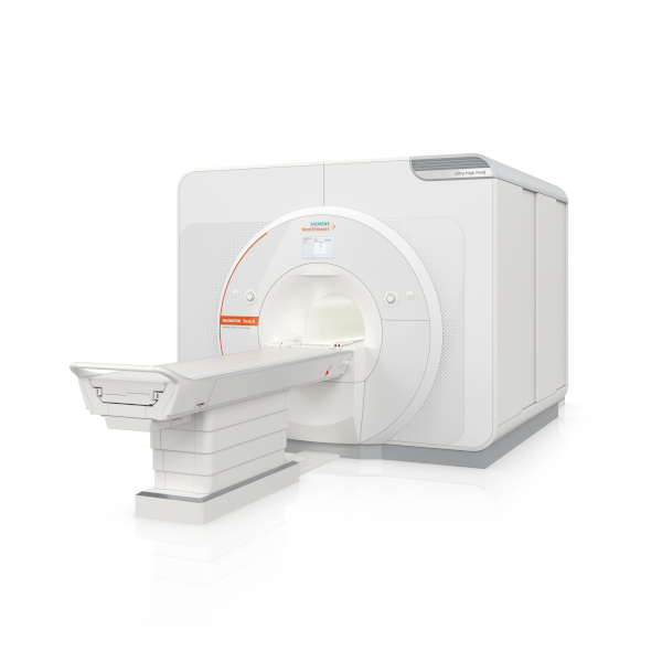
The Magnetom Terra.X: MRI System

The Magnetom Terra.X, a new 7T MRI system, has received 510(k) clearance from the FDA. Manufactured by Siemens Healthineers, it’s a second-generation successor to the Magnetom Terra and offers several enhancements for 7T imaging. Key features include an eight-channel parallel transmit architecture for clinical use, deep learning image reconstruction optimized for 7T, improved diffusion imaging with a high-performance gradient system, and accelerated image acquisition enabling high-resolution brain and knee exams in under 20 minutes. Siemens Healthineers sees this as a significant step in providing better patient care, particularly in neurological and knee imaging. Additionally, the FDA clearance allows existing Magnetom Terra systems to be upgraded to the Magnetom Terra.X.

Image courtesy of Siemens Healthineers

SyMRI 3D for Brain Imaging

SyntheticMR has announced that its latest imaging solution, SyMRI 3D, has received FDA 510(k) clearance for clinical use in the United States. This clearance marks a significant advancement in quantitative MRI technology, offering exceptional resolution and accuracy in brain imaging. SyMRI 3D enables precise volumetric estimations of brain regions, known as parcellation, providing clinicians with deeper insights into brain structure and function. The enhanced resolution facilitates comprehensive lesion analysis, leading to more accurate medical condition assessments. This clearance empowers physicians to make more informed decisions in diagnosis and treatment planning, ultimately improving patient outcomes. SyntheticMR reaffirms its dedication to advancing medical imaging technology and providing innovative tools to enhance patient care through this milestone.


nCommand Lite for Remote Scanning

GE Healthcare has highlighted the FDA clearance of a solution by Ionic Health that enables technologists to remotely supervise patient scans. The system, called “nCommand Lite,” has been tested in Brazil for three years and is vendor-agnostic, allowing remote supervision across MRI, CT, and PET modalities. GE has secured exclusive distribution rights for nCommand in the U.S., aiming to address ongoing workforce shortages in healthcare. Rekha Ranganathan, GE’s chief digital officer for imaging, emphasized the company’s commitment to remote operations and increasing patient access to expert technologists. The system facilitates not only scanning supervision but also training, procedure assessment, and scanning parameter management. GE’s announcement coincides with growing interest in remote scanning, with the American College of Radiology advocating for permanent remote supervision of diagnostic tests. However, technologists have expressed reservations about managing imaging remotely, according to recent survey data from the American Society of Radiologic Technologists.


Sources:

Itnonline.com
Radiologybusiness.com
diagnosticimaging.com
openai.com