Big machines, loud noises, unfamiliar people, funny smells. If you’ve had a medical image taken as an adult, you may not enjoy the experience, but you understand the process. As a child, it’s a totally different story. These factors may frighten or put them in a state of unease during a procedure that will help with their diagnosis or treatment. When it’s time for a little one to receive any medical imaging, there are a few things that can be done to help make them more comfortable and get the best result possible.
First and foremost, the best way to comfort a child before an imaging procedure is to keep them informed. If the child is old enough, of course, a parent, guardian, or medical professional can tell them what to expect during the procedure. Knowing what to wear, how it might feel, and how long it will take ahead of time will help the child feel in control of at least some parts of their day and reduce some stress.
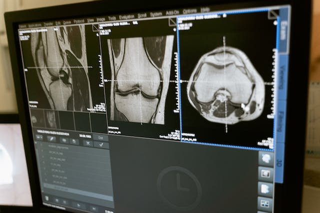
For pediatric x-rays and MRIs, children often need to be in awkward positions to capture the correct image. This can be uncomfortable depending on the age of the child and the injury they have. Studies have shown that a negative experience occurs more often when a child is restrained. One technique that helps put a child more at ease is called comfort positioning. This technique involves positioning the child in a way that feels comforting for them, such as sitting on their parents’ lap or a comfortable chair or bed instead of the examining table. Positions like “tummy to tummy” or “back to chest” can also be used as a calming position with other distractions like a toy or a tablet. These positions help reduce the stress hormone and “minimize the physical symptoms associated with anxiety.” Studies have shown that giving children these options helps with cooperation and gives children more control during their procedures.
In some cases, a child may need to be sedated for the procedure. This can be scary for anyone, especially a child, where they need to stay still for long periods of time in a very tight space. Because of the nature of an MRI, the sound of the machine and the closed tightness, patients are not able to fully communicate their needs, and could therefore become easily anxious and begin to move. In cases like these, studies have shown that music, or the mother’s voice during the procedure may reduce the need for sedation and the amount of drugs needed to sedate.
To help children have a better experience during imaging, companies, such as Phillips, have designed equipment and educational programs to help as well. To prepare children for their scan, they have created the Scan Buddy App which features calming cartoon characters that lead them through the process with games and instruction. When children visit the location where they will get their scan, children are allowed to role play their scan with a “Kitten Scanner,” a child sized MRI machine where they can send stuffed animals through to see what will happen during the scan. In 2021, Phillips launched their pediatric coaching program, where lighting, visuals, and sounds are used to ease the stress of the child.
Calming techniques like these can help turn a very scary experience into one that a child is prepared for, relaxed for, and perhaps even pleasant. A calm child means good imaging results and better outcomes for treatment.