What changed—and why it matters
The FDA has expanded the label for flutemetamol F 18 (Vizamyl), enabling quantification of amyloid plaque burden and long-term therapy monitoring in Alzheimer’s disease. This shift moves amyloid PET beyond a qualitative “positive/negative” decision toward objective, longitudinal assessment that can inform treatment choice, dose intervals, and discontinuation decisions. Business Wire
Professional groups report the update aligns amyloid PET with the clinical era of disease-modifying anti-amyloid therapies (e.g., lecanemab, donanemab), clarifying roles for baseline confirmation, on-treatment monitoring, and response tracking in routine care. Notably, SNMMI stated the FDA granted supplemental indications—including quantitative measurement and use for therapy monitoring—to three amyloid PET agents (flutemetamol F-18/Vizamyl, florbetapir F-18, and florbetaben F-18). SNMMI
Operational updates for radiology leaders
- Protocols & quant pipelines: Build or validate a quant workflow (SUVr or comparable metrics) that’s scanner-calibrated and reproducible across sites. If you operate multi-vendor fleets, document harmonization steps in your SOPs.
- Structured reports: Add fields for quantified burden at baseline, change from baseline, and interpretive guidance tied to therapeutic decisions (initiation, continuation, or discontinuation).
- Scheduling & throughput: Expect rising referral volume from neurology and geriatrics as therapy monitoring enters routine practice; protect access with extended hours or overflow capacity.
- Quality & governance: Define thresholds for biologically meaningful change, reader training for quant review, and reconciliation rules when quant and visual impressions diverge.
For additional context, trade coverage underscores that the updated label formally removes previous limitations around therapy monitoring and permits quant analysis in routine reporting. Empr
How Vesta Teleradiology helps
Vesta’s subspecialty neuro and nuclear medicine radiologists provide:
- Amyloid PET expertise: Visual+quant reads with structured templates aligned to your therapy pathway.
- Coverage when you need it: After-hours, weekends, or daytime overflow—without sacrificing turnaround time.
- Interoperability: Seamless delivery to your PACS/RIS and EMR; clear flags for therapy decisions and recall intervals.
- QA you can see: Peer review, consistency checks across readers, and optional double-reads during program ramp-up.
If you’re standing up or scaling amyloid PET services, we can supply immediate subspecialty coverage and templates tuned to your neurologists’ needs.

 Current Impact on Patients and Healthcare Systems
 Conclusion
 Recent developments underscore the significant benefits of teleradiology for healthcare providers. A report from
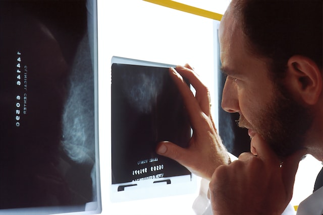
  By focusing on early detection through advanced imaging and leveraging teleradiology, healthcare providers can significantly impact cancer outcomes, offering patients the best chance for successful treatment. National Cancer Prevention Month serves as a reminder of the strides we can make in cancer care through prevention and early detection.
 Implications for Healthcare Providers

 Sources: