Social media is an ever-changing landscape, but it remains one of the best ways for healthcare facilities to reach and engage with current and potential patients. If you’re a healthcare facility looking to get started on social media, here are a few tips to help.
Listen First, Talk Later
Before creating content or engaging with people on social media, listening and observing what’s already happening in your industry is essential. Read up on trending topics, follow influencers, and join conversations relevant to your healthcare facility—this will give you a better understanding of what kinds of content resonates with your target audience.
Once you understand the conversation, you can create content or join existing discussions.

Spread Awareness of Services
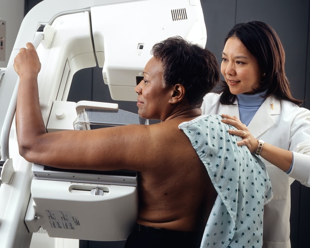
Content doesn’t have to be hard to produce. Does your facility offer mammograms or other diagnostic imaging services? What type of special treatments do you offer patients? Let it be known through your posts! Whether you make a short informative Instagram Reel or a simple graphic, it’s ever crucial to make sure people know what you offer.
Build Relationships
Social media is all about building relationships, so pay attention to how you interact with others online.
Respond thoughtfully to comments and questions from followers; if someone has taken the time to reach out to you via social media, they deserve a well-crafted response that shows them that their voice matters. When responding directly to a patient’s question or comment, always use a professional tone and be sure not to disclose private information.
The beginning response to a patient with a question about diabetes might be:
“ Managing diabetes is no easy task, and if you’re a diabetes patient, it can be challenging to get your condition under control.”
Or:
“I empathize with the daily struggles you must endure keeping diabetes at bay. The good news is that there are ways to help make this easier for you.”
Be Transparent
Social media should be an opportunity for transparency between yourself and your followers.

Share behind-the-scenes photos and videos from your day-to-day operations; this helps build trust by showing that real people work hard at your facility daily. It also allows patients who may not have visited before to get familiarized with the space beforehand, making them more comfortable when they arrive for their appointment or procedure.
Monitor Your Reputation
As a healthcare facility on social media, you must monitor what’s being said about you online – positive and negative.
Be sure to set up alerts for your company name as well as any hashtags associated with it to allow you to keep tabs on mentions of your brand.
If anything negative is posted about you online (such as negative reviews), you can address it quickly before the situation escalates. When a person posts a positive comment, be sure you “like” them, or comment back, so your followers know you respected their opinion.
A respectful response to a negative review about a rude employee at a hospital might be:
“At our hospital, all our staff members strive to ensure the highest level of quality care and customer satisfaction possible. Unfortunately, it’s not always achieved as intended due to human error and environmental stressors.”
Or
“We recognize that patient care starts with respectful communication and interactions between patients and healthcare providers. To improve communication throughout the institution, we are investing in additional staffing resources so that nurses and other team members feel better supported when high volumes present themselves on certain days or times during their shifts.”
Measure Results
Lastly, measure the results of your social media efforts using analytics tools.
Using these tools will allow you to track how many people are visiting your page (and how often), see where they came from (e.g., organic search vs. referral traffic), and track engagement over time (likes/comments/shares). This information will help inform future decisions about what kind of content works best for engaging with patients online.
By measuring results regularly, you can continually refine and improve your strategy over time, so it eventually becomes second nature.
Social media can provide significant benefits for healthcare facilities looking to connect more deeply with current & potential patients—but only if done correctly.
Following these tips will ensure your presence on social media is practical & compliant with relevant regulations & guidelines set forth by governing bodies.
Take some time today & begin implementing these tips into practice right away. Your healthcare facility’s social pages will become vibrant hubs of conversation & connect with the community.
Good luck & happy posting!