Understanding the New Mammography Quality Standards Act (MQSA) Amendment: What It Means for Healthcare Facilities and Patients

In a significant move to enhance breast cancer detection and patient care, the U.S. Food and Drug Administration (FDA) recently enacted a final rule under the Mammography Quality Standards Act (MQSA). Effective as of September 2024, this new amendment mandates that all mammography facilities include information about breast density in their mammography reports and results letters to patients.

This change brings critical updates to mammography practices, promising greater transparency and more personalized healthcare. Let’s explore what this new rule entails and what it means for healthcare facilities and their patients.

What is the Mammography Quality Standards Act ?

The Mammography Quality Standards Act (MQSA) was originally enacted in 1992 to ensure high standards in mammography for the detection of breast cancer. The act requires mammography facilities to meet quality standards, which are regulated by the FDA, to be certified and able to legally operate in the United.  Over the years, the MQSA has played a vital role in improving the accuracy and reliability of mammography, ultimately enhancing early detection and treatment outcomes for breast cancer.

What’s New with the Recent MQSA Amendment?

As of September 2024, the FDA has implemented an important amendment to the MQSA. The new rule requires all mammography facilities to include detailed information about breast density in both the patient’s mammography report and the results letter provided to the patient. This update is a step forward in making breast density a standard part of mammogram interpretations.

Breast density refers to the amount of fibroglandular tissue compared to fatty tissue in the breast, visible on a mammogram. Dense breast tissue, which appears white on a mammogram, can obscure cancerous masses, also white, making it harder to detect breast cancer. Furthermore, women with dense breasts have a higher risk of developing breast cancer.

By including breast density information in mammography reports, healthcare providers and patients can better understand an individual’s specific risk profile and decide on the most appropriate next steps for screening and prevention.

What Does This Mean for Healthcare Facilities?

For healthcare facilities offering mammography services, this new amendment brings several critical changes:

  1. Updated Reporting Requirements: Facilities must now ensure that mammography reports include standardized information about breast density. This includes classifying breast density into one of four categories:
    • Almost entirely fatty
    • Scattered areas of fibroglandular density
    • Heterogeneously dense, which may obscure small masses
    • Extremely dense, which lowers the sensitivity of mammography

These classifications help provide a more precise picture of a patient’s breast tissue composition, allowing for better decision-making in follow-up care.

  1. Enhanced Communication with Patients: The new rule requires that breast density information be clearly communicated in the results letter sent to patients. This step increases transparency, empowers patients to be proactive about their health, and ensures they are aware of potential challenges in detecting cancer due to dense breast tissue.
  2. Compliance and Training: Healthcare facilities must update their protocols to comply with the new requirements. This may involve additional staff training to accurately assess and report breast density, update reporting systems, and educate patients on what breast density means for their health.
  3. Increased Demand for Supplemental Screening: With more awareness of breast density, facilities may see an increased demand for additional screening methods, such as 3D mammograms, breast MRIs, ultrasounds, or contrast-enhanced mammography. Facilities should be prepared to offer these services or provide referrals to facilities that do.

What Does This Mean for Patients?

For patients, particularly women undergoing routine mammography, this amendment provides several benefits:

  1. Greater Awareness of Breast Density: Many women are unaware of their breast density and its impact on cancer detection. By receiving information directly in their mammography results, patients can better understand their personal risk factors and discuss potential concerns with their healthcare providers.
  2. Informed Decision-Making: Knowing their breast density allows patients to make informed decisions about their screening options. For instance, those with dense breasts may opt for additional imaging tests that could provide a more comprehensive evaluation than a standard mammogram alone (American Cancer Society).
  3. Empowerment Through Education: The requirement for breast density information fosters greater patient education. Patients are empowered to ask questions, seek second opinions, or request further testing if they have concerns about the accuracy of their mammogram results.
  4. Improved Early Detection: By understanding their breast density, patients and healthcare providers can work together to develop a tailored screening plan, potentially catching breast cancer earlier when it is most treatable. This could lead to better outcomes and a higher chance of survival.

What Are the Challenges?

While the new MQSA amendment offers many advantages, there are some challenges to consider:

  • Increased Anxiety: Some patients may feel anxious upon learning they have dense breast tissue and are at higher risk for breast cancer. It is essential for healthcare providers to provide clear, compassionate communication to help patients understand their situation without causing unnecessary worry.
  • Potential Costs: Additional screening methods, like breast MRIs or 3D mammography, may involve out-of-pocket costs for some patients, depending on their insurance coverage. Patients and providers must discuss these costs and determine the most appropriate and affordable screening options.
  • Adjustment Period for Facilities: Healthcare facilities will need time to fully implement the new requirements, including training staff, updating reporting procedures, and ensuring compliance with the new rule.

Moving Forward

The recent MQSA amendment represents a significant step toward improving breast cancer detection and patient care in the United States. By mandating the inclusion of breast density information in mammography reports, the FDA aims to enhance early detection and empower patients with critical knowledge about their breast health.

For healthcare facilities, this means updating practices and procedures to meet the new standards, while for patients, it offers a clearer understanding of their risk factors and screening options. While challenges remain, the new rule is ultimately a move toward better, more personalized healthcare and could play a crucial role in the fight against breast cancer.


How Vesta Helps with Mammogram Readings and the Amendment


At Vesta, our experienced radiologists are fully equipped to handle the new MQSA amendments for mammogram interpretations, ensuring that you receive the most accurate and comprehensive information about your breast health. We are here to provide you with the highest standard of care and support every step of the way. Reach out to us to schedule your mammogram or to learn more about what these new standards mean for you.


Sources:

Fda.gov
Acr.org
ncbi.nlm.nih.gov
Healthline.com


Errors in Radiology Interpretations in CT and MR imaging

In a recent study of 10,090 body CT scans, reviewed by attending physicians in the abdominal division, no significant difference in error rates was found between day and night studies. The study identified 44 errors in 2,164 day studies and 226 errors in 7,446 night studies. The research highlights that body CT studies often have higher error rates when interpreted by non-specialists due to the complexity of evaluating multiple organs. Specialists in body imaging, who reviewed the scans, often detected additional observations that generalists missed. To reduce errors, recommendations include using checklists, taking notes during reviews, making all observations before dictating, and thoroughly reviewing reports before finalizing them.


Additional Study

Research published in the Journal of the American College of Radiology highlights that up to 44% of diagnostic errors stem from improperly ordered laboratory tests and radiology exams. This issue is particularly significant for CT and MR imaging. A study led by Dr. Ariadne DeSimone at Brigham and Women’s Hospital found that implementing tailored order options and educational interventions significantly reduced these errors by 83%, from 16% to 3%. The study emphasizes the importance of improving the imaging order process to minimize diagnostic errors and enhance radiologists’ workflow by reducing time spent on correcting incorrect orders.


ChatGPT—Can it Reduce Errors?

chatgpt in interpretations
ChatGPT


A retrospective study explored the potential of ChatGPT-4 in assisting radiologists with error detection in radiology reports. The study involved 200 reports, with 150 common errors intentionally introduced. ChatGPT-4 identified 82.7% of errors, closely matching the performance of senior radiologists (89.3%) and surpassing assistants and residents (80.0%). Notably, ChatGPT-4 was significantly faster, taking just 3.5 seconds per report compared to 25.1 seconds for radiologists, and was more cost-effective. While promising, the study suggests that further prospective validation and specialized training are needed before widespread adoption of AI-assisted re-reading in radiology.


How Vesta can Help


Vesta is a leading teleradiology company offering comprehensive radiology support—partial, full, onsite or remotely. Our team of U.S. Board Certified radiologists encompasses various subspecialties, ensuring that interpretations are accurate and reliable, regardless of the complexity of the case. By leveraging our expertise and advanced technology, we provide seamless and precise radiology services, helping healthcare facilities enhance diagnostic accuracy and patient outcomes.


Sources:

Auntminnie.com
dotmed.com
Medscape.com
openai.com


How Mobile Radiology Companies Can Enhance their Operations with Teleradiology

Mobile radiology companies play a vital role in modern healthcare by bringing diagnostic imaging services directly to patients, whether at home, in nursing facilities, or at remote medical clinics. These mobile units provide convenient access to critical medical tests, often eliminating the need for patients to travel to traditional imaging centers. The U.S. mobile imaging services market was valued at approximately USD 5.3 billion in 2022 and is expected to continue growing significantly.

However, to ensure accurate and timely diagnosis, mobile radiology companies frequently rely on teleradiology services for expert interpretation and analysis of imaging studies. This partnership between mobile radiology and teleradiology companies not only enhances diagnostic capabilities but also streamlines operational efficiency, ensuring that patients receive prompt and reliable medical care. We will explore the benefits of such collaborations, highlighting how they contribute to improved patient outcomes and service delivery in healthcare settings.

Teleradiology offers several benefits to mobile radiology companies, enhancing their operational efficiency and service delivery.

Here are some key benefits supported by studies and industry insights:

Increased Accessibility and Flexibility: Teleradiology enables radiologists to remotely interpret images from various locations, including mobile units and remote sites. This flexibility improves access to radiological expertise, particularly in underserved or remote areas.

Improved Turnaround Times: Studies indicate that teleradiology can significantly reduce turnaround times for reporting and diagnosis. Rapid transmission of images and prompt reporting enhance patient care by accelerating diagnosis and treatment decisions.


imaging interpretation
Radiologist reviews an x-ray

Cost Efficiency: Mobile radiology companies can achieve cost savings through teleradiology by optimizing resource allocation and reducing the need for onsite radiologists. This model minimizes operational expenses while maintaining quality and accessibility.

Scalability and Service Expansion: Teleradiology supports scalability for mobile radiology services, allowing companies to expand their geographic reach and service offerings without geographical constraints. This scalability facilitates broader healthcare access and patient outcomes.

Quality Assurance and Collaboration: Remote consultations and second opinions facilitated by teleradiology promote collaboration among radiologists. This enhances diagnostic accuracy, reduces errors, and ensures quality assurance through peer review and consultations.


Vesta Teleradiology: Mobile Radiology Support

As a leading teleradiology provider, we support mobile radiology companies with comprehensive, accurate interpretations by US board-certified radiologists. Our experts cover a wide range of subspecialties, including musculoskeletal, neuroimaging, pediatric, cardiac, and emergency radiology. We ensure timely, reliable diagnostics, improving patient care and expanding service reach. Trust us to enhance your operations with structured reporting, standardized protocols, and continuous education. Partner with us for seamless integration and exceptional radiology services. Contact us for more information on how we can support your mobile radiology needs.


Sources:

gminsights.com
ncbi.nlm.nih.gov
openail.com


Radiologists Say They’d Accept Lower Pay for Better Work-Life Balance

A recent Medscape survey, published on Friday, reveals that around 60% of radiologists would accept lower pay for a better work-life balance. Many radiologists face challenges balancing family and professional demands, with 39% feeling conflicted as parents and 33% struggling to achieve balance. Michael Ziffra, MD, from Northwestern, noted the persistent time issue for doctors, who wish to spend more time with family while also feeling compelled to prioritize patient care.

The survey found that 65% of radiologists consider prioritizing personal and family time very important, while 30% view it as somewhat important. Regarding exercise, 30% of radiologists work out 2-3 times a week, 24% do so 4-5 times, and only 13% do not exercise at all.

Most radiologists do not prioritize reducing screen time, with 63% not attempting to cut back on personal online hours. For personal internet use, 55% spend 2-4 hours daily, while 46% spend fewer than 2 hours on professional use.

Vacation time also varies, with 45% taking more than six weeks annually, 25% taking 3-4 weeks, and 13% taking 5-6 weeks. Only 5% take less than a week off each year.


Can Yoga Help Radiologists with Burnout?

A study published on January 31 in JAMA Network Open suggests that Yoga, specifically Sudarshan Kriya Yoga (SKY), could help alleviate burnout symptoms in radiologists. Led by Duygu Sag, PhD, and Fahri Saatcioglu, PhD, the research found that SKY reduced psychological distress and burnout while increasing wellness in physicians, including radiologists and radiation oncologists. The study emphasizes SKY as a safe and practical method to manage stress and burnout, which are significant issues in the medical profession.

yoga practice
Can yoga help with burnout?


Burnout negatively impacts physicians’ well-being, personal lives, and patient care, and has been a factor in their decision to leave the workforce. The study, conducted online from November 2021 to March 2022, involved 129 physicians from Turkey, Germany, and Dubai, comparing SKY with a stress management education program. Participants in the SKY group practiced daily and attended weekly follow-up sessions.

The researchers used various scales to measure the programs’ effectiveness and found that the SKY group experienced lower stress, decreased depression, reduced insomnia, increased professional fulfillment, and reduced work exhaustion and burnout compared to the control group. However, there was no significant effect on self-reported medical errors. The authors conclude that SKY could be a practical tool to mitigate stress and burnout in various professions facing similar challenges.


Is this Affecting Radiologist Staffing?

High workloads and mental demands contribute to burnout among radiologists, leading many to retire early or leave the field. The COVID-19 pandemic has exacerbated these issues, accelerating the rate at which radiologists are exiting the profession​


How Teleradiology Helps

Teleradiology offers a vital solution to the current radiologist shortage by enabling remote reading of imaging studies, thus bridging the gap between demand and available workforce. By leveraging advanced technology, teleradiology ensures timely and accurate interpretations, especially for underserved and rural areas that struggle to attract full-time radiologists. As a top teleradiology company, Vesta is committed to providing high-quality remote radiology services, enhancing patient care, and supporting healthcare facilities in managing their workloads efficiently. We support small, medium and large volumes for ERs, private facilities, outpatient imaging and mobile radiology.


Sources:

Radiologybusiness.com
diagnosticimaging.com
openai.com

An Update to the Physician Shortage Problem

The AAMC (Association of American Medical Colleges) has released new projections indicating a physician shortage of up to 86,000 physicians in the United States by 2036. This underscores the critical need for sustained and increased investments in training new physicians to address the country’s healthcare needs. The report, conducted by GlobalData Plc, includes various scenarios based on trends in healthcare delivery and the workforce. While the projected shortfall is smaller than previous estimates, it still highlights the necessity for additional investments in graduate medical education (GME). Demographics, particularly population growth and aging, are driving the increasing demand for physicians. The report also notes a significant portion of the physician workforce nearing retirement age, which will further decrease the physician supply. Addressing underserved communities could require approximately 202,800 more physicians than current estimates. Lifting the federal cap on Medicare support for GME and bipartisan legislation like the Resident Physician Shortage Reduction Act aim to alleviate the shortage, but further efforts are needed to meet future healthcare demands.


Rural Americans’ Healthcare Challenges

Rural Americans face significant healthcare challenges, with fewer available doctors compared to urban areas, exacerbating existing health issues. Dr. Bruce A. Scott, President of the American Medical Association, emphasizes the urgent need for policymakers to address these disparities. Rural communities experience higher rates of various illnesses, exacerbated by economic pressures and limited access to healthy living conditions. The shortage of specialists and the closure of rural hospitals further compound the problem. Insufficient access to primary care physicians is a pressing issue, with inadequate residency spots and decreasing applications from rural areas. The AMA advocates for changes to the Medicare physician payment system, which has seen a decline in rates over the years. Administrative burdens, such as prior authorizations, are also contributing to physician burnout and compromising patient care. To combat the doctor shortage and rural health challenges, the AMA advocates for healthcare reforms, including overhauling the Medicare payment system, expanding telehealth, increasing residency positions, incentivizing rural practice, and addressing workforce stresses.

hospitals in rural America

Radiology Is Being Hit, Too

Radiology departments are grappling with worsening staffing shortages alongside declining reimbursements. During the RSNA 2023 meeting, Ashish Sant from Merge by Merative discussed key trends and challenges. Staffing and cost management remain top concerns due to burnout and insufficient replacements for retiring radiologists. To address these issues, there’s a push towards cloud-based solutions, with a modular approach easing concerns about data security and patient information management. The pandemic has accelerated the shift towards cloud adoption, highlighting benefits such as accessibility and cost reduction. Integrating AI into radiology workflows is another focus, though challenges persist in seamlessly embedding AI solutions. Merge’s partnership with Microsoft Azure aims to provide customers with cloud solutions tailored to their needs.


Radiology Support for the US

Addressing radiology staffing shortages is crucial for ensuring efficient and effective healthcare delivery. Whether you’re a hospital, outpatient center, or part of the Indian Health Service (IHS), Vesta is here to help. Our team can provide on-site radiologists or teleradiologists to meet the specific needs of your facility. By partnering with us, you can ensure timely and accurate radiology services, ultimately improving patient care and outcomes. Don’t let staffing shortages hinder your operations – reach out today to learn how we can support your radiology department.


Sources:

Aamc.org
dtnpf.com
Healthimaging.com
openai.com


AI in Radiology: Biden’s New Executive Order and Latest News

The Biden administration’s recent executive order on artificial intelligence (AI) has significant implications for radiology, as discussed in a review published in JACR. The order aims to ensure responsible use of AI in healthcare and establish a federal program to address unsafe practices. While immediate changes to radiology practice may not be expected, the order signals forthcoming regulatory shifts, particularly in oversight and enforcement by government agencies. This includes scrutiny of computer-aided detection systems and AI for noninterpretative tasks. The FDA premarket review for medical devices like CAD programs is likely to be augmented with additional quality and equity requirements. Health and Human Services will oversee data input into AI algorithms, possibly mandating disclosure of training datasets. Radiologists seeking Medicare reimbursement for AI products will need to prioritize security and compliance with nondiscrimination laws. The order also emphasizes data sharing with the National AI Research Resource and encourages radiologists to engage in policy creation and provide input on regulatory frameworks. However, specific parameters and regulatory details are yet to be defined. Overall, the order serves as a call for federal agencies to mobilize efforts in AI oversight, with radiologists urged to actively participate in shaping policies and best practices.


Predicting Lymph Node Metastasis

A recent study published in Radiology: Imaging Cancer compared the effectiveness of a four-dimensional (4D) convolutional neural network (CNN) model, incorporating clinical and breast MRI findings, with a machine learning model based solely on clinicopathologic features in predicting axillary node status in women with breast cancer. The 4D CNN model achieved a significantly higher area under the curve (AUC) of 87% compared to 63% for the clinical model. It also demonstrated higher sensitivity (89% vs. 75%) and specificity (76% vs. 52%), with a lower false-negative rate (11% vs. 25%). The study suggests that the 4D hybrid model could serve as a valuable tool in selecting patients who may avoid invasive procedures like sentinel lymph node biopsy and aid in treatment decisions for breast cancer patients. However, further external validation of the model is needed, and limitations such as reliance on manual tumor bounding boxes and specific MRI device imaging need to be addressed for broader clinical adoption.


FDA Clearance for Cardiac and Lung AI

Exo, a medical imaging software and device company, has announced FDA clearance for its cardiac and lung artificial intelligence (AI) applications on Exo Iris, their handheld ultrasound device. This expands Exo’s cleared applications to include cardiac, lung, bladder, hip, and thyroid assessments. Iris, powered by AI, facilitates point-of-care ultrasound, particularly benefiting rural or underserved communities, enabling faster diagnosis and treatment. Sandeep Akkaraju, Exo’s CEO, emphasizes the aim of democratizing AI-empowered medical imaging for all caregivers. The AI applications were trained on a diverse dataset and validated across various patient populations and scan types. They enable reliable assessment of pulmonary edema and cardiac function, with additional doppler capabilities for cardiac, abdominal, and vascular applications. Clinicians, including those with limited experience, welcome the efficiency and reliability of Exo’s AI applications, which enhance patient care and healthcare system efficiency.

Sources:
Radiologybusiness.com
mddionline.com
Openai.com

History of the IHS: Indian Health Services

When experts study health across various U.S. demographics, one particular metric often falls into sharp relief: there is a significant health burden weighing on American Indians and Alaska Natives. The AI/AN population accounts for about 9.7 million people in the United States (about 2.9% of the population), and this group routinely ranks near the bottom for life expectancy, insurance coverage, and overall health (both mental and physical).

About 2.6 million of AI/AN people receive healthcare services from the IHS, or Indian Health Services. This program aims (to use their own words), “to raise the physical, mental, social, and spiritual health of American Indians and Alaska Natives to the highest level,” but is the program succeeding? Let’s examine the IHS and its mission, challenges, and efficacy.

The IHS Story

While the U.S. government and federally recognized tribes have worked in partnership to provide AI/AN people with healthcare since the 1700s, the IHS officially began its work in July of 1955. The organization first worked to build hospitals in remote parts of the country that served Native individuals in the area; over the years, the IHS has expanded its efforts to include both health services and public health education.

Today, the IHS is a part of the U.S. Department of Health and Human Services. They operate more than 600 medical facilities on or near Indian reservations across 37 states, and they also work to tackle challenges impacting AI/AN public health.

Challenges Facing the IHS

There’s no arguing that the IHS has laudable goals and that its team of 15,000 employees works hard to improve AI/AN lives. But IHS still faces significant challenges in its efforts. Research shows that about 61% of IHS medical buildings are in “fair” or “poor” conditions, which severely limits medical professionals’ ability to treat their patients. Similarly, many IHS facilities report working with broken or unreliable equipment, which affects the standard of care they can provide.

Furthermore, many IHS buildings are located in remote, rural locations with few amenities like grocery stores, schools, or even adequate housing. This makes recruiting and retaining medical staff especially difficult and limits the pool of quality professionals willing to practice in their network (notably, 50-75% of physicians who contact IHS recruiters have conduct or licensure issues on their record).


Despite these challenges, the IHS continues to make changes that benefit AI/AN peoples across the country. For example, in 2022 the IHS fought to secure $3.5 billion in funding from the government that allowed them to improve water supplies and wastewater disposal systems on tribal lands. Efforts like these help American Indians and Alaskan Natives improve their health and enjoy a better quality of life, and they prove that organizations like the IHS offer a tremendous benefit to the people they serve.


Teleradiology Support for IHS

Ensuring all populations in the US receive adequate care is the goal of your healthcare facility. Vesta is here should you find yourself short staffed for radiologists—we have U.S. Board certified radiologists available for preliminary and final interpretations whenever you need it. In fact, Vesta is already proving teleradiology services to several IHS sites.  Please reach out to us to learn more:


Vesta Teleradiology 1071 S. Sun Dr. Suite 2001 Lake Mary, FL, 32746
Phone: 877-55-VESTA


Key Concerns When Finding a Teleradiology Partner

Finding the right teleradiology partner becomes paramount, especially when faced with staffing shortages at hospitals, urgent care centers, or other healthcare facilities. As these institutions strive to maintain high-quality patient care amidst limited resources, outsourcing radiology services can provide a lifeline and has many benefits. However, the decision to engage a teleradiology partner demands careful consideration. From ensuring rapid turnaround times to guaranteeing impeccable quality and compliance, several crucial factors must be scrutinized to identify the ideal partner. Let’s delve into the essentials of what healthcare providers need to look out for when selecting a teleradiology partner in such critical circumstances.

Guide for Choosing a Radiology Partner

Quality Workflow: Quality assurance in teleradiology involves ensuring that the interpretations provided by the radiologists are accurate and reliable. This includes verifying the qualifications and expertise of the interpreting radiologists, as well as implementing processes for peer review and ongoing quality monitoring.


Subspecialties: If your healthcare center needs specific types of readings like those for EKGs, ECHO, or DXA, it’s good to research if the teleradiology company offers these subspecialties for both preliminary and final readings.


Credentialing and Licensing: It’s crucial to confirm that the radiologists working with the teleradiology partner are appropriately licensed and credentialed to practice in the relevant jurisdictions. This involves verifying their credentials, certifications, and licensure status to ensure compliance with regulatory requirements. Vesta’s radiologists are all U.S. Board Certified.


Turnaround Time: Prompt delivery of radiology reports is essential for timely patient care and treatment decisions. When selecting a teleradiology partner, it’s important to inquire about their average turnaround times and their ability to meet the facility’s specific needs, especially during peak periods or emergencies. Expect fast turnaround times with Vesta. In fact, Vesta can meet emergency STAT needs and provide reports within just 30 minutes with accurate and high-quality reports.

turnaround times
Ask about their turnaround times

Security and Compliance: Teleradiology involves the transmission and storage of sensitive patient information, making data security and compliance with privacy regulations paramount. Healthcare providers should ensure that their teleradiology partner adheres to industry-standard security protocols, such as HIPAA compliance, and employs encryption and other measures to safeguard patient data. Vesta is 100% HIPAA compliant.


Communication and Collaboration: Effective communication channels between the healthcare facility and the teleradiology partner are essential for seamless collaboration. This includes establishing protocols for communication of urgent findings, as well as integrating teleradiology reports into the facility’s electronic health record (EHR) system for easy access by clinicians. Vesta is at your service 24/7/365. We not only retain the services of exceptional Radiologists who are immediately available to your referring physicians, we also employ a knowledgeable staff ready to address any questions.

reporting


Technical Support: Reliable IT infrastructure and technical support are essential for smooth image transmission and workflow efficiency. Healthcare providers should assess the teleradiology partner’s IT capabilities, including their systems for image transfer, storage, and viewing, as well as their responsiveness to technical issues or downtime.


Cost-effectiveness: While quality of service is paramount, healthcare providers must also consider the cost-effectiveness of partnering with a teleradiology provider. This involves evaluating the partner’s pricing structure, including any subscription fees, per-case charges, or additional costs for expedited services, and comparing it with the value provided. Vesta helps healthcare providers whether they have small, medium or even large volumes.


Reputation and Experience: Partnering with a reputable teleradiology provider with a proven track record is crucial for peace of mind and quality assurance. Healthcare providers should research the partner’s reputation, including client testimonials, case studies, and industry recognition, and assess their experience in providing teleradiology services to similar facilities or specialties. Vesta has been in service for over 16 years and has a proven track record of success!


Expert Teleradiology Company in the US: Vesta

Do you need a qualified teleradiology partner? Vesta is here for you whether in full capacity or just partially. Contact us to learn more: 877-558-3782


Sources:

Medium.com
openai..com


New FDA Clearances for Imaging Systems and Solutions

FDA clearance for a diagnostic imaging machine indicates that the device has been deemed safe and effective for its intended use by the Food and Drug Administration (FDA) in the United States. This clearance process involves thorough evaluation of the device’s design, performance, and manufacturing processes to ensure that it meets regulatory standards for quality, safety, and efficacy. Here’s the latest devices that have received FDA clearance.


The Magnetom Terra.X: MRI System

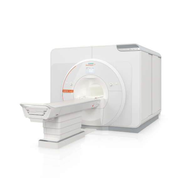
The Magnetom Terra.X, a new 7T MRI system, has received 510(k) clearance from the FDA. Manufactured by Siemens Healthineers, it’s a second-generation successor to the Magnetom Terra and offers several enhancements for 7T imaging. Key features include an eight-channel parallel transmit architecture for clinical use, deep learning image reconstruction optimized for 7T, improved diffusion imaging with a high-performance gradient system, and accelerated image acquisition enabling high-resolution brain and knee exams in under 20 minutes. Siemens Healthineers sees this as a significant step in providing better patient care, particularly in neurological and knee imaging. Additionally, the FDA clearance allows existing Magnetom Terra systems to be upgraded to the Magnetom Terra.X.

Image courtesy of Siemens Healthineers

SyMRI 3D for Brain Imaging

SyntheticMR has announced that its latest imaging solution, SyMRI 3D, has received FDA 510(k) clearance for clinical use in the United States. This clearance marks a significant advancement in quantitative MRI technology, offering exceptional resolution and accuracy in brain imaging. SyMRI 3D enables precise volumetric estimations of brain regions, known as parcellation, providing clinicians with deeper insights into brain structure and function. The enhanced resolution facilitates comprehensive lesion analysis, leading to more accurate medical condition assessments. This clearance empowers physicians to make more informed decisions in diagnosis and treatment planning, ultimately improving patient outcomes. SyntheticMR reaffirms its dedication to advancing medical imaging technology and providing innovative tools to enhance patient care through this milestone.


nCommand Lite for Remote Scanning

GE Healthcare has highlighted the FDA clearance of a solution by Ionic Health that enables technologists to remotely supervise patient scans. The system, called “nCommand Lite,” has been tested in Brazil for three years and is vendor-agnostic, allowing remote supervision across MRI, CT, and PET modalities. GE has secured exclusive distribution rights for nCommand in the U.S., aiming to address ongoing workforce shortages in healthcare. Rekha Ranganathan, GE’s chief digital officer for imaging, emphasized the company’s commitment to remote operations and increasing patient access to expert technologists. The system facilitates not only scanning supervision but also training, procedure assessment, and scanning parameter management. GE’s announcement coincides with growing interest in remote scanning, with the American College of Radiology advocating for permanent remote supervision of diagnostic tests. However, technologists have expressed reservations about managing imaging remotely, according to recent survey data from the American Society of Radiologic Technologists.


Sources:

Itnonline.com
Radiologybusiness.com
diagnosticimaging.com
openai.com


March AI News in Diagnostic Imaging

New Research by Harvard Medical School, MIT and Stanford on AI and Clinician Performance

The potential of medical artificial intelligence (AI) tools to enhance clinicians’ performance in interpreting medical images varies among individual clinicians, as highlighted by recent research led by Harvard Medical School, MIT, and Stanford. Published in Nature Medicine, the study underscores the intricate nature of human-AI interaction, which remains incompletely understood. While some radiologists benefit from AI assistance, others experience interference, affecting diagnostic accuracy.

The findings stress the necessity for personalized AI systems tailored to individual clinicians, emphasizing careful implementation to maximize benefits and minimize harm. Despite variations in AI’s impact, the results shouldn’t deter AI adoption but rather prompt a deeper understanding of human-AI dynamics to design approaches that enhance human performance.

To ensure effective integration of AI in clinical practice, collaboration between AI developers and clinicians is essential, alongside rigorous testing in real-world scenarios. Furthermore, efforts should focus on improving AI accuracy and training radiologists to discern AI inaccuracies, facilitating informed decision-making. Ultimately, understanding the complexities of machine-human interaction is pivotal for optimizing patient care through AI integration in radiology.

radiologist
A radiologist examines an x-ray

AI and Workflows

New research highlights a novel reporting workflow that automatically incorporates artificial intelligence (AI) findings into structured radiology reports, streamlining physicians’ tasks and saving valuable time. German experts shared their experience with the “AI to SR pipeline,” which integrates a commercially available AI tool for chest X-ray pathology detection and localization into structured report templates.

In evaluations conducted at University Medical Center Mainz, expert radiologists found that reports generated using the AI to SR pipeline were faster compared to free-text reporting and conventional structured reporting. Additionally, subjective quality assessments indicated higher ratings for reports created with the pipeline.

In the hospital’s clinical routine, chest X-ray images are sent to the picture archiving and communication system, then automatically forwarded to the AI tool for analysis. The results are output in a DICOM structured reporting format, taking approximately five minutes from image acquisition to final reporting. Radiologists were able to create chest X-ray reports significantly faster with the pipeline compared to free-text and conventional structured reporting, while also rating the AI-generated reports more favorably.

The authors suggest that this AI-driven reporting pipeline offers standardized, time-efficient, and high-quality reporting for chest X-rays, potentially enhancing AI integration into daily clinical practice and maximizing its benefits.


Sources:

Medicalxpress.com
Radiologybusiness.com
Openai.com