Vesta Advances Its Radiologist-Led AI Roadmap

Artificial intelligence is moving rapidly across healthcare, but successful adoption in radiology requires more than adding another technology platform. It requires careful clinical evaluation, thoughtful workflow integration, ongoing quality oversight, and a clear understanding of how the technology supports radiologists.

During the second quarter of 2026, Vesta Teleradiology continued making meaningful progress on the first phase of its AI Innovation Roadmap.

This phase focuses on introducing a limited number of clinically validated AI tools in high-impact areas of radiology. The emphasis remains on quality, patient safety, workflow efficiency, and the physician-led oversight healthcare organizations expect from Vesta.

A Focused Approach to AI-Assisted Teleradiology

Vesta’s approach begins with a practical question: Where can AI provide meaningful assistance within the existing radiology workflow?

Vesta began with select imaging studies where AI-assisted support may help surface time-sensitive findings and improve worklist prioritization. Current areas of support include:

  • Non-contrast CT brain studies
  • Chest X-rays
  • Musculoskeletal X-rays with fracture support

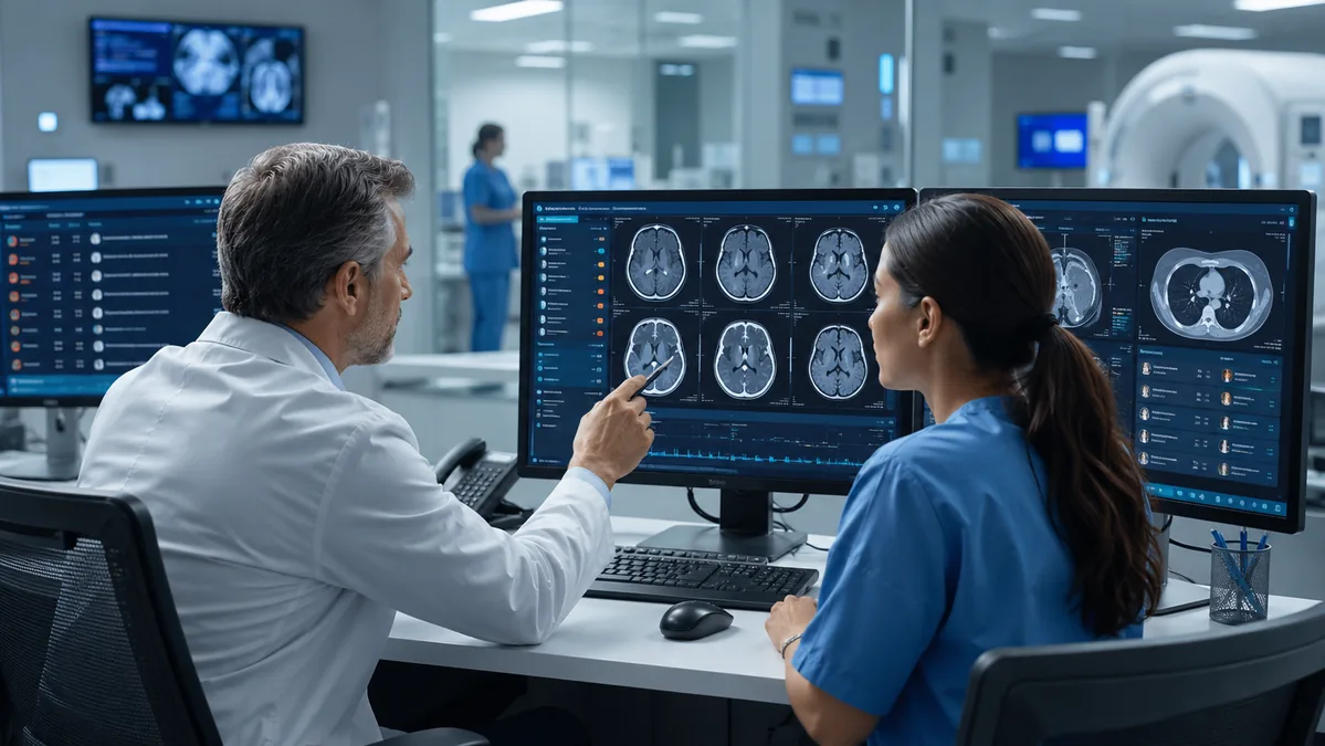
These tools provide advisory information within the reading environment. Each study continues to be reviewed, interpreted, and finalized by a U.S. board-certified Vesta radiologist.

Radiologist-led by design

The radiologist remains responsible for every interpretation and final report.

Supporting Earlier Prioritization of Urgent Studies

One of the most practical applications of imaging AI involves prioritization.

Radiology departments may receive many studies simultaneously, particularly during high-volume periods, overnight shifts, weekends, and unexpected volume surges. AI-assisted tools can help identify select studies that may contain urgent findings and bring them to the radiologist’s attention sooner.

The technology provides another source of information that can help radiologists organize worklists and focus attention where it may be needed first. The radiologist continues to determine the diagnosis and apply clinical judgment.

During Q2, Vesta concentrated on several operational priorities:

  • Earlier visibility into potentially critical findings
  • Greater consistency during high-volume reading periods
  • Faster prioritization of select urgent studies
  • Improved radiologist workflow efficiency
  • Continued physician-led quality oversight
  • Maintaining diagnostic excellence while reducing avoidable delays

These areas reflect how Vesta evaluates innovation. The value of a tool depends on how effectively it supports the people using it and the healthcare organizations relying on the service.

AI That Fits the Existing Workflow

Technology adoption can become difficult when it introduces additional viewers, separate logins, disconnected worklists, or changes to report delivery.

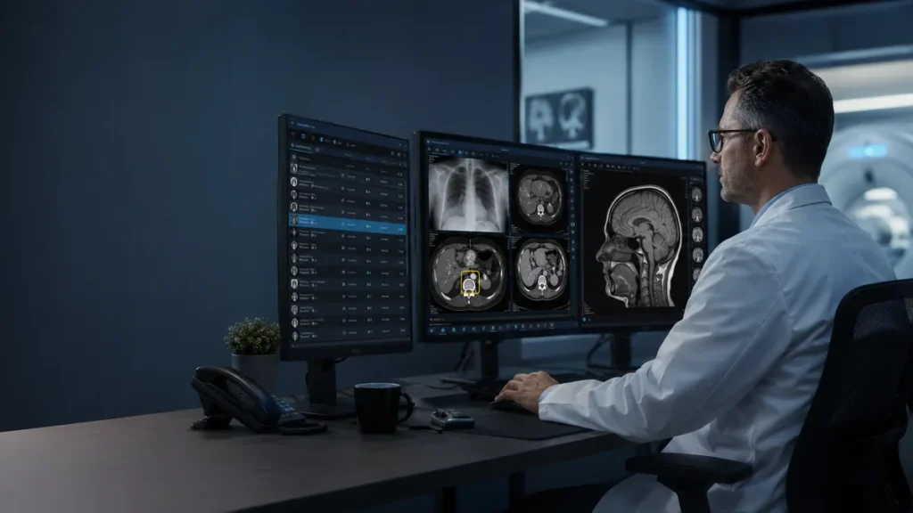
Vesta’s AI-assisted imaging support is designed to operate within the existing clinical reading environment. Select AI outputs are presented directly within the workflow used by Vesta radiologists.

For client facilities, there is no separate AI viewer to manage, no additional account to maintain, and no fundamental change to how reports are delivered.

This integrated approach helps Vesta introduce new capabilities while preserving the familiar processes healthcare teams already use.

Collaborating With Qure.ai

As one component of its broader AI Innovation Roadmap, Vesta is collaborating with Qure.ai, a healthcare AI company that develops medical imaging technologies.

Within Vesta’s current collaboration, Qure.ai supports select AI-assisted workflows involving non-contrast CT brain examinations and chest X-rays. These applications are designed to help identify and prioritize select imaging findings for radiologist review.
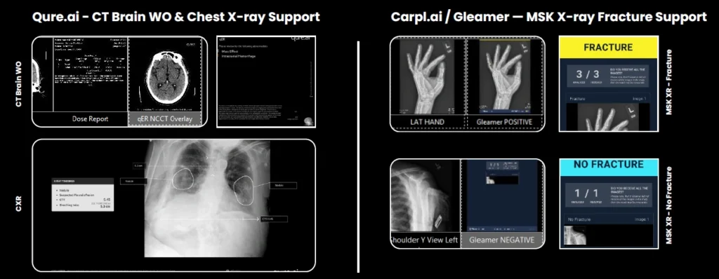
The collaboration supports Vesta’s broader effort to evaluate and implement clinically appropriate AI capabilities within a radiologist-led teleradiology environment. Qure.ai is one of multiple technology partners contributing to Vesta’s wider strategy for improving automation, workflow efficiency, prioritization, and quality.

AI outputs remain advisory. Vesta’s U.S. board-certified radiologists review the complete examination, apply clinical judgment, and remain responsible for every final interpretation and report.

The FDA maintains a public list of AI-enabled medical devices authorized for marketing in the United States, providing healthcare organizations with greater visibility into the evolving medical AI landscape.


Responsible Implementation Matters

Healthcare organizations are increasingly looking beyond whether an AI tool can perform a task. They also want to know how it will be introduced, monitored, governed, and incorporated into clinical decision-making.

In May 2026, the American College of Radiology approved its first practice parameter for imaging AI. The parameter addresses areas such as validation, training, monitoring, workflow integration, and ongoing quality assurance.

These principles align with Vesta’s measured approach.

Technology should support clinical judgment, strengthen workflow consistency, and help healthcare teams deliver dependable imaging services. Every implementation should also include appropriate oversight and continued evaluation.

Client Feedback Will Help Shape the Next Phase

As Vesta expands its AI-assisted imaging capabilities, feedback from hospitals, urgent care facilities, imaging centers, and other healthcare partners will help guide future priorities.

Vesta is especially interested in hearing about:

  • Report quality
  • Turnaround times
  • Clinical workflow
  • Communication
  • Additional AI-assisted capabilities that facilities would find valuable

This feedback helps Vesta focus its innovation roadmap on solutions that address real operational and clinical needs.

Moving Forward With Purpose

Vesta’s Q2 progress represents another step toward integrating AI responsibly into nationwide teleradiology services.

The combination of U.S. board-certified radiologists, advanced technology, dependable clinical processes, and client collaboration can support a stronger imaging workflow. Vesta will continue evaluating opportunities that improve prioritization, quality, efficiency, and access while preserving radiologist-led interpretation.

Your feedback matters

Share your experience with report quality, turnaround times, clinical workflow, communication, and the additional AI capabilities that could create value for your organization and patients.

Contact your Vesta Account Manager or connect with the Vesta team.

Sources

How AI-Assisted Teleradiology Supports Smarter Workflows

Healthcare organizations are managing increasing imaging demand while facing radiologist shortages, after-hours coverage needs, changing patient volumes, and continued pressure to deliver timely reports.

Artificial intelligence has emerged as one resource that may help radiology teams manage these demands more efficiently.

The greatest value often comes from using AI for focused workflow assistance. Within teleradiology, this can include helping prioritize select studies, surfacing potentially important findings, and supporting consistency during high-volume reading periods.

At Vesta Teleradiology, AI-assisted imaging support operates within a radiologist-led model. Technology provides advisory information, while U.S. board-certified radiologists review the complete study, apply clinical judgment, and finalize every report.

What Is AI-Assisted Teleradiology?

AI-assisted teleradiology combines remote radiology interpretation with software designed to analyze select medical images.

Depending on the type of study and authorized use of the technology, an AI tool may:

  • Highlight a region that may require closer review
  • Flag a study that could contain a time-sensitive finding
  • Help organize or prioritize the reading worklist
  • Provide an additional layer of workflow support
  • Assist with consistency during periods of high imaging volume

The output serves as advisory information for the radiologist.

The American College of Radiology’s imaging AI practice parameter emphasizes qualified physician involvement, appropriate validation, user training, performance monitoring, and quality assurance when AI is incorporated into clinical imaging.

How AI Can Support Worklist Prioritization

A radiologist’s worklist may contain studies from emergency departments, inpatient units, urgent care facilities, imaging centers, and outpatient practices.

During busy periods, several urgent and routine studies can arrive within a short time. Traditional priority designations remain important, and AI-assisted analysis may offer another signal that helps bring select studies to a radiologist’s attention.

For example, an AI-assisted tool may flag a non-contrast CT brain study containing features associated with a potentially urgent finding. The study can then receive earlier visibility within the workflow.

The radiologist evaluates the complete examination, reviews the patient’s available clinical information, considers other possible findings, and provides the final report.

This structure combines computational assistance with the context, experience, and judgment of a physician.

Current Areas of AI-Assisted Support at Vesta

Vesta has introduced AI-assisted support for select studies in three high-impact areas.

Non-Contrast CT Brain

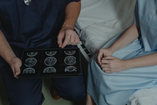
Within Vesta’s collaboration with Qure.ai, AI-assisted support is used for select non-contrast CT brain workflows, including the identification and prioritization of suspected intracranial hemorrhage.

The technology provides advisory information that may help bring a potentially urgent examination to earlier attention. The Vesta radiologist reviews the complete study, evaluates all relevant and incidental findings, and issues the final report.

Qure.ai’s FDA-cleared head CT technology includes applications involving intracranial bleeding, mass effect, midline shift, and cranial fractures. Vesta’s specific implementation should only describe the capabilities that are currently approved and active within Vesta’s workflow.


Chest X-Ray

Chest X-rays represent a significant portion of imaging volume across hospitals, urgent care centers, physician offices, and outpatient imaging facilities.

AI-assisted analysis can help surface select chest findings for radiologist review and support prioritization within a busy worklist.

Within Vesta’s collaboration with Qure.ai, chest X-ray support includes select findings such as pleural effusion, pneumonia, and other applicable chest abnormalities. These outputs are advisory and are reviewed alongside the complete examination by a Vesta radiologist.

Qure.ai has received FDA clearances for multiple chest X-ray applications designed to identify, highlight, categorize, or prioritize select findings for healthcare professionals. Its cleared applications include support involving pleural effusion and pneumothorax, and Qure.ai announced additional chest X-ray indications in 2026

The radiologist remains responsible for evaluating the complete image, considering available clinical information, and providing the final interpretation.


Musculoskeletal X-Ray Fracture Support

Suspected fractures are common in emergency departments, urgent care clinics, orthopedic practices, and outpatient imaging centers.

AI-assisted fracture support can highlight areas that may deserve additional attention. The radiologist reviews the complete image set and determines the findings included in the final interpretation.

Why Workflow Integration Matters

A capable AI tool can still create operational problems when it sits outside the normal reading process.

Separate viewers, additional passwords, new worklists, and disconnected alerts may increase complexity for radiologists and technical teams. They can also make adoption more difficult across a large or distributed radiology practice.

Vesta’s AI-assisted tools are embedded into the existing reading environment for select studies. This approach is designed to avoid unnecessary changes for client facilities.

Healthcare organizations continue receiving reports through their established delivery process. Vesta radiologists receive the available AI-assisted information within their clinical workflow.

The result is a more practical form of implementation that supports innovation while limiting disruption.

Radiologist-Led Interpretation Remains Essential

Medical images contain complex patterns that must be interpreted within the broader clinical picture.

AI can analyze designated features within an image, while the radiologist evaluates the entire examination. The radiologist also considers patient history, prior imaging, technical image quality, clinical indications, incidental findings, and possible alternative explanations.

This is why Vesta keeps physician oversight at the center of every AI-assisted case.

The Joint Commission and the Coalition for Health AI have also emphasized responsible governance, safety, transparency, monitoring, and accountability when healthcare organizations adopt AI.

A successful AI strategy should reinforce established clinical responsibility.

Questions Facilities Should Ask About Imaging AI

Hospitals and imaging organizations evaluating AI-assisted radiology services should consider several practical questions:

  1. Does the technology address a meaningful clinical or operational need?
  2. Has the tool been evaluated for the intended imaging use?
  3. How does the AI output appear within the radiologist’s workflow?
  4. Who remains responsible for the final interpretation?
  5. How is performance monitored after implementation?
  6. Will the facility need new viewers, logins, or reporting processes?
  7. How will clinicians and operational leaders provide feedback?
  8. What quality oversight processes are in place?

These questions help separate meaningful implementation from technology that adds complexity without solving a clear workflow problem.

Radiologist-Led AI Workflow SupportBuilding AI Around Real Healthcare Needs
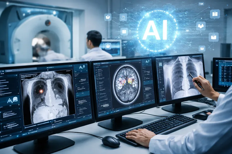
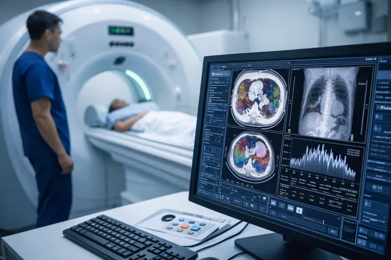
Vesta’s AI Innovation Roadmap is centered on practical use cases that can support quality, prioritization, consistency, and workflow efficiency.

The technology works alongside Vesta’s U.S. board-certified radiologists, 24/7 nationwide coverage, Nighthawk services, subspecialty reads, and customized overflow support. Vesta’s Joint Commission accreditation further reflects its commitment to quality-focused processes and dependable service.

As capabilities expand, client feedback will continue to guide Vesta’s priorities.

Facilities are encouraged to share their experiences with report quality, turnaround times, communication, clinical workflow, and other imaging areas where additional AI-assisted support could provide value.

By combining thoughtful technology adoption with experienced radiologists and strong clinical oversight, Vesta is building an AI-assisted teleradiology model designed for the realities of modern imaging operations.

Explore a practical, radiologist-led approach to AI

Learn how Vesta can support worklist prioritization and workflow efficiency while fitting into your existing imaging processes.

Discuss AI-assisted teleradiology with Vesta.

Sources

2025 Year-End Review: The Radiology & Diagnostic Imaging Headlines That Mattered

Key Takeaways

AI shifted from pilot projects to real workflow infrastructure—with more focus on governance, validation, and safety in daily operations.

Photon-counting CT moved closer to mainstream adoption, strengthening the business case for next-gen CT planning and protocol upgrades.

Reimbursement and policy pressure stayed intense, keeping budgeting, contracting, and service-line ROI under a microscope.

Prior authorization and imaging appropriateness remained major throughput challenges, impacting scheduling, patient access, and operational efficiency.

Cybersecurity and downtime readiness became core imaging priorities, as ransomware and system disruptions increasingly threaten continuity of interpretation.

Radiology didn’t have a single “one story” year—it had a “many small shifts became operational reality” year. In 2025, diagnostic imaging leaders saw AI move from pilots into production workflows, next-gen CT mature from promise to procurement conversations, reimbursement pressures intensify, and cybersecurity become inseparable from patient care. Meanwhile, staffing strain and consolidation continued to reshape how coverage is delivered.

Below is a practical wrap-up of the biggest breakout themes from 2025—and what they signal for 2026 planning.

1) AI moved from point solutions to regulated, workflow-embedded infrastructure

If 2023–2024 was the era of “AI can detect X,” 2025 was the era of “AI has to behave safely inside real clinical systems.” Regulatory claritya and operational expectations became the story as much as the algorithms themselves. RSNA’s coverage highlighted how the FDA has been articulating pathways and challenges for AI-enabled radiology devices—making governance, validation, monitoring, and safety considerations a board-level topic, not just an R&D conversation. Daily Bulletin

At the same time, 2025’s conversation broadened from task-specific tools to foundation models and multimodal systems (images + text) that could impact triage, reporting support, and quality workflows—while also raising new risks around bias, generalizability, and clinical readiness. DirJournal

Operational takeaway for imaging leaders: AI value in 2025 increasingly depended on integration (PACS/RIS/reporting), change management, and clear accountability—especially as adoption expands and expectations shift from novelty to measurable outcomes. The Washington Post

2) Photon-counting CT stepped into the “real adoption” phase

Photon-counting CT (PCCT) wasn’t framed as a future curiosity this year—it showed up as a maturing platform with expanding clinical evidence and increasing operational readiness. RSNA 2025 coverage specifically called out how PCCT is taking center stage as the next CT evolution. Applied Radiology

CT scan in progress with technologist beside scanner and diagnostic imaging workstation displaying CT and chest x-ray resultsAcross 2025 literature and trade coverage, the narrative tightened around what administrators care about: clearer visualization and characterization, potential dose efficiencies, and broader specialty applications as the evidence base grows. ScienceDirect

Operational takeaway: If you’re building 3–5 year replacement plans, 2025 made PCCT a serious line item conversation—especially for high-volume sites where incremental image quality and protocol optimization can compound into throughput, repeat-scan reduction, and clinician confidence.

3) Payment pressure stayed relentless—and policy debates sharpened

For many departments, 2025 felt like a year of doing more with less. The 2025 Medicare Physician Fee Schedule (MPFS) final rule remained a major planning input for imaging groups and hospital finance teams, with ACR publishing a detailed imaging-focused summary of provisions and QPP updates. American College of Radiology

At the end of the year, broader Medicare payment policy debates also made headlines—reinforcing that specialty payment and “efficiency” assumptions are likely to stay politically active topics heading into 2026. Axios

Operational takeaway: Contracting, service line budgeting, and modality ROI assumptions increasingly need “policy sensitivity” built in—especially for outpatient imaging strategy and subspecialty coverage models.

4) Utilization management: prior auth and “right test, right patient” stayed in focus

Utilization controls continued to evolve. CMS prior authorization programs for certain outpatient services remain part of the broader backdrop of controlling unnecessary volume. CMS And late-2025 headlines underscored expanding demonstrations tied to prior authorization in additional settings, which imaging leaders often experience downstream as scheduling friction, referral leakage, or delayed care. Kiplinger

On the imaging appropriateness front, the Medicare AUC program remains a major framework (even as implementation timelines and mechanisms continue to be debated). CMS In 2025, ACR also publicly backed federal legislation (the ROOT Act) positioned as a way to revitalize Medicare imaging appropriateness workflows. American College of Radiology

Operational takeaway: Expect “appropriateness” and “utilization proof” to keep rising as operational requirements—meaning your radiology operation will benefit from tighter ordering communication loops, smarter triage, and documentation hygiene.

5) Breast imaging compliance stayed operationally important—density language included

Breast density notification requirements became routine compliance work after enforcement of MQSA’s amended regulations began in 2024, and 2025 was about living with the operational realities: consistent report language, patient communication workflows, and inspection readiness. U.S. Food and Drug Administration

Notably, 2025 also saw attention on density reporting language options under MQSA—an example of how “small wording changes” can have major downstream effects in templates, patient letters, and audit processes. DenseBreast-info, Inc.

Operational takeaway: Standardization wins here—clear templates, audit trails, and staff training reduce risk while improving patient communication consistency.

6) Workforce strain and burnout remained the constant—and coverage models kept shifting

Radiology’s capacity crunch persisted in 2025. ACR continued to flag ongoing workforce shortages amid rising imaging demand, while national physician burnout tracking suggested improvement from prior peaks but still elevated rates that affect retention and coverage reliability.

Operational takeaway: The “coverage plan” is now a strategic asset. Departments that treat coverage as a system (subspecialty access, peak-demand flex, nights/weekends/holidays, overflow protection, and consistent turnaround governance) are better positioned for 2026.

7) Cybersecurity became inseparable from imaging operations

Cyber risk is no longer “IT’s problem”—it’s a continuity-of-care risk, especially for imaging organizations that depend on always-on networks and data flow. In 2025, radiology-specific alerts and incidents reinforced how real the threat landscape is, from FBI-linked warnings about ransomware targeting healthcare entities to major breach reporting involving large imaging providers. Radiology Business

cyber security risksOperational takeaway: Imaging leaders should be asking: Do we have downtime playbooks? How resilient is PACS access? How are third-party integrations governed? How do we preserve interpretation continuity if local systems are disrupted?

A 2026-ready checklist for imaging leaders

Here’s what 2025’s headlines suggest you prioritize next:

  • AI governance that’s operational, not theoretical: validation, monitoring, and workflow accountability.
  • Modern CT strategy: map where photon-counting CT could change protocols, dose strategy, and long-term equipment planning. Applied Radiology
  • Payment + policy resilience: bake MPFS sensitivity into budgets and service line forecasts.
  • Utilization friction planning: anticipate prior-auth expansion impacts on scheduling and throughput.
  • Compliance consistency in breast imaging: templates, audits, and MQSA-ready workflows.
  • Coverage strategy as a system: subspecialty access + surge/overflow + nights/weekends/holidays planning.
  • Cyber continuity: imaging downtime workflows and vendor access governance.

Where Vesta Teleradiology fits in a “do more with less” reality

For hospitals and imaging centers, one of the most immediate ways to de-risk 2026 is to strengthen coverage—especially when staffing shortages collide with growing imaging demand. Vesta Teleradiology supports facilities with 24/7/365 coverage (including nights, weekends, and holidays) and subspecialty radiology interpretations designed to integrate with your existing technology and workflows.

If you’re planning for 2026 coverage resilience—overflow protection, consistent turnaround times, or expanded subspecialty reads—you can request a quote or schedule a test run here.


Imaging the Individual — In the Trenches: AI, Personalization & Equity at RSNA 2025

RSNA’s 2025 theme, Imaging the Individual, isn’t just about futuristic science—it’s about doing the basics better for each patient, every day. The official Trending Topics preview highlights three threads cutting across subspecialties: AI you can deploy, personalized care you can operationalize, and equity you can measure. This guide translates those themes into practical checkpoints hospitals and imaging centers can use right now. RSNA

1) AI that graduates from pilot to practice

This year’s agenda emphasizes real outcomes over proofs of concept: reader-in-the-loop tools, bias monitoring, and governance. In breast imaging alone, RSNA previews spotlight external validation for image-only risk models and integration of MRI signals into multimodal AI—clear signals that “personalization” is landing in routine workflows. Bring vendor questions that force specifics: external validation cohorts, drift detection, and how metrics (TAT, recalls, rework) appear in your dashboard. RSNA

What to set up before RSNA: define 3–5 outcome metrics and insist every demo shows pre/post performance tied to those measures. Use QIBA concepts to push for standardized inputs/outputs so results are reproducible across scanners and sites. QIBA Wiki

2) Personalization that reaches the reading room

Personalization isn’t only radiogenomics. RSNA’s preview points to risk-stratified pathways you can actually run: e.g., image-only 5-year breast cancer risk at the point of screening to route patients into annual vs. short-interval follow-up or supplemental imaging (CEM/MRI). That pairs well with updated U.S. recommendations: screening beginning at age 40 for average-risk women, then adjusting based on risk and local policy. Build routing rules, templates, and letters now, so RSNA demos can plug into your plan.

Operational checklist:

  • Map risk thresholds → next steps (annual vs. short-interval, CEM/MRI).
  • Standardize templates so risk outputs appear consistently in reports and patient letters.
  • Decide who reviews outlier risk flags and how quickly (SLA).

3) Equity you can instrument—not just endorse

RSNA is foregrounding health equity, with sessions on encoding equity in AI and addressing access gaps for underserved communities. Equity becomes real when you can see it in your data: turnaround times by language, missed-appointment patterns by zip code, recall rates by screening site, and AI performance by subgroup. Build those slices into your analytics now; then ask vendors to show subgroup performance in their dashboards.

Practical moves:

  • Add demographic and language filters to your TAT and recall reports.
  • Require AI vendors to show calibration and error analysis by subgroup.
  • Stand up multilingual patient letter templates to support new screening starts at 40. USPSTF

4) CEM/MRI momentum: choose the lever that fits your service line

RSNA coverage calls out CEM as an increasingly practical adjunct—especially useful for dense-breast populations and diagnostic workups where capacity or cost limits MRI. The RACER trial reported higher accuracy and efficiency for CEM as the primary exam for recalled women vs. conventional imaging—evidence that can justify protocol changes and equipment planning. Meanwhile, MRI retains the sensitivity crown, with renewed attention on background parenchymal enhancement (BPE) as a signal worth documenting consistently.


Action items:

  • Decide where CEM fits: diagnostic recall pathway, dense-breast supplemental strategy, or both.
  • Add BPE level to structured MRI reports and trend it during therapy response clinics.

5) Governance, not guesswork

If personalization is the “what,” governance is the “how.” Use QIBA ideas—claim definitions, acquisition standards, and profile adherence—to control variability across devices and shifts. Tie RSNA learnings to a written governance plan with three parts: 1) protocol book (who owns it, update cadence), 2) quality book (metrics, subgroup views), and 3) AI book (approval process, monitoring, rollback).

6) Where teleradiology extends your capacity

Personalization increases complexity at peaks (recalls, dense-breast seasons, MR backlogs). A teleradiology partner helps you keep individualized pathways moving: standardized templates, subspecialty over-reads, and after-hours coverage that adheres to your risk rules and equity metrics—so “Imaging the Individual” doesn’t stop at 5 p.m.

Headed to RSNA?


Visit Vesta at Booth 1346 (South Hall) to see how we make “Imaging the Individual” work in real clinics—then enter to win a 1-year Medality CME subscription. Don’t wait: email “RSNA CME Entry” to info@vestarad.com now for a reserved entry, and show your confirmation at the booth for a bonus entry.

Powering Quality and Efficiency Through AI

Elevating Radiology. Expanding Access. Enhancing Care.

Vesta Teleradiology is redefining radiology delivery by integrating artificial intelligence (AI) into our diagnostic and operational workflows – helping hospitals of every size achieve higher quality, faster turnaround, and greater consistency in patient care.

Through our newly launched partnerships with Qure.ai and Carpl.ai, Vesta is bringing the benefits of AI assisted imaging to both large health systems and rural or underserved communities across the nation. This innovation enhances the speed, accuracy, and accessibility of radiology services – ensuring clinical excellence reaches every patient, everywhere.

AI Partnerships Driving Clinical Quality and Efficiency

Vesta now integrates Qure.ai’s FDA cleared AI solutions directly into our reading workflow to support both CT and X-ray imaging. For CT Brain (Non-Contrast), the AI automatically detects intracranial hemorrhages, fractures, and mass effect to improve triage and accelerate emergency response times. For Chest X-rays, it identifies nodules, effusions, and acute pulmonary findings to strengthen diagnostic consistency and enable earlier intervention. These tools work as a co-pilot for radiologists – helping prioritize critical studies, standardize interpretations, and deliver higher-quality reports with precision and speed.

Vesta also leverages Carpl.ai’s enterprise grade AI platform for musculoskeletal (MSK) fracture detection, enabling faster identification of subtle skeletal injuries that are often missed under high volume workloads. This integration enhances both radiologist efficiency and patient safety by improving consistency, turnaround times, and workflow throughput.

Expanding AI Across Vesta’s Clinical and Operational Ecosystem

In addition to our partnerships with Qure.ai and Carpl.ai, Vesta continues to implement AI across the organization to enhance both clinical quality and operational efficiency. Through RadPair, Vesta improves dictation accuracy, peer review workflows, and reporting analytics for radiologists – driving consistency and precision across the reading process.

On the operations side, Vesta has developed and launched an AI based support platform that allows staff to instantly retrieve internal protocols, radiologist schedules, credentialing data, and study specialty details from a centralized location. These tools streamline communication, improve turnaround time, and strengthen coordination across departments – supporting faster, more efficient service for clients and radiologists alike.

AI with a Purpose: Clinical Quality Care for All

Vesta’s mission has always been clear – to combine technology, compassion, and clinical excellence to improve access to quality radiology care. By implementing these AI partnerships and innovations, we’re ensuring faster turnaround for emergent and high acuity studies, improved diagnostic accuracy through validated AI support, greater access for rural and underserved hospitals, and consistent quality across every facility, 24/7/365.

These advancements reaffirm Vesta’s leadership as a trusted partner in AI driven radiology innovation, bringing cutting edge technology to the frontlines of patient care while optimizing the systems that support it.

About Vesta Teleradiology

Vesta Teleradiology is a Joint Commission-Accredited, 24/7/365 radiology provider serving hospitals, imaging centers, and healthcare systems nationwide. Our team of board-certified radiologists delivers timely, accurate, and secure interpretations – now further enhanced by AI technology to support faster decisions, higher quality, and better outcomes.

Interested in learning how Vesta’s AI powered radiology can support your hospital or health system?
Contact us at info@vestarad.com or visit www.vestarad.com/contact to schedule a demo or consultation.

Attribution:
Vesta Teleradiology integrates third party AI technologies through collaborations with Qure.ai, Carpl.ai, and RadPair. Descriptions of imaging and workflow capabilities in this publication are based on publicly available clinical use cases and are provided for informational purposes only. All content and messaging on this page are original to Vesta Teleradiology.

FDA’s 2025 AI Draft Guidance: A Buyer’s Checklist for Imaging Leaders

In January 2025, the U.S. Food and Drug Administration released a draft guidance for AI-enabled medical devices that lays out expectations across the total product life cycle—design, validation, bias mitigation, transparency, documentation, and post-market performance monitoring. For imaging leaders, it’s a clear signal to tighten procurement criteria and operational guardrails before piloting AI in CT, MRI, mammo, ultrasound, or PET.

As teams lock in Q4 budgets and head into RSNA season, the FDA’s AI lifecycle draft (Jan 2025) and the now-final PCCP (Dec 2024) have reset what buyers should expect from AI in imaging—devices, software, and workflows. Vendors are updating claims and governance; this issue distills a practical buyer’s checklist—multisite validation with subgroup results, drift monitoring and version control, clear in-viewer transparency—and how pairing those tools with Vesta’s subspecialty coverage and QA turns promise into measurable gains across CT/MRI/US/mammography.

A practical buyer’s checklist

Use this when evaluating AI for your service lines:

  1. Intended use fit: Verify indications, inputs/outputs, and claims match your pathway and patient mix.
  2. Validation depth: Prefer multisite, diverse datasets; stratified results; pre-specified endpoints; documented data lineage and splits.
  3. Bias mitigation: Demand subgroup performance (sex, age, race/ethnicity when available), scanner/vendor variability analyses, and site-transfer testing.
  4. TPLC plan: Require drift monitoring, retraining triggers, versioning, and how updates are communicated.
  5. Human factors & transparency: Ensure limitations, failure modes, and interpretable outputs are presented in-viewer without slowing reads.
  6. Security & support: Patch cadence, vulnerability disclosure, SOC2/ISO posture, uptime SLAs, and rollback paths for version issues.
  7. Governance: Define metrics owners, review cadence, and thresholds to pause or roll back a model.

Implementation playbook: pilot → scale without disruption

Start with a 60–90 day pilot in one high-impact line (e.g., ED stroke CT or mammography triage) and lock in baselines: median TAT, positive/negative agreement, recall rate, PPV/NPV, and discrepancy rate. Set guardrails—when to auto-triage vs. force human review—and document escalation paths for model failures. Require case-level confidence and structured outputs your radiologists can verify quickly. Stand up a model governance huddle (modality lead, QA, IT security, and your teleradiology partner) that meets biweekly to review drift signals, subgroup performance, and near-misses. Bake in a rollback plan (version pinning) and a quiet-hours change window so updates don’t collide with peak volumes. As results stabilize, scale by cohort (e.g., expand to non-contrast head CT, then CTA) and keep training “micro-bursts” for techs/readers—short videos or checklists in-workflow. Tie vendor SLAs to uptime, support response, and clinical KPIs so the AI program stays accountable to operational value.

Where teleradiology fits

AI only delivers when it’s welded to coverage, quality, and speed. A teleradiology partner should provide:

  • 24/7 subspecialty + surge capacity: Vesta absorbs volume peaks so AI never becomes a bottleneck.
  • QA you can see: We benchmark pre/post-AI performance, add targeted second looks for edge cases, and feed variance data back to your team.
  • Standardized outputs: Structured reports that integrate model outputs with radiologist findings—no black-box surprises.
  • Smooth rollout: Pilot by service line (stroke CT, mammo triage, PE workups), then scale with tracked KPIs (TAT, PPV, recalls).
  • Interoperability & security: Seamless PACS/RIS/EMR integration with strict access controls, audit trails, and support for change-controlled updates.

Bottom line: Pairing AI with Vesta Teleradiology gives you round-the-clock subspecialty reads, measurable QA, and operational breathing room while you pilot and scale responsibly. If you’re mapping your AI roadmap under the FDA’s 2025 draft guidance, we’ll be your coverage and quality backbone—so your clinicians see faster answers and your patients see safer care. Visit vestarad.com to get started.


Q1 2025 AI Radiology Roundup: Smarter Screening, Streamlined Referrals, and Intelligent Ultrasound Innovations

The first quarter of 2025 has seen impressive strides in the integration of artificial intelligence across the radiology spectrum. From breast cancer screening and interventional radiology referrals to next-gen ultrasound systems, AI continues to redefine efficiency, accuracy, and clinical outcomes. Below, we highlight three major developments shaping the future of radiology.


  1. Large Language Models Streamline IR Procedure Requests—For Just Pennies

In a study published in the Journal of Vascular and Interventional Radiology, researchers at Duke University Medical Center demonstrated that large language models (LLMs) like GPT-4 can accurately and efficiently route interventional radiology (IR) procedure requests—at a cost of only $0.03 per request.

By training the model on structured rules based on real IR team schedules and procedures, the AI achieved 96.4% accuracy in routing “in-scope” requests and 76% accuracy for out-of-scope queries. The tool helps clinicians connect with the right provider faster, improving coverage efficiency while avoiding unnecessary procedure orders.

With its adaptability to different hospital systems and minimal setup requirements, this LLM-powered tool could soon become a scalable solution for streamlining IR consultations nationwide.

“This approach is highly adaptable… and does not depend on training a dedicated model,” said Dr. Brian P. Triana, lead author.


  1. AI Mammography Boosts Cancer Detection by 29% in Landmark MASAI Trial

A game-changing trial out of Sweden—Mammography Screening with Artificial Intelligence (MASAI)—has reinforced the clinical power of AI in breast cancer screening. Published in The Lancet Digital Health, the randomized study followed over 105,000 women and found that AI-assisted screening increased cancer detection rates by 29% and reduced radiologist workload by 44%.


The AI tool, Transpara, was especially effective in identifying small, invasive cancers and high-grade in situ cancers—without increasing false positives. Radiologists using Transpara received real-time lesion detection and risk scores, helping reduce both overcalls and overlooked cancers.

“AI-supported screening can significantly enhance early detection while optimizing the use of healthcare resources,” said Dr. Kristina Lång of Lund University.

These results underscore AI’s role not just as a support tool but as a potential standard in future breast cancer screening protocols.


  1. Samsung Unveils AI-Powered Ob/Gyn Ultrasound System for U.S. Market

Samsung Medison made waves at the Society for Maternal-Fetal Medicine (SMFM) 2025 with the launch of its new AI-enhanced ob/gyn ultrasound system, the Samsung Z20.

The Z20 features Live ViewAssist, a real-time deep learning tool designed to streamline advanced obstetrical exams. Its capabilities include automatic structure labeling, real-time image quality assessment, and AI-powered measurements—all aimed at improving diagnostic precision and reducing repetitive strain on clinicians.

Addressing challenges in imaging patients with high BMI and promoting ergonomic design, the Z20 represents a leap forward in both performance and provider wellness. Additionally, Samsung showcased Sonio, its cloud-based ultrasound reporting platform, marking a step toward more integrated, AI-driven workflows in women’s health.

From improving clinical throughput to enhancing diagnostic confidence, AI is becoming indispensable in radiology. As Q1 2025 wraps up, the message is clear: artificial intelligence is no longer a futuristic concept in imaging—it’s a present-day solution driving meaningful change.

Stay tuned as we continue to track these innovations and explore how AI will shape the next quarter in diagnostic imaging and beyond.


Top 5 Trends Shaping Radiology in 2025

Radiology is constantly evolving, with advancements and challenges shaping how providers deliver care. As we step into 2025, exciting developments in technology, workforce dynamics, patient engagement, and regulatory compliance are transforming the landscape. In this blog, we’ll dive into the top five trends to watch in radiology this year and explore how they’re influencing the future of the field.


  1. Artificial Intelligence (AI): Revolutionizing Radiology in 2025

AI continues to make waves in radiology, offering improved diagnostic accuracy and efficiency. In 2025, AI tools are more refined than ever, assisting radiologists with cancer detection, anomaly identification, and image interpretation. Advanced algorithms can now process vast amounts of imaging data faster than ever, reducing turnaround times and enhancing patient outcomes.

radiology trendsHowever, challenges remain, including concerns about transparency in AI decision-making and biases in data sets. These hurdles are gradually being addressed with stricter regulations and improved algorithm training. AI isn’t just a tool; it’s becoming a trusted collaborator in radiology practices worldwide.

Read more about AI advancements in radiology here.


  1. Shifts in Diagnostic Imaging: The Rise of Independent Facilities

The trend of moving diagnostic imaging services away from hospitals and into Independent Diagnostic Testing Facilities (IDTFs) continues to grow in 2025. Patients and providers increasingly favor IDTFs for their cost-effectiveness and accessibility.


These facilities are adopting cutting-edge imaging technology, enabling faster and more accurate diagnoses. For healthcare providers, this trend presents an opportunity to collaborate with IDTFs or expand their own outpatient imaging services to meet the rising demand.

Learn more about the rise of IDTFs here.


  1. Addressing Workforce Shortages in Radiology

Workforce challenges remain a key issue in 2025. The demand for radiologists continues to outpace supply, especially as imaging volumes grow due to an aging population and the increased use of advanced diagnostic techniques. These shortages are felt acutely during peak times like the holiday season or in underserved areas.

To mitigate these challenges, healthcare organizations are relying on teleradiology to bridge gaps, ensuring 24/7 coverage without overburdening onsite staff. In addition, many practices are adopting flexible work schedules and prioritizing workplace wellness to attract and retain talent in this competitive market.

Explore workforce challenges and solutions here.


  1. Patient-Centered Care Takes Center Stage

Patient engagement continues to be a major focus in radiology in 2025. Programs like the FDA’s Patient and Caregiver Connection are pushing for more transparency and collaboration in radiology services. These initiatives encourage providers to involve patients in their care by offering clear, timely explanations of imaging results and personalized care recommendations.

Additionally, new tools, such as mobile apps that allow patients to access their imaging records and reports, are empowering individuals to take control of their health. Radiology practices that adopt these technologies are seeing improved patient satisfaction and stronger provider-patient relationships.

Learn more about patient-centered care here.


  1. New Breast Density Legislation in Effect

2025 marks the implementation of new breast density notification laws in many states. These laws require radiologists to inform patients if they have dense breast tissue, which can make it more difficult to detect cancer during mammograms. Dense tissue can also increase the risk of breast cancer, making this information critical for patients and their healthcare providers.

mammogramRadiology practices are adapting to these regulations by enhancing their reporting systems and educating patients about the implications of breast density. This legislation empowers patients to make informed decisions about supplemental screening options, improving early detection and outcomes.

Read more about breast density legislation here.


Looking Forward: Radiology’s Bright Future in 2025

Radiology is more integral to healthcare than ever before, and 2025 promises to be a transformative year. From leveraging AI to addressing workforce shortages, radiology providers are finding innovative ways to enhance care delivery. As patient engagement grows and new regulations take effect, the field is evolving to meet the demands of modern medicine.


For healthcare facilities looking to stay ahead of these trends, partnering with a trusted teleradiology provider can make all the difference. At Vesta Teleradiology, we specialize in sourcing skilled radiologists for both remote and onsite roles. Whether you’re navigating staff shortages, expanding diagnostic capabilities, or seeking flexible coverage, our experienced team can help. Let us be your partner in delivering exceptional care in 2025 and beyond.


Explore how we can support your radiology needs today.

Sources:

apnews.com
stout.com
fda.gov
theimagingwire.com
wikipedia.org
Openai.com

February AI News in Radiology

Brain Tumor Spotted on PET Imaging

An AI algorithm named “JuST_BrainPET” identified a glioblastoma in a patient that had been missed by physicians. This finding, reported in the Journal of Nuclear Medicine, underscores the potential of AI-based decision support in diagnostic and treatment planning. The algorithm automatically segments metabolic tumor volume from healthy tissue on brain PET imaging. In a case study, it detected a lesion in the frontoparietal region, not identified by an expert, which progressed to a small tumor. The AI tool’s early detection could have influenced diagnostic and treatment decisions.


Using Eye-Tracking

Researchers in Lisbon, Portugal, have pioneered a method to enhance AI interpretability in radiology by integrating eye-tracking data into deep learning algorithms. This innovative approach, outlined in the European Journal of Radiology, aims to align AI systems more closely with human understanding, marking a significant leap towards more human-centered AI technologies in radiology. By leveraging eye-gaze data, the researchers sought to bridge the gap between human expertise and AI computational power, anticipating that AI models could learn from the nuanced patterns of image analysis observed by radiologists.


This integration promises AI models that prioritize image characteristics relevant for diagnosis, potentially reducing the disparity between AI decision-making processes and human radiologists’ diagnostic approaches. The potential benefits of this research are vast, potentially leading to AI systems that are not only more effective in identifying pathologies but also more understandable to radiologists, thus fostering trust in AI-assisted diagnostics and accelerating their adoption in healthcare.


Review Paper on AI and Cancer Detection

Professor Pegah Khosravi and her team of researchers explore how artificial intelligence (AI) can enhance anomaly detection in MRI scans to advance precision medicine. Their comprehensive review, published in the Journal of Magnetic Resonance Imaging, focuses on AI techniques like machine learning and deep learning, particularly in identifying tumors in the brain, lungs, breast, and prostate.

The authors discuss several AI strategies for improving tumor detection, including a holistic approach that integrates data from various imaging techniques such as MRI, CT scans, and PET scans, along with genomic information and patient histories. This approach not only enhances anomaly detection accuracy but also facilitates personalized treatments based on comprehensive patient profiles.

Furthermore, the paper explores the use of ensemble methods in AI, which combine different AI models’ strengths to improve anomaly detection. By leveraging these methods, a more thorough analysis of MRI data is ensured. The authors advocate for AI systems that are accurate and transparent in their decision-making processes, fostering trust among healthcare professionals. They also stress the importance of collaboration among researchers, clinicians, and policymakers to effectively implement AI in medical imaging, guiding future advancements in the field.


Sources:

Auntminnie.com
bnnbreaking.com
gc.cuny.edu
openai.com

Top Trends for Radiology and Imaging in 2023

There is no doubt that radiology and imaging will have a few challenges in 2023. With Medicare pay reductions, hospital closures, worker burnout, and a shortage of radiologists, the industry must be creative to continue to provide exceptional patient care.

The industry leaders remain optimistic about their services. There have been more technological advancements, and people are still dedicated to their patients and healthcare. Their efforts are winning combinations for radiology, and they are determined to provide good service despite the challenges.

The current trend for radiology in 2023 will be for more consolidation of radiology practices. Using technology, many urban and outlying hospitals with minimal staffing can transmit information to control centers with professional radiology staffing. The financial burden of training imaging technicians will be, at a minimum allowing the hospital staffing budgets to be used for hands-on patient care.

The Value of Radiology and Imaging

Radiology is a “value-based system” that can reduce medical costs and improve patient outcomes through early disease detection and diagnosis with imaging screening programs. The medical profession, insurance companies, and hospitals recognize this value but struggle with ways to balance their budgets while providing patient care.

Investing in reorganizing the radiology departments has put much of the spendable medical funds to constructive use. The most significant expense for 2023 will be the expansion of artificial intelligence and equipment needed to provide the services.

2023 Equipment, Artificial Intelligence, and Education in Radiology

In November 2022, the Radiological Society of North America (RSNA) held its expo in Chicago, Illinois. Over 900 displays on the expo floor showed just a glimpse of the exciting future of radiology and imaging.

Featured equipment at the expo included MRI contrast that uses 50% less gadolinium, CT scanners with improved image resolution–and less noise, X-ray-based imaging systems, and CT scan with superior structural supports and better equipment warranties.

ct scan
CT Scanners Will See Improvements

Research and development have also made great strides in new X-ray lung airflow imaging technologies. Also, innovative breast imaging technologies that use ultrasound and elastography may reduce the need for some biopsies.

The FDA has now cleared hundreds of artificial intelligence algorithms that will be able to help radiologists with their workflow. These algorithms are applications that can help lessen worker burnout. Networks can integrate these applications, which allows organizations to work together, distributing workload and expertise efficiently.

Artificial intelligence spans many areas of radiology and imaging. Some of the applications not only improve the productivity of radiologists, but AI tools can improve imaging quality to detect disease and even embolisms or hemorrhages.

Artificial intelligence is making strides in education for radiologists and technicians. Research and Innovation are essential to the integrity of the practice, and artificial intelligence is helping to fulfill the academic needs of the staff.

radiology trends
AI in radiology

More to Come

Every year technology advances and enhances healthcare workflows and processes. Most hospitals and clinics still in operation have gone digital, allowing them to operate with advanced equipment and programs designed to provide exceptional patient care.

Radiologists and technicians are still in demand even if the systems incorporate more use of technology. Any radiology shortages can be remedied with the help of established and tech-forward teleradiology companies like Vesta Teleradiology. Education in the radiology and imaging field will need to keep up with the changes to make the technicians better while improving patient diagnoses and patient care