The Latest in Brain Imaging News

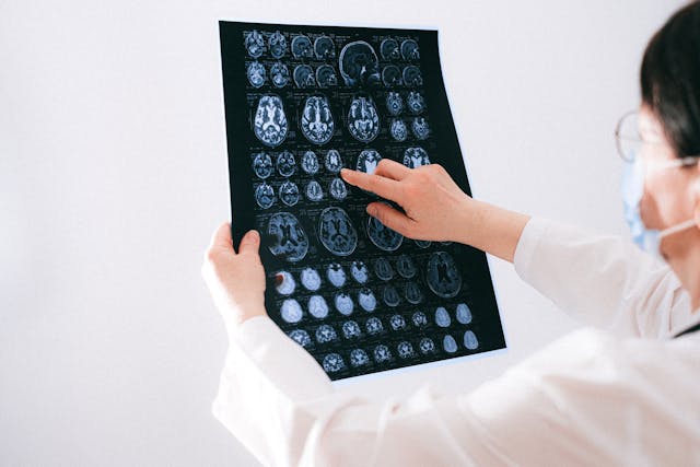
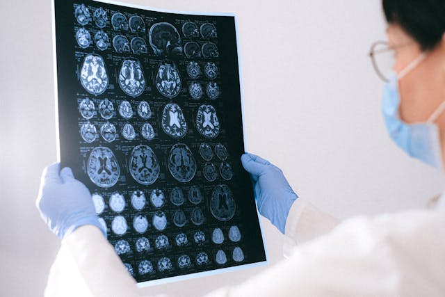
In recent years, awareness surrounding brain injuries has steadily risen, prompting significant strides in diagnostic technologies and treatment modalities. As we delve into the latest developments in this critical area of healthcare, it becomes increasingly apparent that advancements in medical imaging, particularly in the realm of neurological disorders, are poised to revolutionize the landscape of brain injury diagnosis and management.


AI-based Quantitative Brain Imaging System

Philips and Synthetic MR have joined forces to advance the diagnosis of neurological disorders through cutting-edge quantitative brain imaging tools. Their collaboration introduces the Smart Quant Neuro 3D MRI software suite, combining Philips’ SmartSpeed image-reconstruction technology, the 3D SyntAc clinical application, and SyntheticMR’s SyMRI NEURO 3D software. This innovation employs AI to analyze brain tissues, enhancing the detection and analysis of conditions like multiple sclerosis, traumatic brain injuries, and dementia.

The rise of AI in diagnostic imaging, projected to reach $1.2bn by 2027, signifies a transformative shift in improving accuracy and patient outcomes. With the diagnostic imaging market expected to grow to $9.1bn by 2030, fueled by demand for early disease diagnosis and personalized medicine, this partnership underscores the crucial role of AI in enhancing medical imaging.

Read the press release here.


A New Way of Diagnosing Mild TBIs

Researchers have developed a novel brain imaging method to diagnose mild traumatic brain injuries (mTBIs), which are often missed by standard techniques like MRI. This method involves loading gadolinium, a common MRI contrast agent, into micropatches attached to immune cells called macrophages. These cells migrate to areas of brain inflammation caused by mTBIs, enabling MRI detection. The technique, called M-GLAMs, was successfully tested in mice and pigs, showing promise for accurately diagnosing mTBIs. It also allows imaging at lower gadolinium doses, potentially benefiting patients with kidney issues. While unable to pinpoint injury locations, M-GLAMs could aid in identifying and treating brain inflammation. The researchers aim to bring this technology to clinical trials, with support from grants and intellectual property protection.

Read the study here.

New Imaging Tech that Captures Neuronal Activity Across the Brain During Recovery

Researchers at Tufts University School of Medicine have developed a novel imaging technology to monitor neuronal activity throughout the entire brain during the initial weeks of recovery from traumatic brain injury (TBI). Their study, published in Cerebral Cortex, reveals that TBI can induce changes in brain function beyond the injury site. Using a combination of fluorescent sensors and electrodes, they observed altered connectivity patterns in mice post-injury, even in regions distant from the impact. Despite the mice’s ability to perform physical tasks normally, their brain activity during both exercise and rest differed significantly from healthy brains. This impaired ability to switch between states suggests underlying brain state dysfunction post-injury. The findings highlight the brain’s plasticity in response to injury and have potential clinical implications for understanding TBI impacts and tailoring treatments. The researchers aim to further investigate long-term neural activity changes post-recovery and explore the technology’s potential in predicting specific dysfunctions or long-term outcomes of TBI. 

Read the study here.


Sources:

Medicaldevice-network.com
Otd.harvard.edu
Scitechdaily.com
Openai.com


The Latest in Native American Health News: Healthcare Worker Challenges

Physician Shortages

The Indian Health Service (IHS) faces significant physician shortages, with a vacancy rate of 25% in 2018. To address this, the American Medical Association (AMA) recommends creating an office of academic affiliations to establish partnerships with medical schools and residency programs. Currently, the IHS lacks formalized connections with academic medical centers, unlike other federal health systems such as the Veterans Health Administration and the Military Health System. These partnerships could offer training opportunities and help attract physicians to underserved areas. The AMA also suggests raising physician compensation, modernizing facilities, and developing funding streams for rotations and learning opportunities. Additionally, the IHS should evaluate regulatory barriers and provide resources to support physicians serving American Indian, Alaska Native, and Native Hawaiian communities. Overall, the AMA is committed to addressing the physician shortage within the IHS to ensure access to healthcare for these populations.


Cortez Masto’s Legislation for Enhancing Recruitment Efforts

Representatives from the Reno-Sparks Indian Colony Tribal Health Center and the U.S. Department of Health and Human Services advocated for the approval of the IHS Workforce Parity Act before a Senate panel. This legislation, co-sponsored by Senators Catherine Cortez Masto and Markwayne Millen, aims to address healthcare worker recruitment and retention challenges at Indian Health Service (IHS) facilities.

The proposed act would enable part-time providers to access IHS scholarship and loan repayment programs, aligning them with similar programs like the National Health Service Corps (NHSC). This alignment would enhance recruitment efforts in provider-shortage areas, improving access to healthcare in tribal communities.

Testimonies revealed that IHS facilities face significant staffing shortages, with a national vacancy rate of 25%, which can escalate to 50% in rural and frontier tribal clinics in Nevada. The current full-time work requirement for accessing grant and loan repayment benefits acts as a barrier to recruitment and retention efforts.

Understaffing negatively impacts healthcare outcomes in tribal communities, exacerbating conditions such as diabetes, cirrhosis, chronic lung diseases, and behavioral health issues. Failure to address these challenges undermines the U.S. government’s trust responsibility to ensure the healthcare needs of Native communities are met, as outlined in legal agreements between First Nations and the federal government.

New Facilities in Arizona

In Arizona, three new health facilities have opened recently to improve healthcare access for Native American communities, with more projects in progress. Despite strides, Native Americans still face health disparities like diabetes and cardiovascular disease. The Navajo Nation, home to over 244,000 people, operates 12 primary care facilities under the Indian Health Service (IHS), crucial in an underserved area.

Arizona

The Supai Health Station, nestled in the Grand Canyon and reachable only by air, mule, or foot, offers expanded services like primary care and dental. Similarly, the Dilkon Medical Center in the Navajo Nation provides comprehensive healthcare, including in-patient beds and behavioral health support.

Scheduled for May 2024, Sage Memorial Hospital in Ganado will further strengthen healthcare, serving around 23,000 people. Despite progress, challenges persist, including a shortage of hospital beds and healthcare professionals. Recruitment incentives like loan repayment aim to attract Native American individuals to healthcare careers.

Future plans include constructing new facilities in Bodaway Gap, Arizona, and Gallup and Pueblo Pintado, New Mexico, to enhance healthcare access for Native American communities in the region.


Any healthcare facilities needing support in radiology can look to Vesta for accurate and timely interpretations, even for subspecialties. Please contact us to learn more about our 24/7/365 teleradiology services.


Sources:

Nevadacurrent.com
cronkitenews.azpbs.org
ama-assn.org
openai.com

February AI News in Radiology

Brain Tumor Spotted on PET Imaging

An AI algorithm named “JuST_BrainPET” identified a glioblastoma in a patient that had been missed by physicians. This finding, reported in the Journal of Nuclear Medicine, underscores the potential of AI-based decision support in diagnostic and treatment planning. The algorithm automatically segments metabolic tumor volume from healthy tissue on brain PET imaging. In a case study, it detected a lesion in the frontoparietal region, not identified by an expert, which progressed to a small tumor. The AI tool’s early detection could have influenced diagnostic and treatment decisions.


Using Eye-Tracking

Researchers in Lisbon, Portugal, have pioneered a method to enhance AI interpretability in radiology by integrating eye-tracking data into deep learning algorithms. This innovative approach, outlined in the European Journal of Radiology, aims to align AI systems more closely with human understanding, marking a significant leap towards more human-centered AI technologies in radiology. By leveraging eye-gaze data, the researchers sought to bridge the gap between human expertise and AI computational power, anticipating that AI models could learn from the nuanced patterns of image analysis observed by radiologists.


This integration promises AI models that prioritize image characteristics relevant for diagnosis, potentially reducing the disparity between AI decision-making processes and human radiologists’ diagnostic approaches. The potential benefits of this research are vast, potentially leading to AI systems that are not only more effective in identifying pathologies but also more understandable to radiologists, thus fostering trust in AI-assisted diagnostics and accelerating their adoption in healthcare.


Review Paper on AI and Cancer Detection

Professor Pegah Khosravi and her team of researchers explore how artificial intelligence (AI) can enhance anomaly detection in MRI scans to advance precision medicine. Their comprehensive review, published in the Journal of Magnetic Resonance Imaging, focuses on AI techniques like machine learning and deep learning, particularly in identifying tumors in the brain, lungs, breast, and prostate.

The authors discuss several AI strategies for improving tumor detection, including a holistic approach that integrates data from various imaging techniques such as MRI, CT scans, and PET scans, along with genomic information and patient histories. This approach not only enhances anomaly detection accuracy but also facilitates personalized treatments based on comprehensive patient profiles.

Furthermore, the paper explores the use of ensemble methods in AI, which combine different AI models’ strengths to improve anomaly detection. By leveraging these methods, a more thorough analysis of MRI data is ensured. The authors advocate for AI systems that are accurate and transparent in their decision-making processes, fostering trust among healthcare professionals. They also stress the importance of collaboration among researchers, clinicians, and policymakers to effectively implement AI in medical imaging, guiding future advancements in the field.


Sources:

Auntminnie.com
bnnbreaking.com
gc.cuny.edu
openai.com

How to Pick the Best Teleradiology Company

As the demand for healthcare services continues to surge and the shortage of healthcare workers persists, particularly in specialized fields, such as radiology, hospitals and healthcare centers find themselves facing the challenge of ensuring timely and accurate interpretations of medical imaging studies. The critical role of radiologists in diagnosing illnesses and guiding treatment decisions underscores the urgency of addressing this shortage. In response, many institutions are turning to teleradiology companies to bridge the gap and provide remote interpretation services. However, selecting the right teleradiology company is paramount to ensure high-quality patient care and seamless integration into existing workflows. In this discussion, we will explore the criteria for choosing a reputable teleradiology company, considering factors such as expertise, technology infrastructure, turnaround time, and adherence to regulatory standards. By making informed decisions in this regard, healthcare facilities can optimize their radiology services and meet the needs of patients efficiently.

remote radiology company

Checklist for Choosing a Teleradiology Partner

Before selecting a teleradiology company, healthcare providers should consider several key factors to ensure they choose a partner that meets their needs and maintains high standards of service. Here are some important considerations:

  1. Quality and Expertise: Assess the qualifications and experience of the radiologists employed by the teleradiology company. Look for board-certified radiologists with expertise in relevant subspecialties. A recent survey of 2,749 radiologists from 108 countries reveals that while they read across almost five subspecialties daily, many lack confidence in certain areas. About 40% accept studies across all specialties, but less than half feel “very confident” in their current subspecialty, so it is vital to ensure the radiologists you work with have expertise in what you require.
  2. Technology and Infrastructure: Evaluate the teleradiology company’s technology infrastructure, including the software used for image transmission and reporting. Compatibility with existing systems and the ability to securely transmit images while maintaining patient privacy are crucial considerations.
  3. Turnaround Time: Timeliness is critical in radiology reporting. Consider the teleradiology company’s turnaround time for providing interpretations. Ideally, they should offer rapid reporting to facilitate prompt patient care and treatment decisions.
  4. 24/7 Availability: Healthcare facilities may require radiology services round-the-clock. Ensure that the teleradiology company offers 24/7 coverage (like at Vesta Teleradiology) to accommodate emergencies and provide continuous support.
  5. Communication and Collaboration: Effective communication between the teleradiology company and the healthcare facility is essential. Evaluate the company’s communication protocols, including how they handle urgent findings and facilitate collaboration between radiologists and onsite clinicians.
  6. Regulatory Compliance: Verify that the teleradiology company complies with all relevant regulatory standards, such as HIPAA (Health Insurance Portability and Accountability Act) regulations for patient data protection. They should also adhere to industry standards for image quality and reporting accuracy.Regulatory compliance
  7. Scalability and Flexibility: Consider the scalability of the teleradiology service to accommodate fluctuations in imaging volumes. Additionally, assess their flexibility in tailoring services to meet the specific needs of your healthcare facility.
  8. Cost and Value: While cost is a factor, prioritize value over price alone. Evaluate the overall value proposition of the teleradiology company, considering factors such as quality, reliability, and the ability to improve patient outcomes.

By thoroughly evaluating these factors and conducting due diligence, healthcare providers can make an informed decision when choosing a teleradiology company, ultimately enhancing the quality and efficiency of radiology services within their organization.

Partnering with a Top US Teleradiology Company—Vesta

Vesta serves as your dependable ally in radiology, extending support to various subspecialties—whether you’re a busy urban hospital or a private practice. We ensure swift processing for both urgent and routine studies. Recognizing the value of your staff’s time and well-being, our teleradiology services enable them to maintain a healthier work-life balance by covering shifts during nights, weekends, and holidays. We can also accommodate any volumes so please reach out to us to learn more.


Sources:

hcinnovationgroup.com
Radiologybusiness.com
openai.com


Healthcare Services for Native Americans (IHS)

In the pursuit of equitable healthcare in the United States, it is imperative to address the unique needs and challenges faced by all populations, including Native American communities. Despite advancements in healthcare delivery, disparities persist, particularly among Indigenous peoples.

Research additionally indicates that the life expectancy of Native Americans falls short by 5.5 years compared to the national average. Like the general populace, prevalent causes of mortality within Native American communities encompass heart disease, cancer, and accidents. Nevertheless, Native Americans face more than a threefold increase in mortality from diabetes-related complications, are over six times more prone to succumb to alcoholism, and exhibit a mortality rate from liver diseases exceeding four times that of the general population.


Reasons for Improved Healthcare for Natives

Higher Rates of Chronic Diseases: Native Americans experience higher rates of chronic diseases such as diabetes, cardiovascular disease, and obesity compared to the general population. These health issues are often linked to socioeconomic factors, limited access to nutritious foods, and inadequate healthcare services.

Barriers to Healthcare Access: Many Native American communities are located in rural or remote areas with limited access to healthcare facilities. Additionally, cultural and language barriers may deter individuals from seeking medical care.

Cultural Sensitivity: Traditional Western healthcare systems may not always be culturally sensitive to the needs of Native American communities. Culturally tailored healthcare services can improve patient outcomes and foster trust between healthcare providers and patients.


Indian Health Services: Staff Physician Vacancies

The American Medical Association (AMA) acknowledges the severe physician shortage within the Indian Health Service (IHS), with a vacancy rate of 25% in 2018. They advocate for strategies to address this shortage and ensure that American Indians, Alaska Natives, and Native Hawaiians receive adequate healthcare. The AMA’s recommendations include raising physician compensation, modernizing IHS facilities, promoting educational opportunities at IHS facilities, and establishing partnerships with academic medical centers. They stress the importance of addressing regulatory and licensure barriers for physicians interested in serving these communities. Overall, the AMA is committed to long-term solutions to alleviate the physician shortage and improve healthcare access for Indigenous populations.

Teleradiology for Indian Health Services

As a premier teleradiology company, Vesta understands the critical importance of reliable and efficient diagnostic imaging services, especially in regions facing shortages. With our state-of-the-art technology and a team of highly skilled U.S. Board Certified radiologists, we are committed to bridging the gap by offering accurate and timely readings. Whether it’s X-rays, MRIs, mammograms, CT scans, or other subspecialty, our streamlined process ensures swift delivery of results without compromising on quality. By partnering with us, Indian Health Services can confidently meet the demands of patient care, ensuring every individual receives the prompt and precise diagnoses they deserve.


Sources:

Ncbi.nlm.nih.gov
ama-assn.org
Openai.com

Top Imaging News of 2023

As we bid adieu to the final moments of 2023, it’s a great time to reflect on advancements and studies that have redefined the world of imaging this year. In this article, we’ll delve into the hottest news and breakthroughs in imaging, highlighting the remarkable strides that have made the headlines.

Study Suggest that Cancer Death Risk From Low-Dose Radiation Is Underestimated

A recent study featured in the British Medical Journal unveils concerning associations between extended exposure to low-dose radiation, commonly experienced by nuclear industry workers, and amplified cancer-related mortality. Drawing insights from the International Nuclear Workers Study (INWORKS) encompassing data from over 300,000 workers, researchers discovered a stark reality: for each cumulative unit of radiation exposure, the risk of death from solid cancer surged by 52%. Even at the lowest cumulative doses, this risk doubled, challenging the assumption that low-dose exposures present less carcinogenic hazard. While the absolute risk remains small, these findings prompt reconsideration of safety limits for workers and call for further studies to confirm the accelerated risk of cancer with ionizing radiation exposure. The hope is that regulatory bodies will integrate these insights into revising protection standards for individuals exposed to low-dose radiation.

Long COVID

In a study published in Medical Hypotheses, a French group presented a theory regarding the brain fog experienced in long COVID, based on brain patterns identified in patient PET scans. They propose that inflammation triggered by COVID-19 disrupts astrocyte cells’ regulation of glutamate, impacting energy metabolism and leading to cognitive fatigue. The authors suggest targeting this malfunction with therapies focused on astrocytic glutamate regulation as a potential way to alleviate long-COVID neurological symptoms. They highlight the lack of mental clarity, difficulty concentrating, and cognitive strain characterizing long COVID, affecting up to 15% of patients after three months of the initial infection. This study builds on previous findings of hypometabolism patterns in long COVID patients’ brain images and explores cellular mechanisms, including links between glutamate dysregulation and cognitive fatigue from other studies. Drawing parallels with “chemo-fog” in cancer patients and cognitive impairment in Parkinson’s disease, the authors suggest therapeutic strategies targeting the identified brain patterns, citing examples from epilepsy treatments and a recent study using medication to improve cognitive function in long-COVID patients. However, the authors stress the need for further research, proposing PET imaging studies using specific markers to comprehend astrocyte function and glutamate regulation for a comprehensive understanding of long COVID’s underlying mechanisms.

chemo fog
Study on brain fog experienced in long COVID

MRIs and Past Cannabis Users

At the International Society for Magnetic Resonance in Medicine (ISMRM) annual meeting, New Zealand researchers presented findings on heavy cannabis use in adolescence to early adulthood and its correlation with brain structure differences in hippocampus and amygdala subregions. The study, led by medical physicist Rebecca Lee and colleagues from the University of Otago in Christchurch, indicated volumetric disparities in these brain regions among heavy cannabis users compared to non-using controls. Notably, past cannabis users showed smaller volumes in specific hippocampal and amygdala subregions. However, the research did not find detectable differences in cerebral blood flow or white-matter tract integrity related to cannabis use, suggesting potential transient brain changes or no long-term effect on these properties. The study, conducted using MRI techniques, emphasized the need for longitudinal studies to clarify the causation and long-term functional impacts of these structural brain changes associated with heavy cannabis use. Despite revealing structural brain changes linked to cannabis use, the study does not definitively establish a causal relationship between these changes and cannabis consumption. Further prospective longitudinal MRI studies are essential to elucidate causality in this context.

MRI study

All About AI

We’d be remiss to not mention how artificial intelligence has shaped the industry this year. Check out our previous articles highlighting the impact that ChatGPT and Bard have made in 2023.


Sources:

technologynetworks.com
auntminnie.com
Openai.com

A Look at 2023 and ChatGPT In Radiology

ChatGPT has quickly moved beyond its niche beginnings and become an integral part of everyday life. Its reach extends well past casual conversation, now penetrating various industries, notably the intricate world of radiology. As we close out 2023, we take a look at some headlines that show how far ChatGPT has advanced in the realm of diagnostic imaging.

Smart Enough to Pass Exam Questions

In two recent studies published in Radiology, researchers evaluated ChatGPT’s performance in answering radiology board exam questions. While the AI showed potential, it also demonstrated limitations affecting its reliability. ChatGPT, based on GPT-3.5, answered 69% of questions correctly, struggling more with higher-order thinking questions due to its lack of radiology-specific training.

A subsequent study with GPT-4 showcased improvement, answering 81% correctly and excelling in higher-order thinking questions. However, it still faced reliability concerns, answering some questions incorrectly and exhibiting occasional inaccuracies termed “hallucinations.”

Confident language was consistently used, even in incorrect responses, posing a risk, especially for novices who might not recognize inaccuracies.


Decision Making in Cancer Screening: Bard Vs ChatGPT

A study recently published in American Radiology compares ChatGPT-4 and Bard, two large language models, in aiding radiology decisions for breast, ovarian, colorectal, and lung cancer screenings. They tested various prompts, finding both models to perform well overall. ChatGPT-4 showed higher accuracy in certain scenarios, especially with ovarian cancer screening. However, Bard performed better with specific prompts for breast and colorectal cancer. Open-ended prompts improved both models’ performance, suggesting their potential use in unique clinical scenarios. The study acknowledged limitations in scoring subjectivity, limited scorers, and the focus on specific cancer screenings based on ACR guidelines.

bard AI
Can AI assist in diagnostic imaging?

Simplifying Readability of Reports

The study in European Radiology explores using ChatGPT and similar large language models to simplify radiology reports for easier patient comprehension. Researchers had ChatGPT translate complex reports into simpler language for patient understanding. Fifteen radiologists evaluated these simplified reports, finding them generally accurate and complete, yet also identified factual errors and potentially misleading information in a significant portion of the simplified reports. Despite these issues, the study highlights the potential for large language models to enhance patient-centered care in radiology and other medical fields, emphasizing the need for further adaptation and oversight to ensure accuracy and patient safety.


Sources:

Rsna.org
diagnosticimaging.com
Radiologybusiness.com
openai.com


How Does Teleradiology Actually Work?

In today’s rapidly evolving healthcare landscape, the prominence and necessity of teleradiology have surged to the forefront. With advancements in technology and the growing demand for efficient, timely, and accurate diagnostic services, especially in light of staffing shortages, teleradiology has emerged as a pivotal solution. Teleradiology not only addresses the increasing demand for imaging interpretations but also bridges geographical gaps, enabling swift access to specialized radiologists regardless of location. Its rising popularity stems from its ability to enhance healthcare delivery by offering remote interpretations, thereby improving patient outcomes and streamlining diagnostic processes in a progressively interconnected world.

Teleradiology operates by leveraging digital communication technologies to transmit medical images, such as X-rays, MRIs, CT scans, and ultrasounds, from one location to another for interpretation and diagnosis.

Here’s a breakdown of how teleradiology works in real time:

Image Capture: Medical images are taken at a healthcare facility using specialized imaging equipment, creating digital files.

Image Transmission: These digital images are securely transmitted over networks, often utilizing Picture Archiving and Communication Systems (PACS) or secure internet connections.

Remote Interpretation: Radiologists, often located at a different site or working remotely, receive these images. They access the images through specialized software, review them in real time, and provide interpretations, diagnoses, and reports.

Consultation and Reporting: Upon reviewing the images, radiologists generate detailed reports containing their findings, interpretations, and recommendations. They may also engage in consultations with healthcare providers or specialists as needed.

Communication: The reports and findings are transmitted back to the referring healthcare facility, where they become part of the patient’s medical record. This allows for prompt decision-making and treatment planning by the attending physicians.

how teleradiology works

Quality Control: Teleradiology services often have quality assurance measures in place to ensure accurate and timely interpretations, adhering to industry standards and protocols.


Throughout this process, encryption and secure transmission protocols are employed to safeguard patient data and comply with healthcare privacy regulations (such as HIPAA in the United States). The real-time nature of teleradiology facilitates quicker diagnoses, especially in emergency situations, and enables access to specialized expertise irrespective of geographical barriers.


Top Teleradiology Company: Vesta is Here for You 24/7/365

Vesta is here to be your supporting partner in radiology, even for subspecialties. We offer fast turnaround for both STAT and Routine studies. Your staff is important–they don’t always work around the clock and shouldn’t have to. With our teleradiology services, you can allow your staff a better work-life balance while we fill in those gaps whether it’s during night hours, weekends, holidays.


Sources:

Acr.org
openai.com


RSNA 2023 Annual Meeting Highlights and Special Exhibits

The RSNA Annual Meeting stands as a pivotal event in the realm of radiology, serving as a dynamic nexus where cutting-edge advancements, emerging trends, and transformative innovations converge. This annual gathering not only showcases the latest breakthroughs in medical imaging but also fosters a collaborative environment where radiology professionals from various subspecialties can engage with state-of-the-art technologies, exchange insights, and navigate the intricate landscape of healthcare’s future.

Trending topics for RSNA 2023

This year’s meeting will be held on November 26-30. It will feature diverse trends across radiology subspecialties, with a notable emphasis on AI and photon-counting CT applications. Theranostics and the use of large language models are also gaining popularity. The event will offer non-interpretive sessions focusing on skills development and addressing diversity, equity, and inclusion in healthcare. The RSNA Annual Meeting Learning Center will provide a comprehensive experience, including the latest research, CME opportunities, and insights for improving DEI and health equity. Attendees can access a quick-reference guide to navigate trending topics and recommended sessions by subspecialty.

Some notable plenary sessions include:

Leading Through Technology: Valuing Artificial and Human Intelligence led by Elizabeth S. Burnside, MD, MPH; The Future of Healthcare Delivery: Considerations for Patients and Providers led by Vin Gupta, MD and Understanding and Revitalizing the Radiology Workforce led by Jocelyn D. Chertoff, MD, MS.

Technical Exhibits

The RSNA 2023 Technical Exhibits promise an immersive experience at the forefront of medical imaging innovation, boasting nearly 700 leading manufacturers, suppliers, and technology developers. The event showcases the latest advancements in AI through the AI Showcase, featuring over 90 companies presenting AI solutions, software demonstrations, and product displays. Attendees can delve into the RSNA AI Theater to explore hot topics in AI, machine learning, and deep learning, with special recognition for winners of AI challenges. The 3D Printing & Mixed Reality Showcase in the North Hall offers networking opportunities in 3D medical printing, augmented reality, and virtual reality.


Learn more at RSNA.org

Sources:
itnonline.com
rsna.org
openai.com


The Future of Medical Imaging: Latest Augmented Reality Advancements

Medical imaging is crucial in diagnosing and treating various medical conditions. These technologies have transformed the medical field from X-rays to Magnetic Resonance Imaging (MRI) and Computed Tomography (CT) scans.

Augmented Reality (AR) is a technology that overlays digital data, such as images or sounds, onto real-world objects or locations. In the medical field, AR has become an essential tool in patient examinations, surgery, and therapy.

AR-assisted Surgery

One of the significant benefits of AR technology in medicine is its application in surgical procedures. With AR, surgeons can see 3D images of the patient’s internal organs, tissues, and blood vessels during an operation.

This technology has revolutionized how physicians perform surgeries, offering unparalleled precision, accuracy, and visual assistance. Surgeons can perform surgeries with minimal invasion, thus reducing patient recovery time.

Additionally, AR-assisted surgery can benefit doctors in training as they can learn surgical procedures through virtual reality simulations.

Image-guided Intervention and Therapy

Image-guided intervention and therapy is another area that has experienced significant advancements in AR technology. Medical professionals can use AR to display real-time images of the internal organs during a procedure, allowing for precise navigation and guidance.

For example, using AR technology in radiation therapy enables medical professionals to focus radiation beams precisely on the tumor and spare the surrounding healthy tissue.

Patient Education and Rehabilitation

As medical professionals seek to enhance patient education and rehabilitation, AR technology has proven helpful. Patients can use AR to view and understand their medical conditions better.

For instance, AR can help showcase the effects of a particular treatment plan and provide feedback on the treatment progress. AR can also be used to improve rehabilitation sessions, as patients can receive personalized therapy recommendations that suit their needs.

Medical Education and Training

The applications of AR technology in medicine are not limited to diagnosis and treatment. Medical education and training have also benefited tremendously through AR technology.

Through virtual reality simulations, medical students can explore the human body in 3D and get hands-on experience with various procedures. Additionally, trainees can learn how to use medical equipment through AR simulations without endangering patient’s lives.

vr training session

Remote Patient Monitoring

AR technology has the potential to transform remote patient monitoring. Patients can use AR technology to capture images and videos remotely and send them to medical professionals.

This approach saves time and resources, especially for people living in remote areas, enabling them to receive diagnoses and medical advice promptly.

The advancements in augmented reality technology have revolutionized medical imaging by transforming diagnostics and treatment procedures. AR technology has applications in surgery, image-guided interventions and therapy, patient education, medical education and training, and remote patient monitoring.

With continued research and development, AR technology can potentially transform healthcare significantly. The advancements in AR technology are exciting as they offer the possibility of more accurate, efficient, and cost-effective medical procedures, diagnosis, and treatment, and ultimately, improved patient outcomes.