
May marks National Stroke Awareness Month, a time dedicated to raising awareness about stroke prevention, recognition, and treatment. With strokes occurring approximately every 40 seconds in the U.S., timely diagnosis and intervention are paramount to improving patient outcomes
The Critical Window for Stroke Treatment
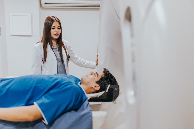
Strokes, whether ischemic or hemorrhagic, require immediate medical attention. The phrase “time is brain” underscores the urgency; delays in diagnosis and treatment can lead to irreversible brain damage or death. Rapid imaging—particularly CT scans and MRIs—is essential to distinguish between stroke types and determine appropriate interventions.
The Emergence of Emergency Teleradiology
Emergency teleradiology has significantly changed how facilities approach stroke diagnosis. By enabling radiologists to interpret imaging studies remotely and in real time, healthcare providers can expedite critical decision-making, even when on-site radiology staff is unavailable. This is particularly valuable in rural or underserved areas where specialist access may be limited.
One recent study reported impressive turnaround times within a global teleradiology stroke network: non-contrast CT scans were interpreted in an average of 9.97 minutes, CT angiograms in 20.57 minutes, and CT perfusion studies in 13.72 minutes (Thieme Connect).
Real-World Impact: Mobile Stroke Units and Teleradiology
Innovations like mobile stroke units (MSUs)—ambulances equipped with onboard CT scanners and teleradiology connections—are delivering care faster than ever. In one comparative study, patients evaluated via MSU had significantly better outcomes and higher thrombolysis rates than those transported via standard ambulance (Radiology Business).
Addressing Disparities in Stroke Care
Timely diagnosis and treatment for stroke are not consistent across regions. Teleradiology helps close these gaps by connecting clinicians in remote or resource-limited locations to expert radiologists quickly. For example, in Queensland, Australia, a regional hospital successfully administered clot-busting drugs after a telestroke consult enabled real-time CT interpretation and neurologist review (Courier Mail).
Vesta Teleradiology: Committed to Rapid Stroke Diagnosis
At Vesta Teleradiology, we recognize the critical importance of timely neuroimaging. Our services provide:
- 24/7/365 emergency teleradiology coverage for stroke-related imaging
- Radiologists with expertise in interpreting CT, CTA, and MRI for stroke diagnosis
- Seamless communication with ER teams for rapid turnaround and actionable reporting
By partnering with Vesta, healthcare providers can strengthen their stroke response systems—improving access, speed, and ultimately, patient outcomes.
Conclusion
As we observe National Stroke Awareness Month, it’s important to spotlight the advancements that are reshaping stroke care. Emergency teleradiology plays a vital role in helping facilities deliver fast, accurate diagnosis when every minute counts. With the right systems and partnerships in place, more lives can be saved—and more patients can recover fully.
Contact Vesta Teleradiology today to learn how our emergency teleradiology services support hospitals, stroke centers, and ER teams across the country.

 This challenge is magnified by a growing shortage of radiologists, particularly those specializing in breast imaging. A 2023 workforce survey from the
 This extension is part of CMS’s ongoing efforts to maintain flexibility in healthcare delivery, particularly in response to the challenges posed by the COVID-19 pandemic. Initially introduced in 2020, the virtual supervision policy has been extended multiple times, reflecting its effectiveness in enhancing access to care, especially in rural and underserved areas.
 Looking Ahead
 And for
 With Vesta Teleradiology’s flexible radiology solutions, healthcare providers can navigate reimbursement challenges while ensuring excellent patient care.
 How Vesta Teleradiology Provides Specialized Radiology Support

 Current Impact on Patients and Healthcare Systems
 Conclusion
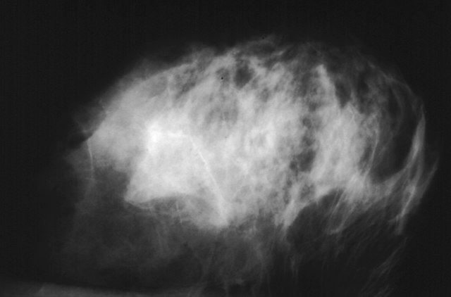
 Recent developments underscore the significant benefits of teleradiology for healthcare providers. A report from
  By focusing on early detection through advanced imaging and leveraging teleradiology, healthcare providers can significantly impact cancer outcomes, offering patients the best chance for successful treatment. National Cancer Prevention Month serves as a reminder of the strides we can make in cancer care through prevention and early detection.