Bone Health with BMD and DEXA Scans

Bone density is the ratio of skeletal weight (mass) to the volume or area of the bones. The heavier the bones, the stronger they will be. It affects physical activity levels, menopause, nerve signals, and more. A bone mineral density (BMD) scan compares your bone mass to an established norm and produces a score unique to you. This is different than a bone scan that looks for infections or cancer, or the presence of a fracture. A BMD scan helps determine the presence of osteopenia, osteoporosis, and the probability of future falls and fractures. A BMD score, combined with personal and family medical history, can help doctors get a complete picture of bone health.


bone density x-ray
bone scan


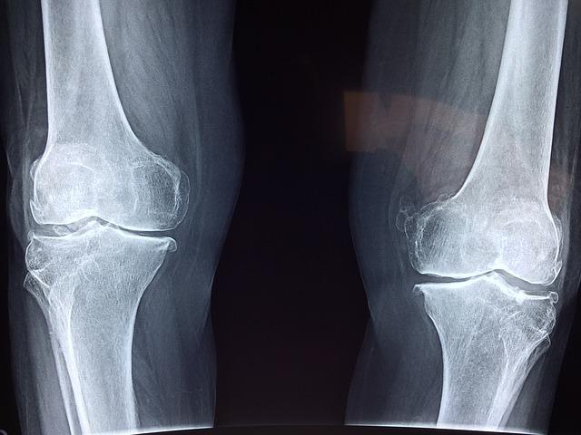
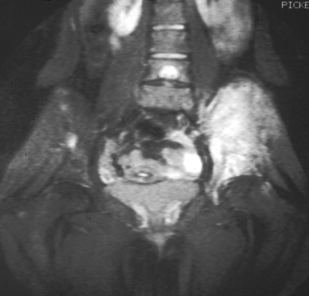
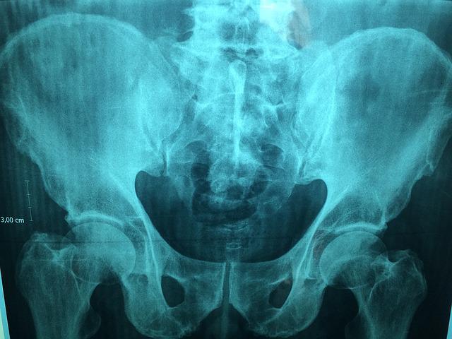
The types of diagnostic imaging used to measure bone density have included ultrasonography, CT and MRI images, and central dual-energy x-ray absorptiometry (DXA or DEXA) tests. In 1988, the dual-energy x-ray absorptiometry (DEXA) scan was approved by the Food and Drug Administration (FDA) for clinical use. Since then, DEXA has become the gold standard for measuring bone mineral density. Its scan of the large bones at the lumbar spine and hips is most used. Shorter scan times and minimal radiation exposure makes it safe. DEXA transmits photons at two energy levels for soft tissue and cortical bone and aids in the diagnosing of osteopenia, osteoporosis, and fracture risk assessment. It is inexpensive and the most accurate imaging modality for assessing bone mass density and health.

Doctors and radiologists use the BMD score to comprise a T-score or Z-score, which is a comparison to a reference group on a standard deviation scale. T-scores are given to adults and are determined by comparison to a young gender-matched group with peak bone mass. Z-scores are given to children and are determined by comparison to an age-matched group. These scores are used in risk fracture assessment, low bone mass or osteoporosis diagnosis, patient criteria for clinical trials, and management guidelines for osteoporosis. It is crucial that BMD measurements are correct, as well as differences in T-score and Z-score population groups. Accurate documentation is necessary for dependable results. Any variation used in this process can affect the actual T-score and Z-score. Improvements in calculation methods are currently ongoing.

Maintaining strong bones is essential. Daily calcium, vitamin D supplements, and weight-bearing exercises can help slow bone loss. In addition, patients should have their BMD checked regularly. Patients should also be counseled on safety measures like fall prevention.

patient and doctor
Doctors should counsel their patients on bone health

Top Teleradiology Company: Vesta

At Vesta Teleradiology, our U.S. Board Certified Radiologists are able to read and interpret DEXA scans. If you need supporting staff to cover nights, weekends and holidays, please reach out to us today: 1-877-55-VESTA.

Sarcoma & Bone Cancer Awareness Month: Newest Technology in Imaging

July is Bone Cancer and Sarcoma Awareness Month. According to the American Association for Cancer Research, in 2021, around 13,000 Americans will be diagnosed with soft tissue sarcoma, while 3,600 will be diagnosed with bone cancer. Luckily, the 5-year survival rate of both types of cancer is fairly high (65 for soft tissue and 66.8 for bone). In a comprehensive study from 1978 to 2004, the survival rate of these types of cancers has improved dramatically, thanks in part to improvements in imaging and early detection.

bone cancer
Sarcoma is a broad term for a group of cancers found in the bone.

Advances in Radiography

While x-ray technology has been used in a health context since 1899, many features surrounding it have changed. X-rays are now more defined with higher resolution, allowing doctors to zoom in to see smaller tumors and other issues. In fact, this past year, scientists at the University of Erlangen-Nuremberg have broken the record for x-ray resolution, down to under 10 nanometers. Furthermore, the process of obtaining can now be digitized, rather than having to use a darkroom to develop scans. These developments make radiology more accessible to patients and provide more immediate results, which helps bone cancer and sarcoma patients get diagnosed earlier when time is of the essence. There are multiple types of radiography used in diagnosing these types of cancer, and specific advances have emerged and evolved the diagnostic process for each type.


Bone Scans

During bone scans, a patient is injected with a tracer that allows oncologists to see abnormalities. Recent changes in bone scans allow doctors to quantify the metastasis of a bone from a scan. Bone scans can sometimes occur as a diagnostic tool before a CT scan, PET scan, or MRI.


CT Scans

CT scans can be ordered with “contrast,” where the patient swallows or takes an IV containing a dye. This can help doctors see certain organs more clearly. These scans can also help doctors see a cross-section of parts of the body and different perspectives of organs than they’d be able to see with a typical x-ray. The latest advancements in CT scans now offerlower radiation exposure by splitting the x-ray beams, making these procedures safer than ever.


PET Scans 

Positron emission tomography (PET) imaging is an emerging tool for bone cancer patients receiving radiation treatments. This tool is now being used increasingly by doctors to monitor tumors between treatments. These advanced scans allow more doctors to receive more information, including receptor expression and metabolism of a tumor. This information helps doctors to understand whether or not a particular type of treatment is working to shrink tumors and to help assess the appropriate dosage.


MRI Scans

Magnetic resonance imaging or MRI scans help oncologists to see the extent of bone and soft tissue cancers by taking cross-sections of affected organs and allowing them to be displayed at different angles. These are taken around a patient while a patient lies still inside a long tube. The most recent changes in MRI technologyhave occurred on the software side. One relevant change includes faster scan times, thereby reducing costs and discomfort for the patient. Furthermore, multiple layers of contrast can now be taken from a single scan, allowing doctors to highlight different issues easily.

bone cancer imaging
Magnetic resonance imaging (MRI) image of human skeleton. MRI diagnoses Ewing’s sarcoma of the right hip. Source: National Cancer Institute

While bone and soft tissue cancers affect many Americans, advancement in imaging tools helps provide clarity and hope for these patients.

Tech Innovation in Teleradiology

Vesta Teleradiology prides itself in being a top innovator in technology and was even awarded this recognition in 2020 by Technology Innovators. We truly understand the importance of keeping up with medical and technological advancements in radiology, imaging and health.


Come to us for teleradiology services for your outpatient imaging centers, nursing homes, hospitals, mobile imaging, wellness centers and urgent care facilities. Expect flexible yet speedy service, with quality interpretations and customizable reports. Please contact us for a quote.