How to Pick the Best Teleradiology Company

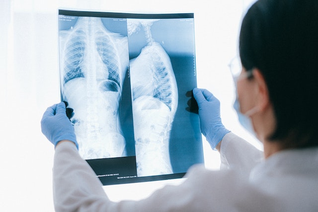
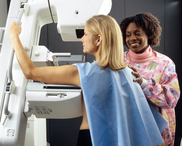
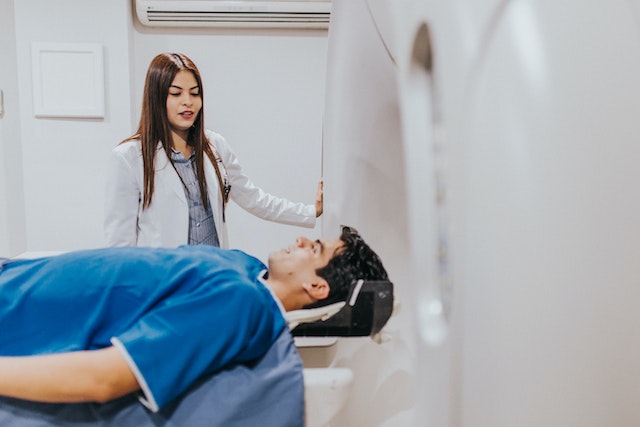
As the demand for healthcare services continues to surge and the shortage of healthcare workers persists, particularly in specialized fields, such as radiology, hospitals and healthcare centers find themselves facing the challenge of ensuring timely and accurate interpretations of medical imaging studies. The critical role of radiologists in diagnosing illnesses and guiding treatment decisions underscores the urgency of addressing this shortage. In response, many institutions are turning to teleradiology companies to bridge the gap and provide remote interpretation services. However, selecting the right teleradiology company is paramount to ensure high-quality patient care and seamless integration into existing workflows. In this discussion, we will explore the criteria for choosing a reputable teleradiology company, considering factors such as expertise, technology infrastructure, turnaround time, and adherence to regulatory standards. By making informed decisions in this regard, healthcare facilities can optimize their radiology services and meet the needs of patients efficiently.

remote radiology company
Rad tech and radiologist

Checklist for Choosing a Teleradiology Partner

Before selecting a teleradiology company, healthcare providers should consider several key factors to ensure they choose a partner that meets their needs and maintains high standards of service. Here are some important considerations:

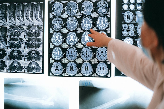
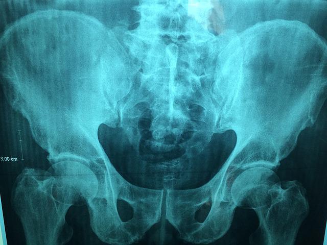
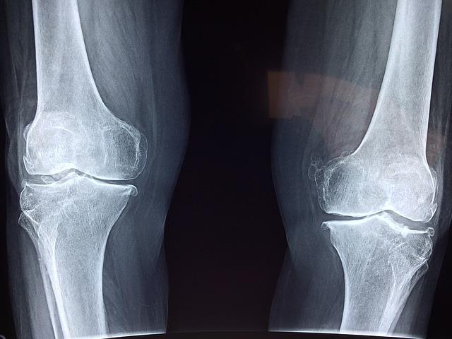
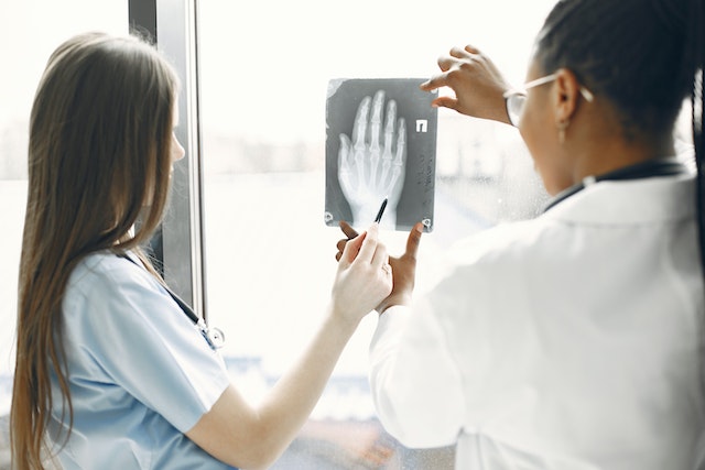
  1. Quality and Expertise: Assess the qualifications and experience of the radiologists employed by the teleradiology company. Look for board-certified radiologists with expertise in relevant subspecialties. A recent survey of 2,749 radiologists from 108 countries reveals that while they read across almost five subspecialties daily, many lack confidence in certain areas. About 40% accept studies across all specialties, but less than half feel “very confident” in their current subspecialty, so it is vital to ensure the radiologists you work with have expertise in what you require.
  2. Technology and Infrastructure: Evaluate the teleradiology company’s technology infrastructure, including the software used for image transmission and reporting. Compatibility with existing systems and the ability to securely transmit images while maintaining patient privacy are crucial considerations.
  3. Turnaround Time: Timeliness is critical in radiology reporting. Consider the teleradiology company’s turnaround time for providing interpretations. Ideally, they should offer rapid reporting to facilitate prompt patient care and treatment decisions.
  4. 24/7 Availability: Healthcare facilities may require radiology services round-the-clock. Ensure that the teleradiology company offers 24/7 coverage (like at Vesta Teleradiology) to accommodate emergencies and provide continuous support.
  5. Communication and Collaboration: Effective communication between the teleradiology company and the healthcare facility is essential. Evaluate the company’s communication protocols, including how they handle urgent findings and facilitate collaboration between radiologists and onsite clinicians.
  6. Regulatory Compliance: Verify that the teleradiology company complies with all relevant regulatory standards, such as HIPAA (Health Insurance Portability and Accountability Act) regulations for patient data protection. They should also adhere to industry standards for image quality and reporting accuracy.Regulatory compliance
  7. Scalability and Flexibility: Consider the scalability of the teleradiology service to accommodate fluctuations in imaging volumes. Additionally, assess their flexibility in tailoring services to meet the specific needs of your healthcare facility.
  8. Cost and Value: While cost is a factor, prioritize value over price alone. Evaluate the overall value proposition of the teleradiology company, considering factors such as quality, reliability, and the ability to improve patient outcomes.

By thoroughly evaluating these factors and conducting due diligence, healthcare providers can make an informed decision when choosing a teleradiology company, ultimately enhancing the quality and efficiency of radiology services within their organization.

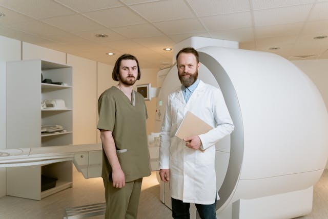
Partnering with a Top US Teleradiology Company—Vesta

Vesta serves as your dependable ally in radiology, extending support to various subspecialties—whether you’re a busy urban hospital or a private practice. We ensure swift processing for both urgent and routine studies. Recognizing the value of your staff’s time and well-being, our teleradiology services enable them to maintain a healthier work-life balance by covering shifts during nights, weekends, and holidays. We can also accommodate any volumes so please reach out to us to learn more.


Sources:

hcinnovationgroup.com
Radiologybusiness.com
openai.com


A Tale of Two Studies: Are Doctors Fulfilling too Many Imaging Requests?

A recent qualitative study aimed to investigate the factors influencing general practitioners’ (GPs) decisions to fulfill patient requests for imaging studies during clinical consultations. Ten GPs from private medical centers in Northwest Sydney were interviewed, and their perspectives were analyzed through content analysis. On the other side of the spectrum, we explore an American study that reveals the importance of encouraging more people to undergo screenings.

Patient Imaging Requests

The study revealed six key themes that emerged from the interviews regarding GPs fulfilling patient imaging requests:

Patient Expectations: Patients’ desires and expectations played a significant role in GPs’ decisions to fulfill imaging requests. Some patients expected certain tests due to their health concerns or beliefs.

‘Therapeutic Scans’: GPs recognized that some patients viewed imaging scans as a form of reassurance or therapeutic action. This influenced GPs to fulfill requests to alleviate patient anxiety.

“I find X-rays have become a therapeutic requirement, [and] that their [patients’] problem often dissipates once the request is given. (Female, 40)”

‘Impressive Labels’: The use of serious-sounding terms in imaging reports contributed to patients’ perception of the severity of their condition. This, in turn, led to increased requests for such scans.

Entitlement: Some patients exhibited an attitude of entitlement, expecting imaging scans without sufficient medical justification. GPs were more likely to decline such requests, especially during initial visits.

Defensive Medicine: GPs expressed concerns about potential legal repercussions if they refused imaging requests. Fear of litigation influenced GPs to fulfill requests to avoid legal issues.

‘New Patients’: GPs were cautious about fulfilling imaging requests from new patients, as they needed time to establish trust and assess the necessity of the requested scans.

The study concluded that GPs faced challenges in balancing their role as gatekeepers of imaging with patient expectations. Clear guidelines, patient education, and GP training were suggested as potential strategies to manage patient expectations and provide appropriate care. The study sheds light on the complexities of patient-GP interactions regarding imaging requests and highlights the need for evidence-based strategies to navigate these challenges. The study was conducted within the Australian Medicare system, where medical imaging is relatively accessible and affordable, and the findings contribute to understanding the dynamics of patient-GP interactions in this context.

Cancer Screenings Saves Millions of Years of Life

Meanwhile an American study conducted by teams from the University of Chicago, University of Michigan, and study sponsor Grail LLC estimates that Americans have gained an additional 12 million years of life due to preventive cancer screenings over the past 25 years, leading to an economic impact of around $6.5 trillion. These screenings, targeting early signs of breast, colon, cervical, and lung cancers among high-risk adults, emphasize the importance of encouraging more individuals to undergo recommended cancer checks and exploring novel screening methods. The study suggests that if all eligible individuals received current screening for these cancers, it could save an extra 3.3 million life-years and add $1.7 trillion in economic impact.

mammograms


The research utilized a mathematical model to assess the impact of cancer screenings endorsed by the U.S. Preventive Services Task Force (USPSTF). This federally authorized panel evaluates evidence for preventive medical services and provides recommendations using letter grades. Since 2010, screenings with “A” or “B” grades have been covered by most insurances without patient costs under the Affordable Care Act.

Although screenings for breast, colorectal, cervical, and lung cancers have saved millions of life-years, their full potential remains untapped. The majority of life-years saved and economic impact come from cervical cancer screening due to its early and consistent screening intervals for individuals aged 18-65. However, the study acknowledges that the availability of the HPV vaccine since 2006 could affect the future value of cervical cancer screening.

While there are validated screening tests lacking for many cancer types, decreased death rates from cancers with available screenings have contributed to the overall reduction in U.S. cancer deaths. To establish more broadly validated cancer screenings, further research is needed, potentially evaluated by the USPSTF. Novel screening tools like blood tests for multiple cancer types are in development, but AI-based tests have yet to undergo review by relevant bodies for efficacy and recommendations.


Sources:
onlinelibrary.wiley.com
michiganmedicine.org
Openai.com

Growth in Demand for Imaging Procedures Will Increase Need for Teleradiology

The healthcare market is experiencing a shift towards outpatient care, driven by reimbursement changes, pandemic effects, and patient preferences, particularly prominent in the U.S. Providers are diversifying into sub-specializations like neurology and oncology, raising the demand for advanced imaging like MRI and CT. This trend has led to increased utilization of outpatient imaging and teleradiology services.

By the NuMbers

Diagnostic imaging is becoming increasingly crucial in healthcare, with the market projected to reach $31.9bn in 2023 and grow at a 4.8% CAGR to $45.8bn in 2030. The rise is driven by chronic diseases, an aging population, and post-Covid-19 demand recovery. To meet this demand, companies are focusing on advanced and accessible technologies, such as handheld ultrasounds. About 1,949 imaging devices are in development, with 112 expected to gain approval in 2023.

imaging device

According to Fortune Business Insights, in 2022, the computed tomography (CT) segment held the largest market share due to a rise in CT scan procedures and higher average pricing. For instance, OECD data for 2021 showed 84.5 million CT scan procedures in the U.S., up by 15.8% from the previous year. The growing geriatric population has also contributed to increased demand for CT scans.

Key Players

Key players include GE Healthcare, Philips, Siemens Healthineers, and more. Challenges include high equipment costs and a shortage of skilled personnel, impacting accessibility and patient care quality. Opportunities arise from the growing demand for imaging services, especially for chronic diseases, and the development of new modalities like 3D mammography and MRI-guided focused ultrasound. Emerging economies like India, China, and Brazil are pivotal, driven by rising chronic diseases. Major players like Siemens Healthineers are expanding in these markets. Additionally, teaching hospitals are increasing demand for advanced imaging methods to enhance patient care.

Your Dedicated Radiology Partner: Vesta

Partnering with Vesta as your radiology partner ensures access to accurate and timely imaging interpretations and readings for subspecialties. Whether you are an outpatient imaging center or traditional hospital, our collaboration offers a seamless and efficient experience. Trust us to be your reliable radiology partner, empowering you with the insights and tools needed for improved healthcare outcomes.


radiology peer reviewSources:

Itnonline.com
Medicaldevices-network.com
fortunebusinessinsights.com
openai.com


How is Teleradiology and AI Impacting the Medical Industry Today?

Artificial Intelligence (AI) is revolutionizing the medical industry, transforming the way healthcare is delivered, diagnosed, and managed. With its ability to analyze vast amounts of data quickly and accurately, AI is reshaping various aspects of healthcare. From aiding in disease diagnosis to personalized treatment recommendations, AI is enhancing the precision and efficiency of medical practices. Moreover, AI-powered technologies are streamlining administrative tasks, optimizing resource allocation, and improving patient outcomes. As AI continues to advance, it holds immense potential to revolutionize healthcare delivery, foster medical innovations, and ultimately improve the quality of patient care on a global scale.

Teleradiology has had a profound impact on healthcare by enabling remote access to radiology expertise, bridging geographical barriers, and ensuring timely diagnoses. It has improved patient care by providing faster turnaround times, facilitating collaboration among radiologists, and increasing access to specialized interpretations, ultimately enhancing diagnostic accuracy and treatment outcomes. Going even further, a latest white paper from One Call describes how teleradiology and AI are helping reduce the strain of the radiology shortage.

artificial intelligence

Teleradiology and AI in Action

Medical imaging vendor, Nanox, is looking to address heath disparities and lack of access care with a new x-ray system which would be offered to countries in Africa, Asian and South American using a pay-per-scan model. The potential of combining cold cathode X-ray technology with teleradiology and artificial intelligence (AI) to enhance diagnostic capabilities and improve healthcare economics. Cold cathode X-ray systems offer advantages such as reduced energy consumption and improved image quality. When integrated with teleradiology, these systems can enable remote interpretation of X-rays, leading to faster diagnoses and improved patient care. Additionally, the use of AI algorithms in conjunction with cold cathode X-ray technology has the potential to enhance image analysis, automate certain tasks, and optimize resource allocation, offering cost-saving opportunities in healthcare settings.

diagnostic imaging
A teleradiologist examines a chest x-ray

There are plans to roll out AI-powered teleradiology by the “Screen for Life” program at the Primary Health Care Corporation in Qatar, aimed at early detection and prevention of cancer in the United Arab Emirates. The program plans to utilize AI algorithms to analyze radiology images, enhancing the accuracy and efficiency of cancer screening. The integration of AI in teleradiology will help automate image interpretation, expedite diagnoses, and reduce the workload on radiologists. The implementation of AI teleradiology in the “Screen for Life” program is expected to improve cancer detection rates, streamline healthcare processes, and ultimately save lives by identifying cancers at earlier stages.

Vesta Teleradiology

Looking to outsource your radiology interpretations using an expert Teleradiology company that is at the forefront of technology including AI?  Please reach out to Vesta to learn more. Vesta Teleradiology can accommodate any type of volume, large, medium and small.

Sources:

Radiologybusiness.com
menafn.com
openai.com
cdc.gov

The Role of a Pediatric Radiologist

In 2011, a large study examined the use of x-rays and other radiation imaging on children—they estimated that the average child would get more than seven radiation scans by the age of 18. No doubt, then, that the role of a pediatric radiologist is important in accurately diagnosing and treating diseases and conditions in children. Their specialized knowledge, expertise in radiation safety, and collaboration with other healthcare professionals contribute to the well-being and optimal care of pediatric patients. It’s estimated that only 3% of all radiologists in the US are pediatric radiologists, making this a small subspeciality. In this article, we will review the role and responsibilities of a pediatric radiologist.

A pediatric radiologist is a specialized medical doctor who focuses on using medical imaging techniques to diagnose and treat diseases and conditions in infants, children, and adolescents. They have expertise in interpreting and analyzing imaging studies specifically related to pediatric patients.

The role of a pediatric radiologist involves the following:


Interpreting Imaging Studies: Pediatric radiologists review and interpret various imaging studies such as X-rays, ultrasounds, computed tomography (CT) scans, magnetic resonance imaging (MRI), and nuclear medicine scans. They analyze the images to identify abnormalities or signs of disease. If you choose to work with Vesta Teleradiology, we have US board certified radiologists that handle subspecialty preliminary and final interpretations in pediatric imaging.

brain scans

Diagnosing Diseases and Conditions: By studying the imaging results, pediatric radiologists can diagnose a wide range of diseases and conditions that affect children. This can include congenital anomalies, infections, tumors, fractures, organ abnormalities, and other medical conditions.

Minimizing Radiation Exposure: Pediatric radiologists are particularly mindful of the potential risks associated with radiation exposure in children. They employ techniques and protocols that minimize the amount of radiation used during imaging procedures, ensuring the safety of young patients while still obtaining the necessary diagnostic information. For example, when appropriate, pediatric radiologists may use lead shields or aprons to protect sensitive areas of a child’s body that are not being imaged. This helps minimize unnecessary radiation exposure to healthy tissues. They also undergo specialized training in pediatric radiology, which includes radiation safety and dose optimization techniques.

Guiding Treatment Decisions: Pediatric radiologists collaborate with other healthcare professionals, including pediatricians, surgeons, and oncologists, to help guide treatment decisions. The imaging findings they provide play a crucial role in determining the most appropriate course of treatment.

Performing Image-Guided Procedures: In addition to interpreting images, pediatric radiologists may also perform image-guided procedures. These procedures can include biopsies, drainages, and minimally invasive interventions. They use imaging technology to guide their instruments and ensure precise placement.

Collaboration and Consultation: Pediatric radiologists work closely with other healthcare professionals, including referring physicians, to discuss imaging findings, provide recommendations, and answer any questions. They may also consult with pediatric subspecialists to provide further expertise in specific areas of pediatric radiology.

Overall, the role of a pediatric radiologist is vital in the accurate diagnosis and appropriate management of medical conditions affecting children. Their specialized knowledge and skills in pediatric imaging contribute to the well-being and treatment outcomes of young patients.


Sources:

https://pubmed.ncbi.nlm.nih.gov/16357371/

OpenAi.com

https://www.cbsnews.com/sacramento/news/average-child-may-get-7-radiation-scans-by-age-18/

Practice Management: How to Soothe Patient Concerns over Diagnostic Imaging

Being told that you need diagnostic imaging can be scary. Healthcare professionals deal with diagnostic imaging every day, so they may become immune to the concerns of patients.

Still, amidst patients’ fear of diagnostic imaging, healthcare professionals can set their minds at ease.

The main thing to remember is to simply treat your patients like humans. Slow down, treat them with kindness, and really listen to their concerns. Doing these simple things can go a long way in setting your patients’ minds at ease.

What Concerns Do Patients Have About Diagnostic Imaging?

One of the most common concerns amongst patients is the claustrophobia caused by MRI machines. Dealing with the tight space of an MRI machine can be very stressful for patients dealing with claustrophobia and anxiety.

Regarding X-rays, patients are often concerned about their radiation exposure. They fear that the imaging may put them at increased risk for cancer.

Treat Your Patient Like a Person

One of the most common complaints from patients is that they don’t feel like the person working with them is treating them with care and understanding.


talking to your patients
Address the patients concerns

Smile and say hello when you first enter the room. Something as small as a smile can instantly put a person’s mind at ease, especially in a clinical setting that is scary for many patients.

Always refer to the patient by their name. Never refer to them as “the patient” while they are in earshot. Doing so sounds cold, while using their name makes the interaction feel more personal.

Maintain eye contact with your patient. Don’t look at their chart so much that you forget to make them feel like they’re being heard. Eye contact can make them less anxious, especially if they’ve been waiting a long time or have specific fears.

Make sure you sit — standing over a seated patient can be intimidating.

Listen & Understand

Health Management emphasizes how important it is to listen to your patient and ask questions. Let them explain what is going on before you begin to speak. Don’t interrupt them. If they have something to add while you’re talking, allow them to interject and listen to what they say.

Giving your patient space to speak can clarify any confusion on both parts.

After they have finished, reiterate what they’ve said to ensure you understand their concerns. Doing so shows the patient that you are interested in helping them and have respect for their situation. It also ensures that both the patient and healthcare provider are on the same page with treatment going forward.

Also, remember to slow down. Many patients feel that healthcare providers are in a rush, that they’re wasting their time, and that the provider would rather be elsewhere. Patients deserve to feel that their time is valued. Slowing down also helps patients to feel like they are heard and understood.

Watch Your Tone

Patients often come to healthcare providers with sensitive information that can make them feel vulnerable. Health Management encourages professionals to speak to patients with a warm, calm tone of voice. It can do a lot to set their mind at ease. Speaking more slowly can do the same.

Educate Your Patients

Lawrence T. Dauer et al. say professionals should educate their patients. They should know exactly what’s happening during the imaging and what effects the imaging may have on their bodies.

For example, many patients fear how much radiation they’re getting. Explain that they are receiving very little radiation exposure.

Don’t lie to them. Professionals know that repeated exposure, a person’s age, and other factors may increase risk. Patients deserve to know that, but you can set their minds at ease by explaining their risks.

Patients should always have informed consent. They should know precisely what they’re going into and be okay with it. Consent is not just about getting the patient to sign a form. It ensures they are adequately educated about their procedure and entirely on board.

Be Clear

As a healthcare professional, you know a lot of medical and technical jargon that the patient likely does not. Speak straightforwardly, using familiar words. It is crucial that the patient understands what is going on with their treatment.

At the same time, Health Management warns you to be careful not to come across as patronizing. Although patients may not know medical jargon, it doesn’t mean they’re stupid.


Reducing MRI Claustrophobia Concerns

The University of Virginia says the main reason patients are fearful of MRI machines is because they lack understanding of them. Many professionals can ease patients’ minds by informing them that MRI machines are well-lit and open at both ends. They are not closed off and dark, as many patients fear.


patient anxiety
Understand some people may have claustrophobia


Professionals can teach their patients easy breathing and meditation techniques to help keep them calm during the procedure. Another option is to tell them to count to keep their minds busy or to go to their “happy place.”

Provide them with a towel or washcloth they can drape over their eyes so they can’t see what’s going on.

Provide patients with headphones and allow them to listen to their music of choice.

Talk to your patients — about anything — get their minds off the procedure.

Conclusion

Diagnostic imaging can be scary for patients, especially those dealing with anxiety. Medical professionals are critical in setting a patient’s mind at ease, and it’s not hard to do.

Slow down, treat your patients with kindness and respect, and ensure they are properly informed. These simple things can help calm your patients and help you build a lasting professional relationship.

What to Expect During a Breast Cancer Screening at an Imaging Center

The best way to detect breast cancer in its early stages is by mammograms–merely an X-ray of the breast. There is no financial strain because health insurance plans are required to cover the costs of mammograms every one or two years.

When you reach your forties, you should begin conversing with your medical providers about when to start and how often you should get a mammogram.

breast cancer awareness
Take care to get your screenings


Specialists usually recommend the procedure for women between 50 to 74 years of age at average risk of breast cancer and performed every one or two years depending on the woman’s risk factors.

How to Prepare for a Mammogram

When your healthcare provider suggests scheduling a mammogram for the first time, there are some considerations.

If possible, you should consider making your appointments at a nearby facility that specializes in mammograms. In doing this, you can have technicians who are very familiar with the process and accurately compare your mammograms yearly.

mammogram technology
Mammogram screening

If you need to change facilities, it’s best to arrange to have all your records sent to the new clinic for X-ray comparison. It is also an excellent idea to bring all the dates and locations of any previous breast procedures done.

Specialists advise that women avoid making mammogram appointments the week before their periods. Breasts are more swollen and sensitive during this time which can cause the mammogram procedure to be uncomfortable for them and may interfere with taking clear X-rays.

Technicians will advise that women wear pants or a skirt with a top that they can easily remove along with their bra. They also recommend that women do not use any deodorant, lotions, or powders that may show up as spots on the X-ray.

When You Arrive at the Imaging Center

Most technicians are very considerate and sensitive to what women experience during mammograms. They will ask you to undress above the waist and will give you a wrap to wear.

Many clinics have private dressing areas where you undress and wait for your technician to escort you into the X-ray room. There will only be the two of you in this area which is helpful to reduce any anxieties you may have.

You will stand in front of the mammogram machine, and the technician will position your body and breast for the most accurate X-rays. Your breast will rest on the device, and a plastic upper plate will lower and compress your breast.

The process takes 10 to 15 seconds for each body position, and there are usually only two or three X-rays taken per breast. The whole procedure only takes about 20 minutes.

Your Mammography Results

Mammography produces black-and-white digital images of your breast tissue that will be sent to a physician specializing in radiology to interpret. An imaging center could also refer to a teleradiology company to have a radiologist do the interpretation. Most clinics offer 2D mammograms for breast screening, but many facilities now offer 3D mammograms.

The radiologist will then report their findings to your healthcare provider. This process will take approximately ten days but could take longer. The radiology report will determine if you will need further X-rays, possible MRIs, or treatments. Physicians call back about 10 to 13% of women for abnormal findings, but most of these findings are not breast cancer.

Regular mammograms are an easy, safe way to screen for breast cancer. The most challenging part about the process may be remembering to make your annual appointment, and many women make their birthdays a reminder to make their yearly appointment.

What can be more celebratory than scheduling what may be a life-saving procedure for your health on the most important day of the year for you, your friends, and your loved ones? 

Bone Health with BMD and DEXA Scans

Bone density is the ratio of skeletal weight (mass) to the volume or area of the bones. The heavier the bones, the stronger they will be. It affects physical activity levels, menopause, nerve signals, and more. A bone mineral density (BMD) scan compares your bone mass to an established norm and produces a score unique to you. This is different than a bone scan that looks for infections or cancer, or the presence of a fracture. A BMD scan helps determine the presence of osteopenia, osteoporosis, and the probability of future falls and fractures. A BMD score, combined with personal and family medical history, can help doctors get a complete picture of bone health.


bone density x-ray
bone scan


The types of diagnostic imaging used to measure bone density have included ultrasonography, CT and MRI images, and central dual-energy x-ray absorptiometry (DXA or DEXA) tests. In 1988, the dual-energy x-ray absorptiometry (DEXA) scan was approved by the Food and Drug Administration (FDA) for clinical use. Since then, DEXA has become the gold standard for measuring bone mineral density. Its scan of the large bones at the lumbar spine and hips is most used. Shorter scan times and minimal radiation exposure makes it safe. DEXA transmits photons at two energy levels for soft tissue and cortical bone and aids in the diagnosing of osteopenia, osteoporosis, and fracture risk assessment. It is inexpensive and the most accurate imaging modality for assessing bone mass density and health.

Doctors and radiologists use the BMD score to comprise a T-score or Z-score, which is a comparison to a reference group on a standard deviation scale. T-scores are given to adults and are determined by comparison to a young gender-matched group with peak bone mass. Z-scores are given to children and are determined by comparison to an age-matched group. These scores are used in risk fracture assessment, low bone mass or osteoporosis diagnosis, patient criteria for clinical trials, and management guidelines for osteoporosis. It is crucial that BMD measurements are correct, as well as differences in T-score and Z-score population groups. Accurate documentation is necessary for dependable results. Any variation used in this process can affect the actual T-score and Z-score. Improvements in calculation methods are currently ongoing.

Maintaining strong bones is essential. Daily calcium, vitamin D supplements, and weight-bearing exercises can help slow bone loss. In addition, patients should have their BMD checked regularly. Patients should also be counseled on safety measures like fall prevention.

patient and doctor
Doctors should counsel their patients on bone health

Top Teleradiology Company: Vesta

At Vesta Teleradiology, our U.S. Board Certified Radiologists are able to read and interpret DEXA scans. If you need supporting staff to cover nights, weekends and holidays, please reach out to us today: 1-877-55-VESTA.

Benefits of Mobile Imaging for Outpatient Healthcare

Most of the healthcare provided by physicians involves some method of imaging. In the past, technicians have performed diagnostic services in the imaging departments of hospitals. Today, imaging services are brought to the patient with the help of mobile imaging.


Since there is such a high demand for x-rays, ultrasounds, MRI, CT scans, and EKGs, in-house hospital departments tend to be overwhelmed, and patients can have long wait times in crowded waiting rooms. Even getting an appointment for the imaging service can take a long time which delays treatment for the patient’s care.


With the emergence of mobile imaging, patients have more accessible, more efficient access to imaging services. With faster access to the patient, healthcare professionals can diagnose their patients more quickly and begin necessary treatments.

mobile radiology for assisted living
A man takes care of a resident in an assisted living facility

Senior Population and Imaging

The best example of the benefits of mobile imaging is with the senior population–many of whom reside in assisted living facilities and nursing homes. Persons over the age of 65 are a fragile population who need imaging services frequently.

The older age group has an increased fall rate, a higher rate of pneumonia and cancers, and compromised immune systems.  They need prompt diagnoses, but transportation to receive medical care is complicated.

The benefits are vast when mobile imaging can accommodate this population’s needs:

  • Overall, costs are reduced for the service because the facility doesn’t need to transport the patient to a hospital imaging department.
  • Understaffed facilities don’t need to assign an employee to accompany the patient for diagnostic imaging in a hospital.
  • There are fewer transfers between facility and hospital.
  • Mobile imaging services reduce anxiety in the elderly because transporting and waiting for imaging services accentuates fear in the undiagnosed.
  • There is a reduced need for hospitalizations and outpatient treatments of the patients because the assisted living facilities and nursing homes can provide prescribed care.
  • Mobile imaging can provide needed diagnostic information to the patient’s attending physician faster than an imaging department can, which expedites a treatment plan for the patient.
  • The patient can remain in familiar surroundings (and with people they know) while receiving diagnostic imaging services. This benefit is significant when the patient has difficulty understanding or processing information.

When people of any age are hurting, fearful, anxious, or lack understanding, their comfort is the most crucial factor in beneficial treatments. Mobile imaging comes to the patient and provides fast, efficient, accessible, and cost-effective diagnostics for the most fragile patients.

 Working with a Teleradiology Company

With Vesta Teleradiology, we work with mobile imaging centers and any health facility that provides this technology to their patients. We work with your workflow and integrate to your technology so sending and receiving scans is a breeze. Learn more about how we can help you now: call us at 1-877-55-VESTA.

teleradiology services
Vesta Teleradiology services include nighthawk, weekend and holiday interpretations

How AI is Making an Impact on Radiology and Imaging

The fields of science and medicine are always progressing. This progression intends to help both patients and providers.

Today, artificial intelligence (AI) is becoming common as a way to diagnose patients. It provides a more efficient way to collect and store information. The software can even analyze imaging to a high level of accuracy. This helps providers catch a problem that they may have missed before.

AI is a field that is advancing quickly. What progress have we seen in the past couple of years? What programs have we begun to put in place?

What Is Artificial Intelligence?

Artificial intelligence refers to highly advanced computers or computer-controlled robots. These computers are capable of performing incredibly complex tasks. Before, we thought these tasks could only be done by intelligent beings.

AI in imaging
AI is making advancements in the medical field

These computers are often associated with human characteristics. They seem to be able to reason and learn from past experiences.

How Is Artificial Intelligence Used For Diagnostic Imaging & Radiation?

Using AI in radiology and imaging has been gaining traction in the medical world. We use it largely to store and analyze data, helping physicians to make a prognosis. AI can store and analyze all a patient’s records. It can then make a diagnosis based on those records. The analysis is often far more accurate than what a human counterpart can do.

The use of AI is also helpful because of its storage capability. AI can have large imaging biobanks to hold more images than standard computers.

It also makes the lives of physicians easier by filtering patients by need. It can recommend appropriate diagnostic imaging based on the patient’s current records. It can also sort patients by priority in the case of an emergency.

What Advancements Have Been Made?

AI means to eliminate problems associated with human limitations. Traditional imaging takes a team of technicians. They must take the imaging as well as interpret it. This can be time-consuming. Plus, AI is able to analyze images with far greater accuracy than the human eye.

Radiomics

Radiomics is a tool that performs a deep analysis of tumors down to the molecular level. AI can perform radiomics with far better accuracy than the human eye or brain.

AI can analyze a specific region and extract over 400 elements. It then takes these features and correlates them with other data to form a diagnosis. The AI can analyze features from radiographs, CT, MRI, or PET studies.

Rapid Brain-Imaging AI Software

Hyperfine is the manufacturer of portable MRI machines. They are now creating these machines with new AI intelligence software. They believe that this new software will be able to perform brain scans in under 3 minutes.

AI-Generated Drugs

In 2020, an AI-created drug went to human clinical trials. The drug intends to treat OCD, and was designed entirely by AI. Exscientia is the manufacturer of the drug. They say that it normally takes about 4.5 years to get a new drug to this stage of testing. With AI generation, the drug got to the human clinical trial stage in under 12 months.

Making A Diagnosis

We stated earlier that AI is being used as a way to more efficiently diagnose patients. Still, relying entirely on AI to do this can complicate things and may be unwise.

So, the researchers of MIT’s Computer Science and Artificial Intelligence Lab worked to combat this. They created a machine learning system that analyzes the data and decides whether to diagnose.

If it “feels” it’s unable to make an accurate prediction, it will defer to a medical professional. It even considers whether to defer to an expert based on who in the medical team is available. It will consider each team member’s availability, level of experience, and specialty.

Conclusion  

AI in diagnostic imaging shows promise to truly advance quality of care for patients. We are excited to see more advancements in this arena. In the meantime, we don’t believe any machine can currently replace a trained human eye when it comes to interpretations. At Vesta, we provide US Board Certified radiologists who work to provide accurate preliminary and final interpretations. Learn how we can support your radiology department– contact us today.