AHRA is close enough now that many hospital imaging leaders are shifting from broad planning to sharper questions about the second half of the year. The annual meeting runs July 12 through 15 in Orlando and brings together imaging management professionals who are dealing with many of the same issues at home: rising demand, staffing pressure, broader modality mix, and growing expectations around efficiency. In that environment, the most useful preparation rarely revolves around a single product or a single staffing opening. It usually starts with a harder look at whether the department’s current structure still fits the work coming through the door.
That question matters because imaging growth has become both a volume story and a complexity story. Vizient has pointed to continued long-term growth in imaging demand, with advanced imaging projected to outpace standard outpatient imaging over the next decade. CT and PET are among the categories drawing particular attention, but the larger takeaway for hospital leaders is broader than one modality. When imaging demand expands, scheduling pressure tends to rise, report turnaround becomes harder to protect, and service lines that once felt manageable can start to strain around the edges.
1. Decide whether your coverage model still matches your modality mix
Many imaging departments carry forward a coverage structure that made sense a few years ago, then discover that the modality mix has changed faster than the support model around it. Growth in CT, MRI, mammography, nuclear medicine, or subspecialty-heavy studies can reshape workflow long before the schedule officially breaks. A department may still be functioning, but leaders often start to see subtle warning signs first: more frequent workarounds, more follow-up calls, more pressure around evenings, and less confidence that the current setup can absorb another jump in volume.
Before AHRA, leaders should take inventory of where the real strain is showing up. Is the pressure concentrated around advanced imaging? Are nights and weekends becoming harder to stabilize? Are subspecialty reads harder to secure when the schedule gets tight? Those questions usually lead to a more honest view of whether the department needs broader support, a different coverage design, or a radiology partner that can help carry a wider range of studies without disrupting the workflow already in place.
2. Treat staffing pressure as an operational issue, not just a recruiting issue
Staffing remains one of the biggest planning issues heading into this summer. The American College of Radiology’s 2026 workforce update reported continued concern around radiologist supply and highlighted higher attrition in practices with rural sites. That finding carries weight even for departments outside rural markets. Coverage instability in one part of the system often ripples outward through call schedules, reading availability, and access to subspecialty support.
For imaging leaders, the practical question goes beyond whether open positions exist. The more useful question is how staffing pressure is already affecting throughput, quality, or service consistency. In many departments, the challenge shows up as heavier call burden, slower reads during peak periods, or too much dependence on a narrow group of radiologists to cover complex studies. Looking at staffing through that operational lens often leads to stronger conversations about flexibility, overnight structure, and how to protect performance as volumes keep moving upward.
3. Focus on workflow improvement that actually reduces friction
A department can have capable radiologists and still fight avoidable bottlenecks. That is one reason workflow has become such a major leadership topic. Imaging teams are under pressure to prioritize urgent studies well, communicate clearly, and move work through the system with fewer handoff problems. Coverage matters, but coverage alone does not guarantee a smooth operation.
This is where AI keeps entering the conversation. The FDA’s public list of AI-enabled medical devices continues to expand, and radiology remains one of the most active categories. For hospital imaging leaders, that trend opens the door to useful questions. Does a tool help surface time-sensitive studies sooner? Does it fit the existing reading workflow? Does it support radiologists rather than create one more screen, one more login, or one more step? The departments getting the most value from workflow technology are usually the ones that stay disciplined about practical fit instead of chasing novelty.
4. Plan for steadiness, not just speed
Turnaround time will always matter, but leadership conversations have moved past speed alone. Imaging departments also need consistency. That includes dependable overnight coverage, clear communication pathways, stable reporting quality, and enough flexibility to handle high-volume periods without rewriting the playbook every few months. Leaders preparing for AHRA should think carefully about whether their current model supports steadiness across ordinary days and difficult ones alike.
That kind of steadiness often depends on partnership strategy as much as staffing strategy. A radiology support model should strengthen the department across growth, overflow, and modality expansion. It should help the team absorb complexity with less disruption, not more. Heading into AHRA, the most productive mindset may be this: look honestly at where pressure is building, identify which workflow and coverage issues carry the most operational cost, and use that clarity to guide the next round of decisions.
FAQs
What is AHRA 2026? AHRA’s 2026 Annual Meeting is scheduled for July 12 through 15 in Orlando and is designed for medical imaging management professionals.
Why does modality mix matter so much right now? As advanced imaging volume grows, departments often need broader reading support, stronger subspecialty access, and a workflow that can handle more complex studies without adding friction.
Why are imaging leaders paying close attention to workflow tools? Because efficiency gains only matter when the tools fit the existing reading environment and help teams prioritize work without complicating the process.
Sources
https://ahra.org/education-events/upcoming-events/annual-meeting
https://ahra2026.eventscribe.net/


 Vesta has invested in


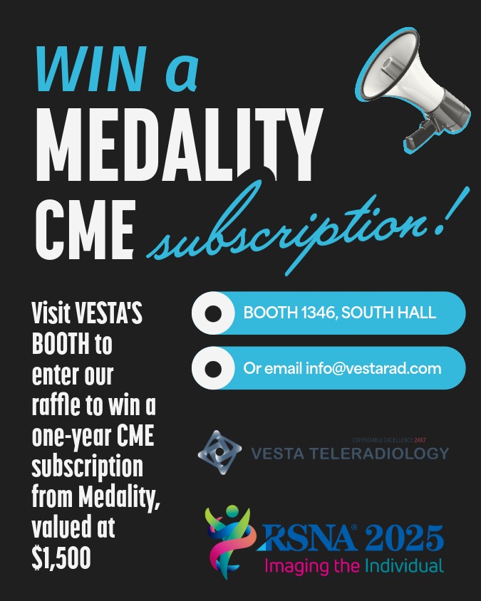 Why this RSNA prize matters for teams—not just individuals
 From Quantitative Imaging to Clinical Translation