FDA’s 2025 AI Draft Guidance: A Buyer’s Checklist for Imaging Leaders

In January 2025, the U.S. Food and Drug Administration released a draft guidance for AI-enabled medical devices that lays out expectations across the total product life cycle—design, validation, bias mitigation, transparency, documentation, and post-market performance monitoring. For imaging leaders, it’s a clear signal to tighten procurement criteria and operational guardrails before piloting AI in CT, MRI, mammo, ultrasound, or PET.

As teams lock in Q4 budgets and head into RSNA season, the FDA’s AI lifecycle draft (Jan 2025) and the now-final PCCP (Dec 2024) have reset what buyers should expect from AI in imaging—devices, software, and workflows. Vendors are updating claims and governance; this issue distills a practical buyer’s checklist—multisite validation with subgroup results, drift monitoring and version control, clear in-viewer transparency—and how pairing those tools with Vesta’s subspecialty coverage and QA turns promise into measurable gains across CT/MRI/US/mammography.

A practical buyer’s checklist

Use this when evaluating AI for your service lines:

  1. Intended use fit: Verify indications, inputs/outputs, and claims match your pathway and patient mix.
  2. Validation depth: Prefer multisite, diverse datasets; stratified results; pre-specified endpoints; documented data lineage and splits.
  3. Bias mitigation: Demand subgroup performance (sex, age, race/ethnicity when available), scanner/vendor variability analyses, and site-transfer testing.
  4. TPLC plan: Require drift monitoring, retraining triggers, versioning, and how updates are communicated.
  5. Human factors & transparency: Ensure limitations, failure modes, and interpretable outputs are presented in-viewer without slowing reads.
  6. Security & support: Patch cadence, vulnerability disclosure, SOC2/ISO posture, uptime SLAs, and rollback paths for version issues.
  7. Governance: Define metrics owners, review cadence, and thresholds to pause or roll back a model.

Implementation playbook: pilot → scale without disruption

Start with a 60–90 day pilot in one high-impact line (e.g., ED stroke CT or mammography triage) and lock in baselines: median TAT, positive/negative agreement, recall rate, PPV/NPV, and discrepancy rate. Set guardrails—when to auto-triage vs. force human review—and document escalation paths for model failures. Require case-level confidence and structured outputs your radiologists can verify quickly. Stand up a model governance huddle (modality lead, QA, IT security, and your teleradiology partner) that meets biweekly to review drift signals, subgroup performance, and near-misses. Bake in a rollback plan (version pinning) and a quiet-hours change window so updates don’t collide with peak volumes. As results stabilize, scale by cohort (e.g., expand to non-contrast head CT, then CTA) and keep training “micro-bursts” for techs/readers—short videos or checklists in-workflow. Tie vendor SLAs to uptime, support response, and clinical KPIs so the AI program stays accountable to operational value.

Where teleradiology fits

AI only delivers when it’s welded to coverage, quality, and speed. A teleradiology partner should provide:

  • 24/7 subspecialty + surge capacity: Vesta absorbs volume peaks so AI never becomes a bottleneck.
  • QA you can see: We benchmark pre/post-AI performance, add targeted second looks for edge cases, and feed variance data back to your team.
  • Standardized outputs: Structured reports that integrate model outputs with radiologist findings—no black-box surprises.
  • Smooth rollout: Pilot by service line (stroke CT, mammo triage, PE workups), then scale with tracked KPIs (TAT, PPV, recalls).
  • Interoperability & security: Seamless PACS/RIS/EMR integration with strict access controls, audit trails, and support for change-controlled updates.

Bottom line: Pairing AI with Vesta Teleradiology gives you round-the-clock subspecialty reads, measurable QA, and operational breathing room while you pilot and scale responsibly. If you’re mapping your AI roadmap under the FDA’s 2025 draft guidance, we’ll be your coverage and quality backbone—so your clinicians see faster answers and your patients see safer care. Visit vestarad.com to get started.


Summer 2025 Imaging Roundup: AI, New Modalities & Trends

The summer of 2025 has been packed with advancements in diagnostic imaging, from cutting-edge AI systems improving detection rates to emerging modalities pushing the boundaries of precision and speed. Here’s a look back at the most important developments from June through August that are shaping the future of radiology.

AI Is Reshaping Radiology Workflows

Generative AI Productivity Boost

In June, Northwestern Medicine unveiled a generative AI system capable of reducing radiologist reading time by up to 40% while identifying life-threatening conditions in milliseconds. This tool not only improves workflow efficiency but also offers a potential solution to the ongoing radiologist shortage (Northwestern Medicine).

ProFound AI for Mammography

A peer-reviewed study confirmed that iCAD’s ProFound AI significantly increases cancer detection rates, boosts diagnostic accuracy, and improves workflow for mammography screenings (ITN Online).

Aidoc’s $150M Expansion

July saw AI platform Aidoc raise $150 million in funding, led by NVIDIA and other major investors, aimed at expanding its reach into more hospitals and imaging centers globally (Aidoc).

Emerging Imaging Modalities and Research

Top Content Trends

Radiology publications in July spotlighted rising interest in abbreviated breast MRI, MRI-guided ultrasound for Parkinson’s disease, and dual-energy CT for understanding Long COVID-related lung changes (Diagnostic Imaging).

Photon-Counting CT and Whole-Body MRI

Photon-counting CT continues to gain attention for its ability to deliver higher resolution at lower doses, while whole-body MRI is increasingly used for cancer staging and early detection in high-risk populations (Radiology Business).

Multimodality Imaging at ACC.25

Cardiologists and radiologists at the ACC.25 conference explored how quantitative CT, functional cardiac MRI, and AI-enhanced echocardiography can bridge the gap between diagnostics and real-time therapy planning (American College of Cardiology).

August: A Month of Imaging Breakthroughs

AI-Native Imaging Viewers

Tech company New Lantern launched AI-native viewer modes for mammography and PET/CT, delivering sub-second load times and workflow automation (TMCNet).

Digital Radiography Gets Smarter

Advances in digital radiography are enhancing precision and speed, with newer systems providing better image quality at lower radiation doses (USA News).

ProCUSNet Ultrasound AI

Researchers at Stanford developed ProCUSNet, an AI tool that improved lesion detection by 44% and caught 82% of clinically significant prostate cancers on ultrasound—outperforming human interpretation (Becker’s Hospital Review).

DiffUS for Intraoperative Imaging

A new AI-based technique called DiffUS can create realistic ultrasound images from 3D MRI data, aiding in surgical planning and intraoperative navigation (arXiv).

Next-Gen PET Tracer

A novel PET tracer, Ga-68 Trivehexin, has shown promise in more accurately detecting breast cancer lesions and fibrotic lung tissue compared to traditional tracers (Journal of Nuclear Medicine).

Looking Ahead

The pace of innovation in diagnostic imaging this summer reinforces a clear trend: AI is no longer just an assistive tool—it’s becoming deeply embedded in clinical workflows. Coupled with emerging modalities like photon-counting CT and new PET tracers, radiology is entering an era of higher precision, speed, and accessibility.

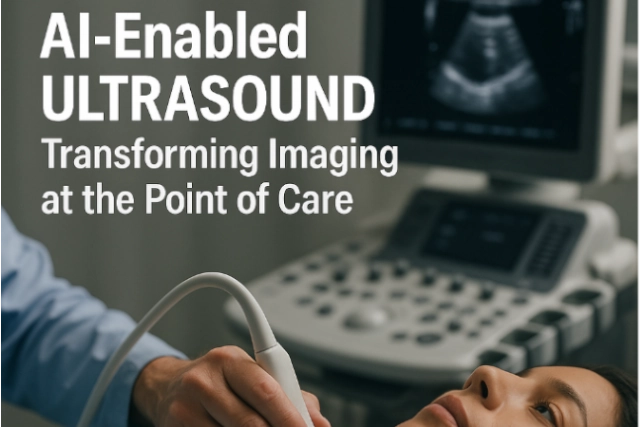
AI-Enabled Ultrasound: Transforming Imaging at the Point of Care


In today’s fast-paced healthcare environment, ultrasound is increasingly recognized not just for prenatal or cardiac assessment, but as a versatile diagnostic tool across specialties. Now, artificial intelligence (AI) is accelerating ultrasound’s impact — reducing operator dependency, improving diagnostic confidence, and enabling faster bedside care. For imaging leaders, especially in rural or underserved settings, AI-powered ultrasound technology paired with teleradiology support offers a compelling path for enhanced access and precision.

Innovations in AI-Ultrasound You Should Know

  1. FDA Clearance for AI Thyroid Ultrasound
    In 2024, See-Mode Technologies received FDA clearance for an AI-powered thyroid ultrasound system that can detect and classify nodules using the ACR TI-RADS scale. It has shown promising results in standardizing reporting and reducing unnecessary biopsies and follow-ups.
    Source: https://www.auntminnie.com
  2. Projected Market Growth
    The global AI ultrasound market is projected to grow at a compound annual growth rate (CAGR) of 22% through 2029. This rapid growth is fueled by the rising burden of chronic disease, limited radiologist availability, and the push for faster, more accessible diagnostics.

    Source: https://www.pharmiweb.com/

  3. Rural Potential with Point-of-Care AI
    A JAMA Cardiology viewpoint outlines how AI-assisted point-of-care ultrasound (POCUS) can enable more accurate cardiovascular assessments even when performed by generalists—especially valuable in remote areas without imaging specialists.
    Source: https://jamanetwork.com
  4. Clinician Enthusiasm and Challenges
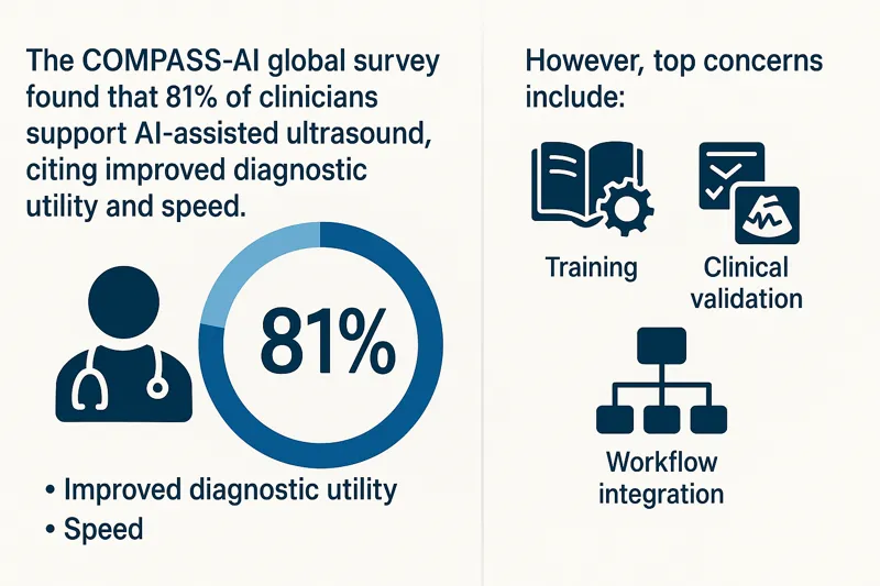
    The COMPASS-AI global survey found that 81% of clinicians support AI-assisted ultrasound, citing improved diagnostic utility and speed. However, top concerns include training, clinical validation, and workflow integration.

    Source: https://theultrasoundjournal.springeropen.com/

Infographic showing COMPASS-AI survey results on clinician support for AI-enabled ultrasound, benefits, and concernsWhy It Matters for Facilities and Radiology Teams

  • Reduces staffing burden: AI ultrasound reduces variability among operators, ideal for high-turnover or remote settings.
  • Speeds up decision-making: Frontline providers can quickly gather meaningful imaging data, while teleradiologists handle the interpretation.
  • Expands imaging reach: Portable, AI-powered ultrasound extends diagnostic capabilities to underserved regions.
  • Supports standardization: AI helps standardize image acquisition and reporting, improving overall workflow efficiency.

How Vesta Teleradiology Enhances AI-Ultrasound Value

While AI augments imaging workflows, expert interpretation is still essential. Vesta provides:

  • Subspecialty reads across thyroid, vascular, MSK, and more
  • 24/7 coverage with fast turnaround times
  • Seamless PACS/RIS integration for AI-acquired ultrasound data

Our radiologists help bridge the gap between frontline imaging and specialist analysis—ensuring that every AI-enabled ultrasound scan contributes to timely, confident patient care.

Bringing AI and Teleradiology Together

Whether you’re running a rural health center, a large outpatient clinic, or an emergency department, AI ultrasound paired with expert teleradiology interpretation helps:

  • Increase imaging access without compromising accuracy
  • Alleviate staffing constraints
  • Deliver faster diagnoses
  • Improve patient outcomes

AI in ultrasound is not replacing radiologists — it’s helping them focus on what matters most. With Vesta’s support, healthcare organizations can embrace innovation while maintaining high-quality, consistent imaging interpretation.


The Future of AI + Human Collaboration in Radiology

Artificial intelligence (AI) is playing an increasingly important role in radiology and diagnostic imaging. From workflow optimization to automated image analysis, AI tools are now assisting radiologists in more imaging departments than ever before. Right now, AI tools are assisting with tasks like automatically prioritizing critical cases, generating draft reports, and flagging potential abnormalities in studies such as chest X-rays, mammograms, and CT scans.”

At the same time, it’s clear that AI’s role is best seen as complementary to human expertise, not a replacement. In fact, a 2023 study published in JAMA Network Open found that radiologists using AI frequently sometimes experienced higher burnout rates—especially when workflows were not well integrated or added new demands.

This highlights an important lesson: for AI to truly benefit radiology, it must be thoughtfully implemented, supporting radiologists rather than complicating their work.

Why Human Expertise Remains Essential

While AI offers exciting capabilities—such as triaging cases, flagging abnormalities, or standardizing reports—there is no substitute for the experience and clinical judgment of a radiologist.

Subspecialty areas like:

  •         Neuroradiology
  •         Musculoskeletal imaging
  •         Cardiac imaging
  •         Pediatric radiology

…require nuanced interpretation that today’s AI tools simply cannot match.

Vesta Teleradiology supports healthcare facilities by ensuring that every read is performed by a board-certified U.S.-based radiologist—with subspecialty expertise available across all major modalities.


Balancing AI + Workflow: A Smarter Approach

Many imaging departments today are navigating how to integrate AI without adding unnecessary complexity.


At Vesta, we work with partner facilities to provide flexible teleradiology services that complement their existing workflows—whether or not they are using AI tools internally.


Our approach emphasizes:

✅ Efficient, reliable human reads

✅ Subspecialty expertise when needed

✅ Consistent communication with referring providers

✅ Flexibility to support 24/7 coverage and manage fluctuations in volume


By helping facilities maintain high-quality interpretations with efficient turnaround, Vesta supports radiology teams as they adopt new technologies and respond to growing imaging demand.


Looking Ahead: The Collaborative Future of Radiology

AI’s role in radiology will continue to evolve. The most effective imaging departments will combine:


  •         Advanced AI tools where they add value
  •         Skilled radiologists providing expert interpretation
  •         Clear, integrated workflows that reduce friction
  •         Strategic partnerships to ensure coverage and subspecialty access


At Vesta Teleradiology, we believe that human expertise will remain the foundation of diagnostic imaging—and that thoughtful integration of AI can enhance, not replace, that expertise.


We’re committed to working with healthcare facilities to build balanced solutions that support radiologists, improve patient care, and keep pace with the demands of modern imaging.


If your team is looking for flexible, expert support—whether for subspecialty reads, after-hours coverage, or help managing increased imaging demand—Vesta Teleradiology is here to help.


Contact us to learn more.


Mid-Year Radiology Trends: What’s Shaping Diagnostic Imaging in 2025

The pace of change in radiology and diagnostic imaging only accelerated in 2025. From emerging technologies to new ways of working, the field is evolving rapidly to meet both growing patient demand and the ongoing challenge of radiologist shortages.

Here’s a look at the key mid-year trends shaping radiology so far this year—and how facilities can stay ahead with the right partners.


  1. AI Is Evolving—But Radiologists Remain at the Center

AI tools in radiology are becoming more sophisticated, particularly in automating administrative tasks like report generation, triage, and workflow optimization.

A recent article from Business Insider noted that many radiologists now use generative AI to streamline productivity—not replace their diagnostic expertise. The key is finding the right balance: AI assists, but human interpretation remains critical.

At Vesta Teleradiology, our board-certified radiologists embrace AI tools that improve speed and accuracy while maintaining clinical oversight and patient safety.


  1. Staffing Pressures Continue—and Teleradiology Bridges the Gap

Radiologist shortages are still a frontline issue in 2025. The Neiman Health Policy Institute projects the shortage will persist through 2055 without proactive changes. This strain is particularly acute in oncology and rural hospitals, where delays in imaging results can directly impact outcomes.

Teleradiology is now an essential solution for many facilities. At Vesta, we provide:
✅ 24/7/365 STAT & routine reads
✅ Subspecialty support (Neuro, MSK, Cardiac, Pediatrics, and more)
✅ No minimum read requirements
✅ Customizable workflows to fit your needs


  1. Photon-Counting CT: A Game-Changer for Imaging

Photon-counting CT (PCCT) is gaining traction in 2025, offering higher resolution images with lower radiation doses. Early adopters are seeing promising results in cardiovascular and oncologic imaging.

As new modalities enter clinical use, having expert radiologists trained in advanced imaging techniques is vital. Vesta’s subspecialty readers are ready to interpret the most complex cases with precision.

  1. The Rise of Digital Twins in Imaging

Digital twins—virtual models of patients—are becoming more practical in healthcare. Radiology plays a key role by providing the high-fidelity imaging needed to create these models for personalized medicine, treatment planning, and disease monitoring.

As these technologies develop, facilities will need radiologists with the expertise to interpret increasingly complex imaging data—and flexible partners to help scale their capabilities.


  1. Growing Focus on Turnaround Times and Patient Experience

With patients and referring physicians expecting faster results, facilities are under pressure to reduce turnaround times—especially for oncology, trauma, and screening programs.

Vesta Teleradiology helps meet this demand with:

  • 24/7 availability to prevent backlogs
  • Real-time communication for critical findings
  • Customizable reporting to fit your workflow and brand


Conclusion: How to Stay Ahead in a Fast-Moving Year

The radiology landscape is dynamic—and staying ahead requires agility, expertise, and trusted partners. Whether you’re looking to bridge staffing gaps, scale subspecialty reads, or handle advanced imaging modalities, Vesta Teleradiology is here to help.

Our U.S.-based, board-certified radiologists deliver precision reads with flexible, scalable solutions for hospitals, imaging centers, and healthcare systems nationwide.

Let’s connect today to customize a radiology solution that fits your 2025 needs—and beyond.

Contact Vesta Teleradiology.


Sources:

Business Insider
arXiv.org 
arXiv.org 
The Imaging Wire 

Q1 2025 AI Radiology Roundup: Smarter Screening, Streamlined Referrals, and Intelligent Ultrasound Innovations

The first quarter of 2025 has seen impressive strides in the integration of artificial intelligence across the radiology spectrum. From breast cancer screening and interventional radiology referrals to next-gen ultrasound systems, AI continues to redefine efficiency, accuracy, and clinical outcomes. Below, we highlight three major developments shaping the future of radiology.


  1. Large Language Models Streamline IR Procedure Requests—For Just Pennies

In a study published in the Journal of Vascular and Interventional Radiology, researchers at Duke University Medical Center demonstrated that large language models (LLMs) like GPT-4 can accurately and efficiently route interventional radiology (IR) procedure requests—at a cost of only $0.03 per request.

By training the model on structured rules based on real IR team schedules and procedures, the AI achieved 96.4% accuracy in routing “in-scope” requests and 76% accuracy for out-of-scope queries. The tool helps clinicians connect with the right provider faster, improving coverage efficiency while avoiding unnecessary procedure orders.

With its adaptability to different hospital systems and minimal setup requirements, this LLM-powered tool could soon become a scalable solution for streamlining IR consultations nationwide.

“This approach is highly adaptable… and does not depend on training a dedicated model,” said Dr. Brian P. Triana, lead author.


  1. AI Mammography Boosts Cancer Detection by 29% in Landmark MASAI Trial

A game-changing trial out of Sweden—Mammography Screening with Artificial Intelligence (MASAI)—has reinforced the clinical power of AI in breast cancer screening. Published in The Lancet Digital Health, the randomized study followed over 105,000 women and found that AI-assisted screening increased cancer detection rates by 29% and reduced radiologist workload by 44%.


The AI tool, Transpara, was especially effective in identifying small, invasive cancers and high-grade in situ cancers—without increasing false positives. Radiologists using Transpara received real-time lesion detection and risk scores, helping reduce both overcalls and overlooked cancers.

“AI-supported screening can significantly enhance early detection while optimizing the use of healthcare resources,” said Dr. Kristina Lång of Lund University.

These results underscore AI’s role not just as a support tool but as a potential standard in future breast cancer screening protocols.


  1. Samsung Unveils AI-Powered Ob/Gyn Ultrasound System for U.S. Market

Samsung Medison made waves at the Society for Maternal-Fetal Medicine (SMFM) 2025 with the launch of its new AI-enhanced ob/gyn ultrasound system, the Samsung Z20.

The Z20 features Live ViewAssist, a real-time deep learning tool designed to streamline advanced obstetrical exams. Its capabilities include automatic structure labeling, real-time image quality assessment, and AI-powered measurements—all aimed at improving diagnostic precision and reducing repetitive strain on clinicians.

Addressing challenges in imaging patients with high BMI and promoting ergonomic design, the Z20 represents a leap forward in both performance and provider wellness. Additionally, Samsung showcased Sonio, its cloud-based ultrasound reporting platform, marking a step toward more integrated, AI-driven workflows in women’s health.

From improving clinical throughput to enhancing diagnostic confidence, AI is becoming indispensable in radiology. As Q1 2025 wraps up, the message is clear: artificial intelligence is no longer a futuristic concept in imaging—it’s a present-day solution driving meaningful change.

Stay tuned as we continue to track these innovations and explore how AI will shape the next quarter in diagnostic imaging and beyond.


Addressing the Persistent Radiologist Shortage: Challenges and Solutions for the Future

The ongoing imbalance between radiologist supply and medical imaging demand in the U.S. is projected to continue through 2055 without significant intervention, according to recent research by the Neiman Health Policy Institute, (NHPI), published in the Journal of the American College of Radiology on February 12. As the population grows and ages, and imaging utilization increases, the shortage of radiologists poses a significant challenge for healthcare systems nationwide.

Projected Growth in Radiologist Supply

The NHPI study anticipates a nearly 26% increase in the supply of radiologists over the next 30 years, assuming residency numbers remain unchanged. However, even this growth may not be sufficient to meet rising imaging demands. If residency positions increase, the radiologist workforce could see a 40% expansion by 2055. Yet, attrition rates—especially post-COVID—pose a threat to this growth, highlighting the need for initiatives aimed at improving workplace well-being and retaining experienced radiologists.

Increasing Demand for Imaging Services

The demand for imaging services is expected to rise between 17% and 27% by 2055, driven largely by population growth and aging. Specific modalities like CT scans may see utilization increases as high as 59%, while others, such as nuclear medicine, may experience a decline. These projections underscore the urgency of balancing supply and demand to prevent prolonged patient wait times and compromised care.

Current Impact on Patients and Healthcare Systems

Patients across the U.S., including those in West Michigan, are already feeling the impact of the radiologist shortage. Delays in receiving imaging results have caused frustration, particularly for individuals with pressing health concerns such as fibroids and breast cancer risk. Healthcare providers, from radiologists to patient care technicians, are also facing mounting pressure to deliver timely care amidst workforce shortages.

Potential Solutions to Mitigate the Shortage

To address this crisis, experts emphasize the need to increase radiology residency slots and curb inappropriate imaging use. Monitoring attrition patterns and enhancing workplace conditions are also crucial. Technological advancements, such as AI for improving radiologist efficiency and clinical decision support systems, present promising avenues for alleviating some of the burden on the current workforce.

Conclusion

The radiologist shortage in the U.S. is a complex issue that requires multifaceted solutions. Increasing residency positions, enhancing workplace well-being, and leveraging technology are essential steps to ensure patients receive timely and accurate imaging services.

Top Radiology Company: Onsite and Remote

At Vesta Teleradiology, we are committed to bridging the gap caused by radiologist shortages. Our team of U.S. board-certified radiologists offers both on-site and remote services, providing reliable imaging interpretations to meet your facility’s needs efficiently. Let us help you navigate the challenges of radiologist shortages with our expert solutions.


RSNA 2024 Highlights: AI, Imaging Advancements, and Industry Recognition

The Radiological Society of North America’s (RSNA) 2024 annual meeting showcased significant advancements in medical imaging, including artificial intelligence (AI), innovations in computed tomography (CT) and magnetic resonance imaging (MRI), and strategies to address the ongoing radiology staffing shortage. The event also honored leaders in the field for their exceptional contributions.

Advancements in Radiology Technology

AI Integration

Artificial intelligence was a major focus, with over 200 exhibitors showcasing AI-related innovations. The FDA has approved nearly 1,000 clinical AI algorithms, 80% of which pertain to medical imaging. AI’s expanding role includes rapid stroke detection, workflow orchestration, and FFR-CT assessments, now included in national guidelines. However, discussions emphasized the need for rigorous monitoring to prevent bias and performance degradation in these algorithms.

CT and MRI Innovations

The conference highlighted advancements in imaging technologies, unveiling three new AI-enhanced CT systems aimed at improving diagnostics and patient comfort. These innovations promise faster, more accurate imaging, enhancing both clinical outcomes and workflow efficiency.

Addressing the Staffing Shortage

The radiology workforce shortage remains a critical challenge. Proposed solutions include expanding residency programs, adopting AI to reduce workloads, and ensuring fair compensation. While AI shows promise in easing demands, experts caution it is not a standalone solution to the staffing crisis. Teleradiology companies like Vesta can play a vital role in addressing these shortages by providing access to qualified radiologists, ensuring timely interpretations, and supporting healthcare facilities in maintaining efficient workflows.

Industry Recognition

RSNA 2024 also celebrated the accomplishments of industry leaders:

  • Gold Medal Awards: Recognizing excellence in leadership and innovation, the awards were presented to James P. Borgstede, MD, Elizabeth S. Burnside, MD, MPH, and Beverly G. Coleman, MD, for their groundbreaking contributions to radiology and healthcare.
  • Alexander R. Margulis Award for Scientific Excellence: This award honored the authors of a 20-year study on low-dose CT screening for lung cancer, emphasizing its life-saving potential through early detection.
  • Outstanding Researcher Award: Jeffrey G. Jarvik, MD, MPH, was recognized for his impactful work in spine imaging and back pain research.

Conclusion

RSNA 2024 reinforced radiology’s pivotal role in healthcare, spotlighting transformative technologies, addressing workforce challenges, and honoring outstanding achievements. The integration of AI, combined with ongoing innovation in imaging and efforts to bolster the radiology workforce, ensures the field continues to thrive and evolve.


Sources:

Radiologbusiness.com
rsna.org
openai.com


AI in Radiology: Biden’s New Executive Order and Latest News

The Biden administration’s recent executive order on artificial intelligence (AI) has significant implications for radiology, as discussed in a review published in JACR. The order aims to ensure responsible use of AI in healthcare and establish a federal program to address unsafe practices. While immediate changes to radiology practice may not be expected, the order signals forthcoming regulatory shifts, particularly in oversight and enforcement by government agencies. This includes scrutiny of computer-aided detection systems and AI for noninterpretative tasks. The FDA premarket review for medical devices like CAD programs is likely to be augmented with additional quality and equity requirements. Health and Human Services will oversee data input into AI algorithms, possibly mandating disclosure of training datasets. Radiologists seeking Medicare reimbursement for AI products will need to prioritize security and compliance with nondiscrimination laws. The order also emphasizes data sharing with the National AI Research Resource and encourages radiologists to engage in policy creation and provide input on regulatory frameworks. However, specific parameters and regulatory details are yet to be defined. Overall, the order serves as a call for federal agencies to mobilize efforts in AI oversight, with radiologists urged to actively participate in shaping policies and best practices.


Predicting Lymph Node Metastasis

A recent study published in Radiology: Imaging Cancer compared the effectiveness of a four-dimensional (4D) convolutional neural network (CNN) model, incorporating clinical and breast MRI findings, with a machine learning model based solely on clinicopathologic features in predicting axillary node status in women with breast cancer. The 4D CNN model achieved a significantly higher area under the curve (AUC) of 87% compared to 63% for the clinical model. It also demonstrated higher sensitivity (89% vs. 75%) and specificity (76% vs. 52%), with a lower false-negative rate (11% vs. 25%). The study suggests that the 4D hybrid model could serve as a valuable tool in selecting patients who may avoid invasive procedures like sentinel lymph node biopsy and aid in treatment decisions for breast cancer patients. However, further external validation of the model is needed, and limitations such as reliance on manual tumor bounding boxes and specific MRI device imaging need to be addressed for broader clinical adoption.


FDA Clearance for Cardiac and Lung AI

Exo, a medical imaging software and device company, has announced FDA clearance for its cardiac and lung artificial intelligence (AI) applications on Exo Iris, their handheld ultrasound device. This expands Exo’s cleared applications to include cardiac, lung, bladder, hip, and thyroid assessments. Iris, powered by AI, facilitates point-of-care ultrasound, particularly benefiting rural or underserved communities, enabling faster diagnosis and treatment. Sandeep Akkaraju, Exo’s CEO, emphasizes the aim of democratizing AI-empowered medical imaging for all caregivers. The AI applications were trained on a diverse dataset and validated across various patient populations and scan types. They enable reliable assessment of pulmonary edema and cardiac function, with additional doppler capabilities for cardiac, abdominal, and vascular applications. Clinicians, including those with limited experience, welcome the efficiency and reliability of Exo’s AI applications, which enhance patient care and healthcare system efficiency.

Sources:
Radiologybusiness.com
mddionline.com
Openai.com

March AI News in Diagnostic Imaging

New Research by Harvard Medical School, MIT and Stanford on AI and Clinician Performance

The potential of medical artificial intelligence (AI) tools to enhance clinicians’ performance in interpreting medical images varies among individual clinicians, as highlighted by recent research led by Harvard Medical School, MIT, and Stanford. Published in Nature Medicine, the study underscores the intricate nature of human-AI interaction, which remains incompletely understood. While some radiologists benefit from AI assistance, others experience interference, affecting diagnostic accuracy.

The findings stress the necessity for personalized AI systems tailored to individual clinicians, emphasizing careful implementation to maximize benefits and minimize harm. Despite variations in AI’s impact, the results shouldn’t deter AI adoption but rather prompt a deeper understanding of human-AI dynamics to design approaches that enhance human performance.

To ensure effective integration of AI in clinical practice, collaboration between AI developers and clinicians is essential, alongside rigorous testing in real-world scenarios. Furthermore, efforts should focus on improving AI accuracy and training radiologists to discern AI inaccuracies, facilitating informed decision-making. Ultimately, understanding the complexities of machine-human interaction is pivotal for optimizing patient care through AI integration in radiology.

radiologist
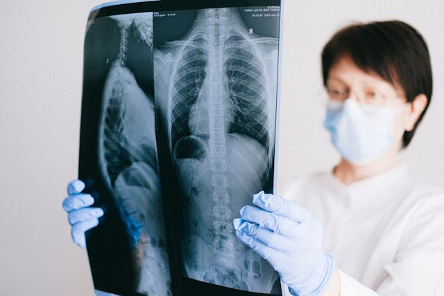
A radiologist examines an x-ray

AI and Workflows

New research highlights a novel reporting workflow that automatically incorporates artificial intelligence (AI) findings into structured radiology reports, streamlining physicians’ tasks and saving valuable time. German experts shared their experience with the “AI to SR pipeline,” which integrates a commercially available AI tool for chest X-ray pathology detection and localization into structured report templates.

In evaluations conducted at University Medical Center Mainz, expert radiologists found that reports generated using the AI to SR pipeline were faster compared to free-text reporting and conventional structured reporting. Additionally, subjective quality assessments indicated higher ratings for reports created with the pipeline.

In the hospital’s clinical routine, chest X-ray images are sent to the picture archiving and communication system, then automatically forwarded to the AI tool for analysis. The results are output in a DICOM structured reporting format, taking approximately five minutes from image acquisition to final reporting. Radiologists were able to create chest X-ray reports significantly faster with the pipeline compared to free-text and conventional structured reporting, while also rating the AI-generated reports more favorably.

The authors suggest that this AI-driven reporting pipeline offers standardized, time-efficient, and high-quality reporting for chest X-rays, potentially enhancing AI integration into daily clinical practice and maximizing its benefits.


Sources:

Medicalxpress.com
Radiologybusiness.com
Openai.com