The first quarter of 2025 has seen impressive strides in the integration of artificial intelligence across the radiology spectrum. From breast cancer screening and interventional radiology referrals to next-gen ultrasound systems, AI continues to redefine efficiency, accuracy, and clinical outcomes. Below, we highlight three major developments shaping the future of radiology.
-
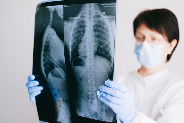
Large Language Models Streamline IR Procedure Requests—For Just Pennies
In a study published in the Journal of Vascular and Interventional Radiology, researchers at Duke University Medical Center demonstrated that large language models (LLMs) like GPT-4 can accurately and efficiently route interventional radiology (IR) procedure requests—at a cost of only $0.03 per request.
By training the model on structured rules based on real IR team schedules and procedures, the AI achieved 96.4% accuracy in routing “in-scope” requests and 76% accuracy for out-of-scope queries. The tool helps clinicians connect with the right provider faster, improving coverage efficiency while avoiding unnecessary procedure orders.
With its adaptability to different hospital systems and minimal setup requirements, this LLM-powered tool could soon become a scalable solution for streamlining IR consultations nationwide.
“This approach is highly adaptable… and does not depend on training a dedicated model,” said Dr. Brian P. Triana, lead author.
-
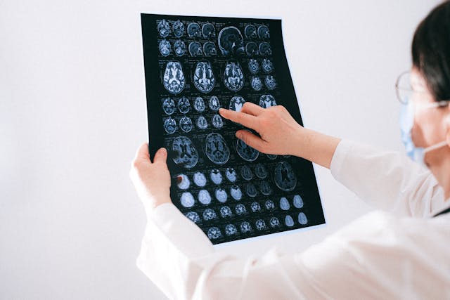
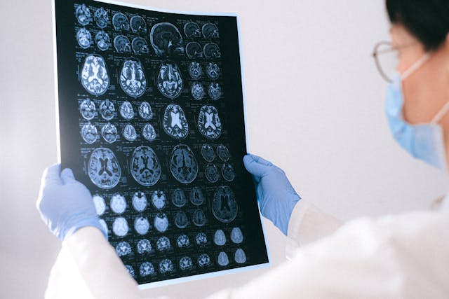
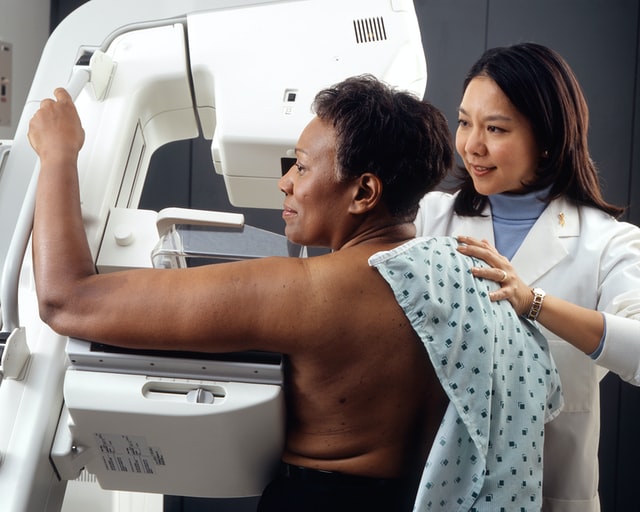
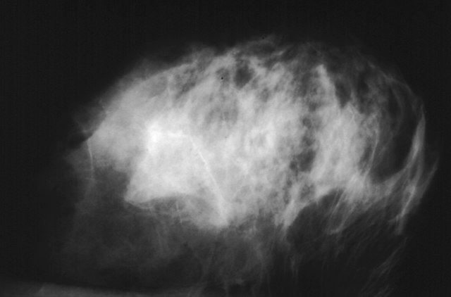
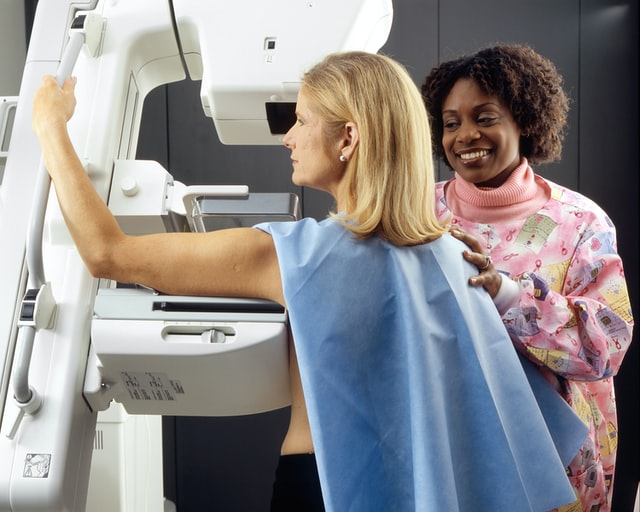
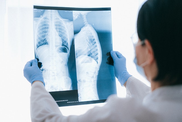
AI Mammography Boosts Cancer Detection by 29% in Landmark MASAI Trial
A game-changing trial out of Sweden—Mammography Screening with Artificial Intelligence (MASAI)—has reinforced the clinical power of AI in breast cancer screening. Published in The Lancet Digital Health, the randomized study followed over 105,000 women and found that AI-assisted screening increased cancer detection rates by 29% and reduced radiologist workload by 44%.
The AI tool, Transpara, was especially effective in identifying small, invasive cancers and high-grade in situ cancers—without increasing false positives. Radiologists using Transpara received real-time lesion detection and risk scores, helping reduce both overcalls and overlooked cancers.
“AI-supported screening can significantly enhance early detection while optimizing the use of healthcare resources,” said Dr. Kristina Lång of Lund University.
These results underscore AI’s role not just as a support tool but as a potential standard in future breast cancer screening protocols.
-
Samsung Unveils AI-Powered Ob/Gyn Ultrasound System for U.S. Market
Samsung Medison made waves at the Society for Maternal-Fetal Medicine (SMFM) 2025 with the launch of its new AI-enhanced ob/gyn ultrasound system, the Samsung Z20.
The Z20 features Live ViewAssist, a real-time deep learning tool designed to streamline advanced obstetrical exams. Its capabilities include automatic structure labeling, real-time image quality assessment, and AI-powered measurements—all aimed at improving diagnostic precision and reducing repetitive strain on clinicians.
Addressing challenges in imaging patients with high BMI and promoting ergonomic design, the Z20 represents a leap forward in both performance and provider wellness. Additionally, Samsung showcased Sonio, its cloud-based ultrasound reporting platform, marking a step toward more integrated, AI-driven workflows in women’s health.
From improving clinical throughput to enhancing diagnostic confidence, AI is becoming indispensable in radiology. As Q1 2025 wraps up, the message is clear: artificial intelligence is no longer a futuristic concept in imaging—it’s a present-day solution driving meaningful change.
Stay tuned as we continue to track these innovations and explore how AI will shape the next quarter in diagnostic imaging and beyond.