
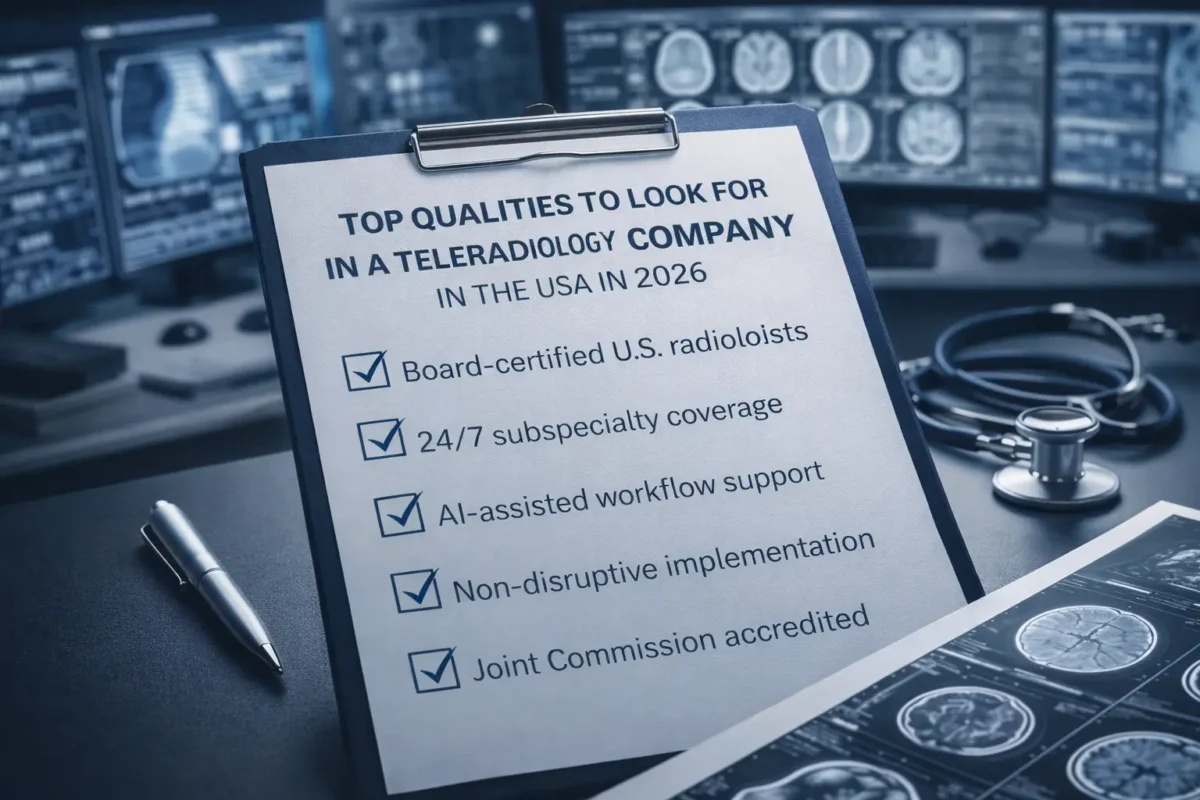
Why imaging centers need a more specific kind of partner
Not every imaging center needs the same radiology arrangement. Some centers need overflow help during busy periods. Others need low-volume overnight support, stronger subspecialty access, or more consistent turnaround across a broader modality mix. For brick-and-mortar imaging centers, the real priority is finding a teleradiology partner that fits how the center actually operates.
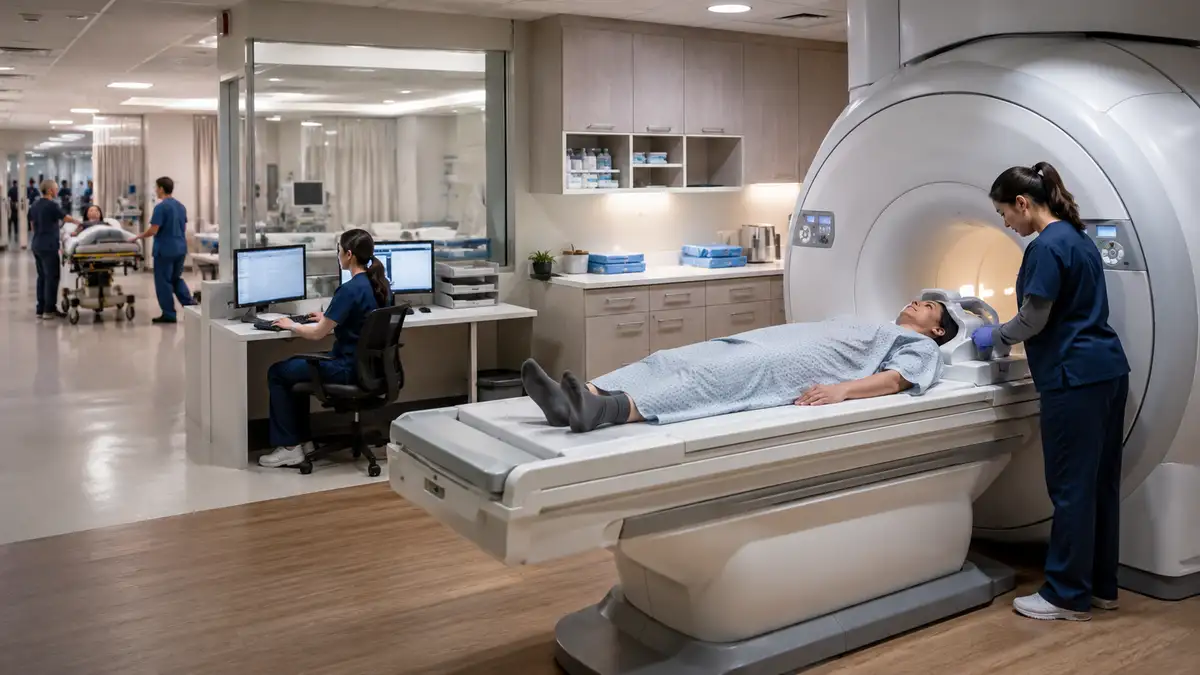
That is an important distinction because many conversations in the market still center on urgent care or mobile imaging use cases, where the study mix often leans heavily toward X-ray and ultrasound. Traditional imaging centers tend to have broader needs. CT, MRI, mammography, and sometimes nuclear medicine all bring different workflow and interpretation demands.
Modality depth should be one of the first questions
A group that mainly supports basic X-ray and ultrasound may not be the right fit for a center built around advanced imaging. The more useful question is whether the radiology partner can support the center’s current modality mix and continue to do so as the center grows.
That matters even more as outpatient imaging expands. Vizient reported that outpatient settings now account for a large share of imaging volume and projected long-term growth in advanced imaging, especially CT and PET. As that demand rises, imaging centers need coverage models that can support both volume and complexity.
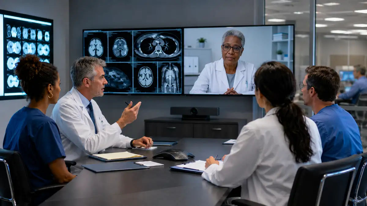
Subspecialty support can strengthen both quality and referrals
Not every case requires a subspecialist, but some studies clearly benefit from one. Centers that offer more advanced workups or want to strengthen referrer confidence often value access to neuroradiology, musculoskeletal radiology, breast imaging expertise, or other subspecialty support.
This can have practical business value. Referring physicians notice when reports are timely, clear, and clinically useful. They also notice when a center can support a broader range of studies without avoidable delays.
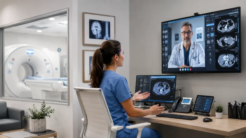
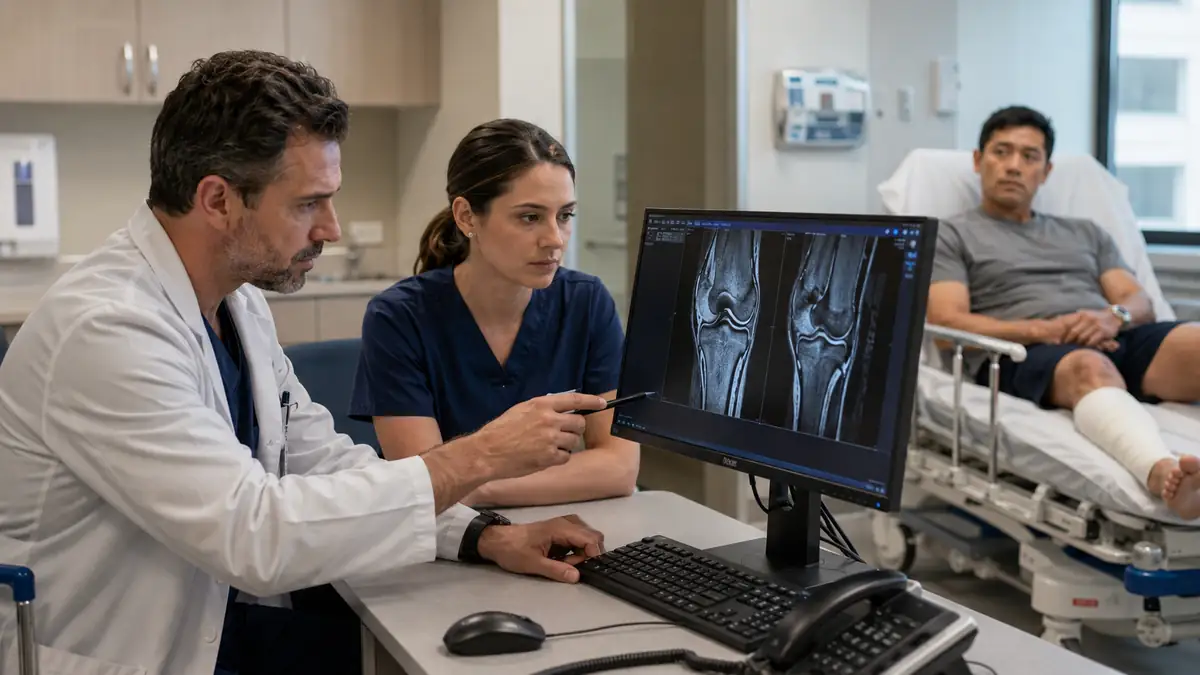
Reporting workflow and communication matter just as much
Turnaround time always matters, but reporting consistency matters too. Imaging center leaders want reports that are readable and dependable, and they want communication pathways that work when something urgent appears. A strong radiology partner should fit the center’s existing workflow rather than forcing staff to work around unnecessary friction.
Technology decisions increasingly affect that experience. The FDA’s list of AI-enabled medical devices continues to grow, and radiology remains one of the leading categories. For imaging centers, the takeaway is not to chase every new tool. It is to work with partners that can support practical workflow improvements without complicating reporting, communication, or case prioritization.
Flexibility is essential for growing centers
Volume rarely stays perfectly steady. Referral patterns shift. Staffing changes. Some months are busier than expected, while others are more uneven. The right teleradiology partner should be able to absorb those swings without leaving the center overcommitted when volume softens or under-supported when it spikes.
That is especially important for centers that want to offer a broad menu of imaging services while keeping operations efficient. A flexible, full-service partner can help the center scale intelligently rather than reactively.
What the best partnerships look like
The strongest radiology partnerships for imaging centers tend to feel operationally integrated. They support the center across modalities, maintain dependable turnaround, provide access to subspecialty reads, and make workflow easier rather than harder.
For brick-and-mortar imaging centers, that kind of fit is often the difference between basic coverage and a partnership that actually strengthens the business.
FAQs
Why does modality coverage matter when choosing a teleradiology partner? Because many imaging centers perform more than basic X-ray and ultrasound. A strong partner should be able to support CT, MRI, mammography, and other modalities relevant to the center.
Should imaging centers look for subspecialty reads? Yes, especially if they perform advanced studies or want to improve quality, referrer confidence, and clinical depth.
How important is technology compatibility? It is very important. Reporting, communication, and workflow tools should support efficiency without creating unnecessary complexity for staff or referring providers.
Vesta is Your Partner
For brick-and-mortar imaging centers looking to strengthen coverage, improve turnaround, and support a broader range of modalities, the right radiology partner can make a meaningful difference. Vesta Teleradiology works with imaging centers in key markets including Texas, California, Florida, Georgia, Illinois, Ohio, North Carolina, and Kentucky, offering full-service radiology support designed around real operational needs. From CT and MRI to mammography, ultrasound, X-ray, and more, Vesta provides flexible on-site and remote coverage that helps imaging centers grow with confidence.


 What hospitals can do now (short-term, operations-first)
 3) Smooth scheduling around your true capacity


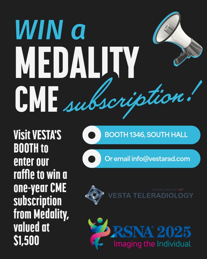 Why this RSNA prize matters for teams—not just individuals