New FDA Clearances for Imaging Systems and Solutions

FDA clearance for a diagnostic imaging machine indicates that the device has been deemed safe and effective for its intended use by the Food and Drug Administration (FDA) in the United States. This clearance process involves thorough evaluation of the device’s design, performance, and manufacturing processes to ensure that it meets regulatory standards for quality, safety, and efficacy. Here’s the latest devices that have received FDA clearance.


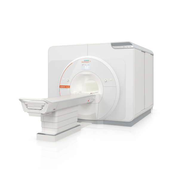
The Magnetom Terra.X: MRI System

The Magnetom Terra.X, a new 7T MRI system, has received 510(k) clearance from the FDA. Manufactured by Siemens Healthineers, it’s a second-generation successor to the Magnetom Terra and offers several enhancements for 7T imaging. Key features include an eight-channel parallel transmit architecture for clinical use, deep learning image reconstruction optimized for 7T, improved diffusion imaging with a high-performance gradient system, and accelerated image acquisition enabling high-resolution brain and knee exams in under 20 minutes. Siemens Healthineers sees this as a significant step in providing better patient care, particularly in neurological and knee imaging. Additionally, the FDA clearance allows existing Magnetom Terra systems to be upgraded to the Magnetom Terra.X.

Image courtesy of Siemens Healthineers

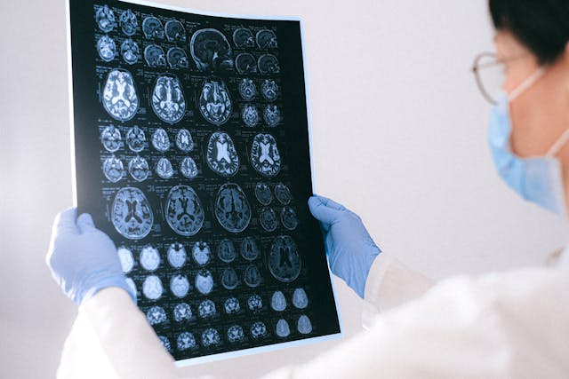
SyMRI 3D for Brain Imaging

SyntheticMR has announced that its latest imaging solution, SyMRI 3D, has received FDA 510(k) clearance for clinical use in the United States. This clearance marks a significant advancement in quantitative MRI technology, offering exceptional resolution and accuracy in brain imaging. SyMRI 3D enables precise volumetric estimations of brain regions, known as parcellation, providing clinicians with deeper insights into brain structure and function. The enhanced resolution facilitates comprehensive lesion analysis, leading to more accurate medical condition assessments. This clearance empowers physicians to make more informed decisions in diagnosis and treatment planning, ultimately improving patient outcomes. SyntheticMR reaffirms its dedication to advancing medical imaging technology and providing innovative tools to enhance patient care through this milestone.


nCommand Lite for Remote Scanning

GE Healthcare has highlighted the FDA clearance of a solution by Ionic Health that enables technologists to remotely supervise patient scans. The system, called “nCommand Lite,” has been tested in Brazil for three years and is vendor-agnostic, allowing remote supervision across MRI, CT, and PET modalities. GE has secured exclusive distribution rights for nCommand in the U.S., aiming to address ongoing workforce shortages in healthcare. Rekha Ranganathan, GE’s chief digital officer for imaging, emphasized the company’s commitment to remote operations and increasing patient access to expert technologists. The system facilitates not only scanning supervision but also training, procedure assessment, and scanning parameter management. GE’s announcement coincides with growing interest in remote scanning, with the American College of Radiology advocating for permanent remote supervision of diagnostic tests. However, technologists have expressed reservations about managing imaging remotely, according to recent survey data from the American Society of Radiologic Technologists.


Sources:

Itnonline.com
Radiologybusiness.com
diagnosticimaging.com
openai.com


The Latest in Brain Imaging News

In recent years, awareness surrounding brain injuries has steadily risen, prompting significant strides in diagnostic technologies and treatment modalities. As we delve into the latest developments in this critical area of healthcare, it becomes increasingly apparent that advancements in medical imaging, particularly in the realm of neurological disorders, are poised to revolutionize the landscape of brain injury diagnosis and management.


AI-based Quantitative Brain Imaging System

Philips and Synthetic MR have joined forces to advance the diagnosis of neurological disorders through cutting-edge quantitative brain imaging tools. Their collaboration introduces the Smart Quant Neuro 3D MRI software suite, combining Philips’ SmartSpeed image-reconstruction technology, the 3D SyntAc clinical application, and SyntheticMR’s SyMRI NEURO 3D software. This innovation employs AI to analyze brain tissues, enhancing the detection and analysis of conditions like multiple sclerosis, traumatic brain injuries, and dementia.

The rise of AI in diagnostic imaging, projected to reach $1.2bn by 2027, signifies a transformative shift in improving accuracy and patient outcomes. With the diagnostic imaging market expected to grow to $9.1bn by 2030, fueled by demand for early disease diagnosis and personalized medicine, this partnership underscores the crucial role of AI in enhancing medical imaging.

Read the press release here.


A New Way of Diagnosing Mild TBIs

Researchers have developed a novel brain imaging method to diagnose mild traumatic brain injuries (mTBIs), which are often missed by standard techniques like MRI. This method involves loading gadolinium, a common MRI contrast agent, into micropatches attached to immune cells called macrophages. These cells migrate to areas of brain inflammation caused by mTBIs, enabling MRI detection. The technique, called M-GLAMs, was successfully tested in mice and pigs, showing promise for accurately diagnosing mTBIs. It also allows imaging at lower gadolinium doses, potentially benefiting patients with kidney issues. While unable to pinpoint injury locations, M-GLAMs could aid in identifying and treating brain inflammation. The researchers aim to bring this technology to clinical trials, with support from grants and intellectual property protection.

Read the study here.

tbi

New Imaging Tech that Captures Neuronal Activity Across the Brain During Recovery

Researchers at Tufts University School of Medicine have developed a novel imaging technology to monitor neuronal activity throughout the entire brain during the initial weeks of recovery from traumatic brain injury (TBI). Their study, published in Cerebral Cortex, reveals that TBI can induce changes in brain function beyond the injury site. Using a combination of fluorescent sensors and electrodes, they observed altered connectivity patterns in mice post-injury, even in regions distant from the impact. Despite the mice’s ability to perform physical tasks normally, their brain activity during both exercise and rest differed significantly from healthy brains. This impaired ability to switch between states suggests underlying brain state dysfunction post-injury. The findings highlight the brain’s plasticity in response to injury and have potential clinical implications for understanding TBI impacts and tailoring treatments. The researchers aim to further investigate long-term neural activity changes post-recovery and explore the technology’s potential in predicting specific dysfunctions or long-term outcomes of TBI. 

Read the study here.


Sources:

Medicaldevice-network.com
Otd.harvard.edu
Scitechdaily.com
Openai.com


How AI is Making an Impact on Radiology and Imaging

The fields of science and medicine are always progressing. This progression intends to help both patients and providers.

Today, artificial intelligence (AI) is becoming common as a way to diagnose patients. It provides a more efficient way to collect and store information. The software can even analyze imaging to a high level of accuracy. This helps providers catch a problem that they may have missed before.

AI is a field that is advancing quickly. What progress have we seen in the past couple of years? What programs have we begun to put in place?

What Is Artificial Intelligence?

Artificial intelligence refers to highly advanced computers or computer-controlled robots. These computers are capable of performing incredibly complex tasks. Before, we thought these tasks could only be done by intelligent beings.

AI in imaging
AI is making advancements in the medical field

These computers are often associated with human characteristics. They seem to be able to reason and learn from past experiences.

How Is Artificial Intelligence Used For Diagnostic Imaging & Radiation?

Using AI in radiology and imaging has been gaining traction in the medical world. We use it largely to store and analyze data, helping physicians to make a prognosis. AI can store and analyze all a patient’s records. It can then make a diagnosis based on those records. The analysis is often far more accurate than what a human counterpart can do.

The use of AI is also helpful because of its storage capability. AI can have large imaging biobanks to hold more images than standard computers.

It also makes the lives of physicians easier by filtering patients by need. It can recommend appropriate diagnostic imaging based on the patient’s current records. It can also sort patients by priority in the case of an emergency.

What Advancements Have Been Made?

AI means to eliminate problems associated with human limitations. Traditional imaging takes a team of technicians. They must take the imaging as well as interpret it. This can be time-consuming. Plus, AI is able to analyze images with far greater accuracy than the human eye.

Radiomics

Radiomics is a tool that performs a deep analysis of tumors down to the molecular level. AI can perform radiomics with far better accuracy than the human eye or brain.

AI can analyze a specific region and extract over 400 elements. It then takes these features and correlates them with other data to form a diagnosis. The AI can analyze features from radiographs, CT, MRI, or PET studies.

Rapid Brain-Imaging AI Software

Hyperfine is the manufacturer of portable MRI machines. They are now creating these machines with new AI intelligence software. They believe that this new software will be able to perform brain scans in under 3 minutes.

AI-Generated Drugs

In 2020, an AI-created drug went to human clinical trials. The drug intends to treat OCD, and was designed entirely by AI. Exscientia is the manufacturer of the drug. They say that it normally takes about 4.5 years to get a new drug to this stage of testing. With AI generation, the drug got to the human clinical trial stage in under 12 months.

Making A Diagnosis

We stated earlier that AI is being used as a way to more efficiently diagnose patients. Still, relying entirely on AI to do this can complicate things and may be unwise.

So, the researchers of MIT’s Computer Science and Artificial Intelligence Lab worked to combat this. They created a machine learning system that analyzes the data and decides whether to diagnose.

If it “feels” it’s unable to make an accurate prediction, it will defer to a medical professional. It even considers whether to defer to an expert based on who in the medical team is available. It will consider each team member’s availability, level of experience, and specialty.

Conclusion  

AI in diagnostic imaging shows promise to truly advance quality of care for patients. We are excited to see more advancements in this arena. In the meantime, we don’t believe any machine can currently replace a trained human eye when it comes to interpretations. At Vesta, we provide US Board Certified radiologists who work to provide accurate preliminary and final interpretations. Learn how we can support your radiology department– contact us today.