History of the IHS: Indian Health Services

When experts study health across various U.S. demographics, one particular metric often falls into sharp relief: there is a significant health burden weighing on American Indians and Alaska Natives. The AI/AN population accounts for about 9.7 million people in the United States (about 2.9% of the population), and this group routinely ranks near the bottom for life expectancy, insurance coverage, and overall health (both mental and physical).

About 2.6 million of AI/AN people receive healthcare services from the IHS, or Indian Health Services. This program aims (to use their own words), “to raise the physical, mental, social, and spiritual health of American Indians and Alaska Natives to the highest level,” but is the program succeeding? Let’s examine the IHS and its mission, challenges, and efficacy.

The IHS Story

While the U.S. government and federally recognized tribes have worked in partnership to provide AI/AN people with healthcare since the 1700s, the IHS officially began its work in July of 1955. The organization first worked to build hospitals in remote parts of the country that served Native individuals in the area; over the years, the IHS has expanded its efforts to include both health services and public health education.

Today, the IHS is a part of the U.S. Department of Health and Human Services. They operate more than 600 medical facilities on or near Indian reservations across 37 states, and they also work to tackle challenges impacting AI/AN public health.

Challenges Facing the IHS

There’s no arguing that the IHS has laudable goals and that its team of 15,000 employees works hard to improve AI/AN lives. But IHS still faces significant challenges in its efforts. Research shows that about 61% of IHS medical buildings are in “fair” or “poor” conditions, which severely limits medical professionals’ ability to treat their patients. Similarly, many IHS facilities report working with broken or unreliable equipment, which affects the standard of care they can provide.

Furthermore, many IHS buildings are located in remote, rural locations with few amenities like grocery stores, schools, or even adequate housing. This makes recruiting and retaining medical staff especially difficult and limits the pool of quality professionals willing to practice in their network (notably, 50-75% of physicians who contact IHS recruiters have conduct or licensure issues on their record).


Despite these challenges, the IHS continues to make changes that benefit AI/AN peoples across the country. For example, in 2022 the IHS fought to secure $3.5 billion in funding from the government that allowed them to improve water supplies and wastewater disposal systems on tribal lands. Efforts like these help American Indians and Alaskan Natives improve their health and enjoy a better quality of life, and they prove that organizations like the IHS offer a tremendous benefit to the people they serve.


Teleradiology Support for IHS

Ensuring all populations in the US receive adequate care is the goal of your healthcare facility. Vesta is here should you find yourself short staffed for radiologists—we have U.S. Board certified radiologists available for preliminary and final interpretations whenever you need it. In fact, Vesta is already proving teleradiology services to several IHS sites.  Please reach out to us to learn more:


Vesta Teleradiology 1071 S. Sun Dr. Suite 2001 Lake Mary, FL, 32746
Phone: 877-55-VESTA


Key Concerns When Finding a Teleradiology Partner

Finding the right teleradiology partner becomes paramount, especially when faced with staffing shortages at hospitals, urgent care centers, or other healthcare facilities. As these institutions strive to maintain high-quality patient care amidst limited resources, outsourcing radiology services can provide a lifeline and has many benefits. However, the decision to engage a teleradiology partner demands careful consideration. From ensuring rapid turnaround times to guaranteeing impeccable quality and compliance, several crucial factors must be scrutinized to identify the ideal partner. Let’s delve into the essentials of what healthcare providers need to look out for when selecting a teleradiology partner in such critical circumstances.

Guide for Choosing a Radiology Partner

Quality Workflow: Quality assurance in teleradiology involves ensuring that the interpretations provided by the radiologists are accurate and reliable. This includes verifying the qualifications and expertise of the interpreting radiologists, as well as implementing processes for peer review and ongoing quality monitoring.


Subspecialties: If your healthcare center needs specific types of readings like those for EKGs, ECHO, or DXA, it’s good to research if the teleradiology company offers these subspecialties for both preliminary and final readings.


Credentialing and Licensing: It’s crucial to confirm that the radiologists working with the teleradiology partner are appropriately licensed and credentialed to practice in the relevant jurisdictions. This involves verifying their credentials, certifications, and licensure status to ensure compliance with regulatory requirements. Vesta’s radiologists are all U.S. Board Certified.


Turnaround Time: Prompt delivery of radiology reports is essential for timely patient care and treatment decisions. When selecting a teleradiology partner, it’s important to inquire about their average turnaround times and their ability to meet the facility’s specific needs, especially during peak periods or emergencies. Expect fast turnaround times with Vesta. In fact, Vesta can meet emergency STAT needs and provide reports within just 30 minutes with accurate and high-quality reports.

turnaround times
Ask about their turnaround times

Security and Compliance: Teleradiology involves the transmission and storage of sensitive patient information, making data security and compliance with privacy regulations paramount. Healthcare providers should ensure that their teleradiology partner adheres to industry-standard security protocols, such as HIPAA compliance, and employs encryption and other measures to safeguard patient data. Vesta is 100% HIPAA compliant.


Communication and Collaboration: Effective communication channels between the healthcare facility and the teleradiology partner are essential for seamless collaboration. This includes establishing protocols for communication of urgent findings, as well as integrating teleradiology reports into the facility’s electronic health record (EHR) system for easy access by clinicians. Vesta is at your service 24/7/365. We not only retain the services of exceptional Radiologists who are immediately available to your referring physicians, we also employ a knowledgeable staff ready to address any questions.

reporting


Technical Support: Reliable IT infrastructure and technical support are essential for smooth image transmission and workflow efficiency. Healthcare providers should assess the teleradiology partner’s IT capabilities, including their systems for image transfer, storage, and viewing, as well as their responsiveness to technical issues or downtime.


Cost-effectiveness: While quality of service is paramount, healthcare providers must also consider the cost-effectiveness of partnering with a teleradiology provider. This involves evaluating the partner’s pricing structure, including any subscription fees, per-case charges, or additional costs for expedited services, and comparing it with the value provided. Vesta helps healthcare providers whether they have small, medium or even large volumes.


Reputation and Experience: Partnering with a reputable teleradiology provider with a proven track record is crucial for peace of mind and quality assurance. Healthcare providers should research the partner’s reputation, including client testimonials, case studies, and industry recognition, and assess their experience in providing teleradiology services to similar facilities or specialties. Vesta has been in service for over 16 years and has a proven track record of success!


Expert Teleradiology Company in the US: Vesta

Do you need a qualified teleradiology partner? Vesta is here for you whether in full capacity or just partially. Contact us to learn more: 877-558-3782


Sources:

Medium.com
openai..com


New FDA Clearances for Imaging Systems and Solutions

FDA clearance for a diagnostic imaging machine indicates that the device has been deemed safe and effective for its intended use by the Food and Drug Administration (FDA) in the United States. This clearance process involves thorough evaluation of the device’s design, performance, and manufacturing processes to ensure that it meets regulatory standards for quality, safety, and efficacy. Here’s the latest devices that have received FDA clearance.


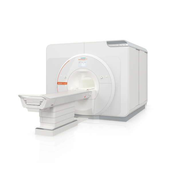
The Magnetom Terra.X: MRI System

The Magnetom Terra.X, a new 7T MRI system, has received 510(k) clearance from the FDA. Manufactured by Siemens Healthineers, it’s a second-generation successor to the Magnetom Terra and offers several enhancements for 7T imaging. Key features include an eight-channel parallel transmit architecture for clinical use, deep learning image reconstruction optimized for 7T, improved diffusion imaging with a high-performance gradient system, and accelerated image acquisition enabling high-resolution brain and knee exams in under 20 minutes. Siemens Healthineers sees this as a significant step in providing better patient care, particularly in neurological and knee imaging. Additionally, the FDA clearance allows existing Magnetom Terra systems to be upgraded to the Magnetom Terra.X.

Image courtesy of Siemens Healthineers

SyMRI 3D for Brain Imaging

SyntheticMR has announced that its latest imaging solution, SyMRI 3D, has received FDA 510(k) clearance for clinical use in the United States. This clearance marks a significant advancement in quantitative MRI technology, offering exceptional resolution and accuracy in brain imaging. SyMRI 3D enables precise volumetric estimations of brain regions, known as parcellation, providing clinicians with deeper insights into brain structure and function. The enhanced resolution facilitates comprehensive lesion analysis, leading to more accurate medical condition assessments. This clearance empowers physicians to make more informed decisions in diagnosis and treatment planning, ultimately improving patient outcomes. SyntheticMR reaffirms its dedication to advancing medical imaging technology and providing innovative tools to enhance patient care through this milestone.


nCommand Lite for Remote Scanning

GE Healthcare has highlighted the FDA clearance of a solution by Ionic Health that enables technologists to remotely supervise patient scans. The system, called “nCommand Lite,” has been tested in Brazil for three years and is vendor-agnostic, allowing remote supervision across MRI, CT, and PET modalities. GE has secured exclusive distribution rights for nCommand in the U.S., aiming to address ongoing workforce shortages in healthcare. Rekha Ranganathan, GE’s chief digital officer for imaging, emphasized the company’s commitment to remote operations and increasing patient access to expert technologists. The system facilitates not only scanning supervision but also training, procedure assessment, and scanning parameter management. GE’s announcement coincides with growing interest in remote scanning, with the American College of Radiology advocating for permanent remote supervision of diagnostic tests. However, technologists have expressed reservations about managing imaging remotely, according to recent survey data from the American Society of Radiologic Technologists.


Sources:

Itnonline.com
Radiologybusiness.com
diagnosticimaging.com
openai.com


March AI News in Diagnostic Imaging

New Research by Harvard Medical School, MIT and Stanford on AI and Clinician Performance

The potential of medical artificial intelligence (AI) tools to enhance clinicians’ performance in interpreting medical images varies among individual clinicians, as highlighted by recent research led by Harvard Medical School, MIT, and Stanford. Published in Nature Medicine, the study underscores the intricate nature of human-AI interaction, which remains incompletely understood. While some radiologists benefit from AI assistance, others experience interference, affecting diagnostic accuracy.

The findings stress the necessity for personalized AI systems tailored to individual clinicians, emphasizing careful implementation to maximize benefits and minimize harm. Despite variations in AI’s impact, the results shouldn’t deter AI adoption but rather prompt a deeper understanding of human-AI dynamics to design approaches that enhance human performance.

To ensure effective integration of AI in clinical practice, collaboration between AI developers and clinicians is essential, alongside rigorous testing in real-world scenarios. Furthermore, efforts should focus on improving AI accuracy and training radiologists to discern AI inaccuracies, facilitating informed decision-making. Ultimately, understanding the complexities of machine-human interaction is pivotal for optimizing patient care through AI integration in radiology.

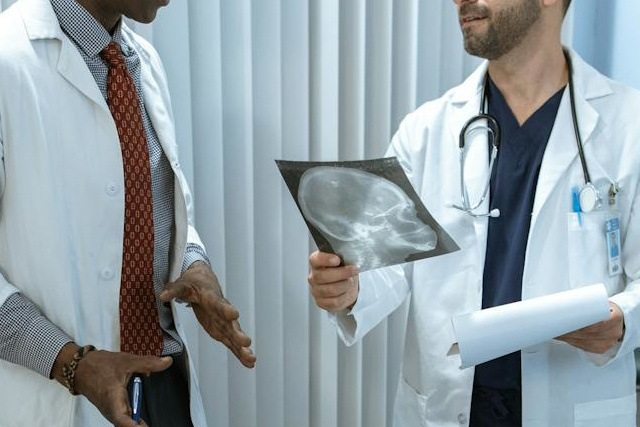
radiologist
A radiologist examines an x-ray

AI and Workflows

New research highlights a novel reporting workflow that automatically incorporates artificial intelligence (AI) findings into structured radiology reports, streamlining physicians’ tasks and saving valuable time. German experts shared their experience with the “AI to SR pipeline,” which integrates a commercially available AI tool for chest X-ray pathology detection and localization into structured report templates.

In evaluations conducted at University Medical Center Mainz, expert radiologists found that reports generated using the AI to SR pipeline were faster compared to free-text reporting and conventional structured reporting. Additionally, subjective quality assessments indicated higher ratings for reports created with the pipeline.

In the hospital’s clinical routine, chest X-ray images are sent to the picture archiving and communication system, then automatically forwarded to the AI tool for analysis. The results are output in a DICOM structured reporting format, taking approximately five minutes from image acquisition to final reporting. Radiologists were able to create chest X-ray reports significantly faster with the pipeline compared to free-text and conventional structured reporting, while also rating the AI-generated reports more favorably.

The authors suggest that this AI-driven reporting pipeline offers standardized, time-efficient, and high-quality reporting for chest X-rays, potentially enhancing AI integration into daily clinical practice and maximizing its benefits.


Sources:

Medicalxpress.com
Radiologybusiness.com
Openai.com


The Latest in Native American Health News: Healthcare Worker Challenges

Physician Shortages

The Indian Health Service (IHS) faces significant physician shortages, with a vacancy rate of 25% in 2018. To address this, the American Medical Association (AMA) recommends creating an office of academic affiliations to establish partnerships with medical schools and residency programs. Currently, the IHS lacks formalized connections with academic medical centers, unlike other federal health systems such as the Veterans Health Administration and the Military Health System. These partnerships could offer training opportunities and help attract physicians to underserved areas. The AMA also suggests raising physician compensation, modernizing facilities, and developing funding streams for rotations and learning opportunities. Additionally, the IHS should evaluate regulatory barriers and provide resources to support physicians serving American Indian, Alaska Native, and Native Hawaiian communities. Overall, the AMA is committed to addressing the physician shortage within the IHS to ensure access to healthcare for these populations.


Cortez Masto’s Legislation for Enhancing Recruitment Efforts

Representatives from the Reno-Sparks Indian Colony Tribal Health Center and the U.S. Department of Health and Human Services advocated for the approval of the IHS Workforce Parity Act before a Senate panel. This legislation, co-sponsored by Senators Catherine Cortez Masto and Markwayne Millen, aims to address healthcare worker recruitment and retention challenges at Indian Health Service (IHS) facilities.

The proposed act would enable part-time providers to access IHS scholarship and loan repayment programs, aligning them with similar programs like the National Health Service Corps (NHSC). This alignment would enhance recruitment efforts in provider-shortage areas, improving access to healthcare in tribal communities.

Testimonies revealed that IHS facilities face significant staffing shortages, with a national vacancy rate of 25%, which can escalate to 50% in rural and frontier tribal clinics in Nevada. The current full-time work requirement for accessing grant and loan repayment benefits acts as a barrier to recruitment and retention efforts.

Understaffing negatively impacts healthcare outcomes in tribal communities, exacerbating conditions such as diabetes, cirrhosis, chronic lung diseases, and behavioral health issues. Failure to address these challenges undermines the U.S. government’s trust responsibility to ensure the healthcare needs of Native communities are met, as outlined in legal agreements between First Nations and the federal government.

New Facilities in Arizona

In Arizona, three new health facilities have opened recently to improve healthcare access for Native American communities, with more projects in progress. Despite strides, Native Americans still face health disparities like diabetes and cardiovascular disease. The Navajo Nation, home to over 244,000 people, operates 12 primary care facilities under the Indian Health Service (IHS), crucial in an underserved area.

Arizona

The Supai Health Station, nestled in the Grand Canyon and reachable only by air, mule, or foot, offers expanded services like primary care and dental. Similarly, the Dilkon Medical Center in the Navajo Nation provides comprehensive healthcare, including in-patient beds and behavioral health support.

Scheduled for May 2024, Sage Memorial Hospital in Ganado will further strengthen healthcare, serving around 23,000 people. Despite progress, challenges persist, including a shortage of hospital beds and healthcare professionals. Recruitment incentives like loan repayment aim to attract Native American individuals to healthcare careers.

Future plans include constructing new facilities in Bodaway Gap, Arizona, and Gallup and Pueblo Pintado, New Mexico, to enhance healthcare access for Native American communities in the region.


Any healthcare facilities needing support in radiology can look to Vesta for accurate and timely interpretations, even for subspecialties. Please contact us to learn more about our 24/7/365 teleradiology services.


Sources:

Nevadacurrent.com
cronkitenews.azpbs.org
ama-assn.org
openai.com

February AI News in Radiology

Brain Tumor Spotted on PET Imaging

An AI algorithm named “JuST_BrainPET” identified a glioblastoma in a patient that had been missed by physicians. This finding, reported in the Journal of Nuclear Medicine, underscores the potential of AI-based decision support in diagnostic and treatment planning. The algorithm automatically segments metabolic tumor volume from healthy tissue on brain PET imaging. In a case study, it detected a lesion in the frontoparietal region, not identified by an expert, which progressed to a small tumor. The AI tool’s early detection could have influenced diagnostic and treatment decisions.


Using Eye-Tracking

Researchers in Lisbon, Portugal, have pioneered a method to enhance AI interpretability in radiology by integrating eye-tracking data into deep learning algorithms. This innovative approach, outlined in the European Journal of Radiology, aims to align AI systems more closely with human understanding, marking a significant leap towards more human-centered AI technologies in radiology. By leveraging eye-gaze data, the researchers sought to bridge the gap between human expertise and AI computational power, anticipating that AI models could learn from the nuanced patterns of image analysis observed by radiologists.


This integration promises AI models that prioritize image characteristics relevant for diagnosis, potentially reducing the disparity between AI decision-making processes and human radiologists’ diagnostic approaches. The potential benefits of this research are vast, potentially leading to AI systems that are not only more effective in identifying pathologies but also more understandable to radiologists, thus fostering trust in AI-assisted diagnostics and accelerating their adoption in healthcare.


Review Paper on AI and Cancer Detection

Professor Pegah Khosravi and her team of researchers explore how artificial intelligence (AI) can enhance anomaly detection in MRI scans to advance precision medicine. Their comprehensive review, published in the Journal of Magnetic Resonance Imaging, focuses on AI techniques like machine learning and deep learning, particularly in identifying tumors in the brain, lungs, breast, and prostate.

The authors discuss several AI strategies for improving tumor detection, including a holistic approach that integrates data from various imaging techniques such as MRI, CT scans, and PET scans, along with genomic information and patient histories. This approach not only enhances anomaly detection accuracy but also facilitates personalized treatments based on comprehensive patient profiles.

Furthermore, the paper explores the use of ensemble methods in AI, which combine different AI models’ strengths to improve anomaly detection. By leveraging these methods, a more thorough analysis of MRI data is ensured. The authors advocate for AI systems that are accurate and transparent in their decision-making processes, fostering trust among healthcare professionals. They also stress the importance of collaboration among researchers, clinicians, and policymakers to effectively implement AI in medical imaging, guiding future advancements in the field.


Sources:

Auntminnie.com
bnnbreaking.com
gc.cuny.edu
openai.com

How to Pick the Best Teleradiology Company

As the demand for healthcare services continues to surge and the shortage of healthcare workers persists, particularly in specialized fields, such as radiology, hospitals and healthcare centers find themselves facing the challenge of ensuring timely and accurate interpretations of medical imaging studies. The critical role of radiologists in diagnosing illnesses and guiding treatment decisions underscores the urgency of addressing this shortage. In response, many institutions are turning to teleradiology companies to bridge the gap and provide remote interpretation services. However, selecting the right teleradiology company is paramount to ensure high-quality patient care and seamless integration into existing workflows. In this discussion, we will explore the criteria for choosing a reputable teleradiology company, considering factors such as expertise, technology infrastructure, turnaround time, and adherence to regulatory standards. By making informed decisions in this regard, healthcare facilities can optimize their radiology services and meet the needs of patients efficiently.

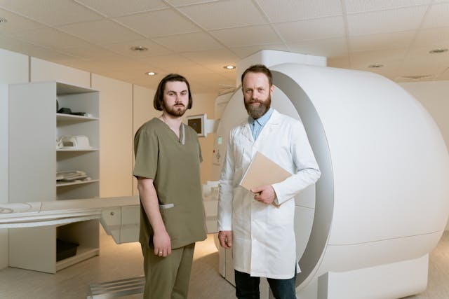
remote radiology company
Rad tech and radiologist

Checklist for Choosing a Teleradiology Partner

Before selecting a teleradiology company, healthcare providers should consider several key factors to ensure they choose a partner that meets their needs and maintains high standards of service. Here are some important considerations:

  1. Quality and Expertise: Assess the qualifications and experience of the radiologists employed by the teleradiology company. Look for board-certified radiologists with expertise in relevant subspecialties. A recent survey of 2,749 radiologists from 108 countries reveals that while they read across almost five subspecialties daily, many lack confidence in certain areas. About 40% accept studies across all specialties, but less than half feel “very confident” in their current subspecialty, so it is vital to ensure the radiologists you work with have expertise in what you require.
  2. Technology and Infrastructure: Evaluate the teleradiology company’s technology infrastructure, including the software used for image transmission and reporting. Compatibility with existing systems and the ability to securely transmit images while maintaining patient privacy are crucial considerations.
  3. Turnaround Time: Timeliness is critical in radiology reporting. Consider the teleradiology company’s turnaround time for providing interpretations. Ideally, they should offer rapid reporting to facilitate prompt patient care and treatment decisions.
  4. 24/7 Availability: Healthcare facilities may require radiology services round-the-clock. Ensure that the teleradiology company offers 24/7 coverage (like at Vesta Teleradiology) to accommodate emergencies and provide continuous support.
  5. Communication and Collaboration: Effective communication between the teleradiology company and the healthcare facility is essential. Evaluate the company’s communication protocols, including how they handle urgent findings and facilitate collaboration between radiologists and onsite clinicians.
  6. Regulatory Compliance: Verify that the teleradiology company complies with all relevant regulatory standards, such as HIPAA (Health Insurance Portability and Accountability Act) regulations for patient data protection. They should also adhere to industry standards for image quality and reporting accuracy.Regulatory compliance
  7. Scalability and Flexibility: Consider the scalability of the teleradiology service to accommodate fluctuations in imaging volumes. Additionally, assess their flexibility in tailoring services to meet the specific needs of your healthcare facility.
  8. Cost and Value: While cost is a factor, prioritize value over price alone. Evaluate the overall value proposition of the teleradiology company, considering factors such as quality, reliability, and the ability to improve patient outcomes.

By thoroughly evaluating these factors and conducting due diligence, healthcare providers can make an informed decision when choosing a teleradiology company, ultimately enhancing the quality and efficiency of radiology services within their organization.

Partnering with a Top US Teleradiology Company—Vesta

Vesta serves as your dependable ally in radiology, extending support to various subspecialties—whether you’re a busy urban hospital or a private practice. We ensure swift processing for both urgent and routine studies. Recognizing the value of your staff’s time and well-being, our teleradiology services enable them to maintain a healthier work-life balance by covering shifts during nights, weekends, and holidays. We can also accommodate any volumes so please reach out to us to learn more.


Sources:

hcinnovationgroup.com
Radiologybusiness.com
openai.com


Top Imaging News of 2023

As we bid adieu to the final moments of 2023, it’s a great time to reflect on advancements and studies that have redefined the world of imaging this year. In this article, we’ll delve into the hottest news and breakthroughs in imaging, highlighting the remarkable strides that have made the headlines.

Study Suggest that Cancer Death Risk From Low-Dose Radiation Is Underestimated

A recent study featured in the British Medical Journal unveils concerning associations between extended exposure to low-dose radiation, commonly experienced by nuclear industry workers, and amplified cancer-related mortality. Drawing insights from the International Nuclear Workers Study (INWORKS) encompassing data from over 300,000 workers, researchers discovered a stark reality: for each cumulative unit of radiation exposure, the risk of death from solid cancer surged by 52%. Even at the lowest cumulative doses, this risk doubled, challenging the assumption that low-dose exposures present less carcinogenic hazard. While the absolute risk remains small, these findings prompt reconsideration of safety limits for workers and call for further studies to confirm the accelerated risk of cancer with ionizing radiation exposure. The hope is that regulatory bodies will integrate these insights into revising protection standards for individuals exposed to low-dose radiation.

Long COVID

In a study published in Medical Hypotheses, a French group presented a theory regarding the brain fog experienced in long COVID, based on brain patterns identified in patient PET scans. They propose that inflammation triggered by COVID-19 disrupts astrocyte cells’ regulation of glutamate, impacting energy metabolism and leading to cognitive fatigue. The authors suggest targeting this malfunction with therapies focused on astrocytic glutamate regulation as a potential way to alleviate long-COVID neurological symptoms. They highlight the lack of mental clarity, difficulty concentrating, and cognitive strain characterizing long COVID, affecting up to 15% of patients after three months of the initial infection. This study builds on previous findings of hypometabolism patterns in long COVID patients’ brain images and explores cellular mechanisms, including links between glutamate dysregulation and cognitive fatigue from other studies. Drawing parallels with “chemo-fog” in cancer patients and cognitive impairment in Parkinson’s disease, the authors suggest therapeutic strategies targeting the identified brain patterns, citing examples from epilepsy treatments and a recent study using medication to improve cognitive function in long-COVID patients. However, the authors stress the need for further research, proposing PET imaging studies using specific markers to comprehend astrocyte function and glutamate regulation for a comprehensive understanding of long COVID’s underlying mechanisms.

chemo fog
Study on brain fog experienced in long COVID

MRIs and Past Cannabis Users

At the International Society for Magnetic Resonance in Medicine (ISMRM) annual meeting, New Zealand researchers presented findings on heavy cannabis use in adolescence to early adulthood and its correlation with brain structure differences in hippocampus and amygdala subregions. The study, led by medical physicist Rebecca Lee and colleagues from the University of Otago in Christchurch, indicated volumetric disparities in these brain regions among heavy cannabis users compared to non-using controls. Notably, past cannabis users showed smaller volumes in specific hippocampal and amygdala subregions. However, the research did not find detectable differences in cerebral blood flow or white-matter tract integrity related to cannabis use, suggesting potential transient brain changes or no long-term effect on these properties. The study, conducted using MRI techniques, emphasized the need for longitudinal studies to clarify the causation and long-term functional impacts of these structural brain changes associated with heavy cannabis use. Despite revealing structural brain changes linked to cannabis use, the study does not definitively establish a causal relationship between these changes and cannabis consumption. Further prospective longitudinal MRI studies are essential to elucidate causality in this context.

MRI study

All About AI

We’d be remiss to not mention how artificial intelligence has shaped the industry this year. Check out our previous articles highlighting the impact that ChatGPT and Bard have made in 2023.


Sources:

technologynetworks.com
auntminnie.com
Openai.com

A Look at 2023 and ChatGPT In Radiology

ChatGPT has quickly moved beyond its niche beginnings and become an integral part of everyday life. Its reach extends well past casual conversation, now penetrating various industries, notably the intricate world of radiology. As we close out 2023, we take a look at some headlines that show how far ChatGPT has advanced in the realm of diagnostic imaging.

Smart Enough to Pass Exam Questions

In two recent studies published in Radiology, researchers evaluated ChatGPT’s performance in answering radiology board exam questions. While the AI showed potential, it also demonstrated limitations affecting its reliability. ChatGPT, based on GPT-3.5, answered 69% of questions correctly, struggling more with higher-order thinking questions due to its lack of radiology-specific training.

A subsequent study with GPT-4 showcased improvement, answering 81% correctly and excelling in higher-order thinking questions. However, it still faced reliability concerns, answering some questions incorrectly and exhibiting occasional inaccuracies termed “hallucinations.”

Confident language was consistently used, even in incorrect responses, posing a risk, especially for novices who might not recognize inaccuracies.


Decision Making in Cancer Screening: Bard Vs ChatGPT

A study recently published in American Radiology compares ChatGPT-4 and Bard, two large language models, in aiding radiology decisions for breast, ovarian, colorectal, and lung cancer screenings. They tested various prompts, finding both models to perform well overall. ChatGPT-4 showed higher accuracy in certain scenarios, especially with ovarian cancer screening. However, Bard performed better with specific prompts for breast and colorectal cancer. Open-ended prompts improved both models’ performance, suggesting their potential use in unique clinical scenarios. The study acknowledged limitations in scoring subjectivity, limited scorers, and the focus on specific cancer screenings based on ACR guidelines.

bard AI
Can AI assist in diagnostic imaging?

Simplifying Readability of Reports

The study in European Radiology explores using ChatGPT and similar large language models to simplify radiology reports for easier patient comprehension. Researchers had ChatGPT translate complex reports into simpler language for patient understanding. Fifteen radiologists evaluated these simplified reports, finding them generally accurate and complete, yet also identified factual errors and potentially misleading information in a significant portion of the simplified reports. Despite these issues, the study highlights the potential for large language models to enhance patient-centered care in radiology and other medical fields, emphasizing the need for further adaptation and oversight to ensure accuracy and patient safety.


Sources:

Rsna.org
diagnosticimaging.com
Radiologybusiness.com
openai.com


The Future of Medical Imaging: Latest Augmented Reality Advancements

Medical imaging is crucial in diagnosing and treating various medical conditions. These technologies have transformed the medical field from X-rays to Magnetic Resonance Imaging (MRI) and Computed Tomography (CT) scans.

Augmented Reality (AR) is a technology that overlays digital data, such as images or sounds, onto real-world objects or locations. In the medical field, AR has become an essential tool in patient examinations, surgery, and therapy.

AR-assisted Surgery

One of the significant benefits of AR technology in medicine is its application in surgical procedures. With AR, surgeons can see 3D images of the patient’s internal organs, tissues, and blood vessels during an operation.

This technology has revolutionized how physicians perform surgeries, offering unparalleled precision, accuracy, and visual assistance. Surgeons can perform surgeries with minimal invasion, thus reducing patient recovery time.

Additionally, AR-assisted surgery can benefit doctors in training as they can learn surgical procedures through virtual reality simulations.

Image-guided Intervention and Therapy

Image-guided intervention and therapy is another area that has experienced significant advancements in AR technology. Medical professionals can use AR to display real-time images of the internal organs during a procedure, allowing for precise navigation and guidance.

For example, using AR technology in radiation therapy enables medical professionals to focus radiation beams precisely on the tumor and spare the surrounding healthy tissue.

Patient Education and Rehabilitation

As medical professionals seek to enhance patient education and rehabilitation, AR technology has proven helpful. Patients can use AR to view and understand their medical conditions better.

For instance, AR can help showcase the effects of a particular treatment plan and provide feedback on the treatment progress. AR can also be used to improve rehabilitation sessions, as patients can receive personalized therapy recommendations that suit their needs.

Medical Education and Training

The applications of AR technology in medicine are not limited to diagnosis and treatment. Medical education and training have also benefited tremendously through AR technology.

Through virtual reality simulations, medical students can explore the human body in 3D and get hands-on experience with various procedures. Additionally, trainees can learn how to use medical equipment through AR simulations without endangering patient’s lives.

vr training session

Remote Patient Monitoring

AR technology has the potential to transform remote patient monitoring. Patients can use AR technology to capture images and videos remotely and send them to medical professionals.

This approach saves time and resources, especially for people living in remote areas, enabling them to receive diagnoses and medical advice promptly.

The advancements in augmented reality technology have revolutionized medical imaging by transforming diagnostics and treatment procedures. AR technology has applications in surgery, image-guided interventions and therapy, patient education, medical education and training, and remote patient monitoring.

With continued research and development, AR technology can potentially transform healthcare significantly. The advancements in AR technology are exciting as they offer the possibility of more accurate, efficient, and cost-effective medical procedures, diagnosis, and treatment, and ultimately, improved patient outcomes.