Breast Cancer Awareness Month Kicks Off Now: The Latest in Breast Cancer Studies

October is Breast Cancer Awareness Month, a time when individuals and organizations around the world unite to raise awareness about one of the most prevalent and potentially life-threatening diseases affecting women and, in some cases, men. Throughout this month, campaigns, events, and educational initiatives aim to promote early detection, support those impacted by breast cancer, and advance research efforts. In this article, we will delve into the latest news and developments in the field of breast cancer awareness and research, highlighting the ongoing efforts to combat this disease and improve the lives of those affected by it.


Migraine and Breast Cancer: Is There a Link?

Migraine, a debilitating neurological disorder affecting 14-15% of the global population, has been associated with various health risks, including stroke, high blood pressure, epilepsy, tinnitus, and irritable bowel syndrome (IBS). Recent research has explored a potential link between migraines and breast cancer, both influenced by estrogen levels. While some studies suggest a higher breast cancer risk in individuals with migraines, others indicate the opposite or mixed results.

woman with a migraine

A study by researchers from the Cancer Center at West China Hospital of Sichuan University in China delved into this connection, utilizing genetic data from genome-wide association studies (GWAS). Their Mendelian randomization analysis revealed that women with any type of migraine face an increased risk of overall breast cancer and estrogen receptor (ER)-positive breast cancer. Notably, women experiencing migraine headaches without aura showed a heightened risk of ER-negative breast cancer, with suggestive associations for overall breast cancer risk.

However, medical experts caution that this study is retrospective and associative, requiring replication in diverse populations to establish a causal relationship. The degree of increased risk is relatively small compared to other genetic factors influencing breast cancer risk. Nevertheless, this research opens the door to future investigations into the complex interplay between migraines, genetics, and breast cancer, shedding light on potential contributors to this disease.


Are Older Women At Risk for Overdiagnosis?

A study involving 54,635 women aged 70 and older found that continuing breast cancer screening after age 70 carries a significant risk of overdiagnosis, which is the detection and treatment of cancers that would not have caused harm in a person’s lifetime. Over 80% of women aged 70-84 and over 60% of women aged 85 and older continued screening. The study showed that overdiagnosis estimates ranged from 31% of breast cancer cases in the 70-74 age group to 54% in the 85 and older group. However, there was no statistically significant reduction in breast cancer-specific death associated with screening in any age group. Overdiagnosis was primarily driven by detecting in situ and localized invasive breast cancer, not advanced cases. The study emphasizes the importance of considering patient preferences, risk tolerance, comfort with uncertainty, and willingness to undergo treatment when making screening decisions for older women. The study’s limitations include the potential misclassification of diagnostic mammograms as screening and the inability to adjust for certain breast cancer risk factors.

Vesta Teleradiology: Mammogram Interpretations, Day and Night

In conclusion, as healthcare practices navigate the intricacies of mammogram interpretations, our company is here to provide unwavering support. We understand the importance of accurate diagnoses in breast health, which is why our dedicated team is available day and night, even during holidays, to assist healthcare professionals. Your commitment to patient care is our priority, and we’re here to ensure that you have the expertise and assistance you need for precise mammogram interpretations.


A Tale of Two Studies: Are Doctors Fulfilling too Many Imaging Requests?

A recent qualitative study aimed to investigate the factors influencing general practitioners’ (GPs) decisions to fulfill patient requests for imaging studies during clinical consultations. Ten GPs from private medical centers in Northwest Sydney were interviewed, and their perspectives were analyzed through content analysis. On the other side of the spectrum, we explore an American study that reveals the importance of encouraging more people to undergo screenings.

Patient Imaging Requests

The study revealed six key themes that emerged from the interviews regarding GPs fulfilling patient imaging requests:

Patient Expectations: Patients’ desires and expectations played a significant role in GPs’ decisions to fulfill imaging requests. Some patients expected certain tests due to their health concerns or beliefs.

‘Therapeutic Scans’: GPs recognized that some patients viewed imaging scans as a form of reassurance or therapeutic action. This influenced GPs to fulfill requests to alleviate patient anxiety.

“I find X-rays have become a therapeutic requirement, [and] that their [patients’] problem often dissipates once the request is given. (Female, 40)”

‘Impressive Labels’: The use of serious-sounding terms in imaging reports contributed to patients’ perception of the severity of their condition. This, in turn, led to increased requests for such scans.

Entitlement: Some patients exhibited an attitude of entitlement, expecting imaging scans without sufficient medical justification. GPs were more likely to decline such requests, especially during initial visits.

Defensive Medicine: GPs expressed concerns about potential legal repercussions if they refused imaging requests. Fear of litigation influenced GPs to fulfill requests to avoid legal issues.

‘New Patients’: GPs were cautious about fulfilling imaging requests from new patients, as they needed time to establish trust and assess the necessity of the requested scans.

The study concluded that GPs faced challenges in balancing their role as gatekeepers of imaging with patient expectations. Clear guidelines, patient education, and GP training were suggested as potential strategies to manage patient expectations and provide appropriate care. The study sheds light on the complexities of patient-GP interactions regarding imaging requests and highlights the need for evidence-based strategies to navigate these challenges. The study was conducted within the Australian Medicare system, where medical imaging is relatively accessible and affordable, and the findings contribute to understanding the dynamics of patient-GP interactions in this context.

Cancer Screenings Saves Millions of Years of Life

Meanwhile an American study conducted by teams from the University of Chicago, University of Michigan, and study sponsor Grail LLC estimates that Americans have gained an additional 12 million years of life due to preventive cancer screenings over the past 25 years, leading to an economic impact of around $6.5 trillion. These screenings, targeting early signs of breast, colon, cervical, and lung cancers among high-risk adults, emphasize the importance of encouraging more individuals to undergo recommended cancer checks and exploring novel screening methods. The study suggests that if all eligible individuals received current screening for these cancers, it could save an extra 3.3 million life-years and add $1.7 trillion in economic impact.

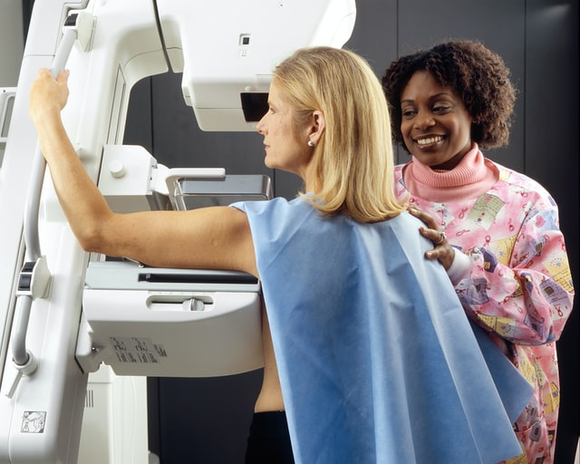
mammograms


The research utilized a mathematical model to assess the impact of cancer screenings endorsed by the U.S. Preventive Services Task Force (USPSTF). This federally authorized panel evaluates evidence for preventive medical services and provides recommendations using letter grades. Since 2010, screenings with “A” or “B” grades have been covered by most insurances without patient costs under the Affordable Care Act.

Although screenings for breast, colorectal, cervical, and lung cancers have saved millions of life-years, their full potential remains untapped. The majority of life-years saved and economic impact come from cervical cancer screening due to its early and consistent screening intervals for individuals aged 18-65. However, the study acknowledges that the availability of the HPV vaccine since 2006 could affect the future value of cervical cancer screening.

While there are validated screening tests lacking for many cancer types, decreased death rates from cancers with available screenings have contributed to the overall reduction in U.S. cancer deaths. To establish more broadly validated cancer screenings, further research is needed, potentially evaluated by the USPSTF. Novel screening tools like blood tests for multiple cancer types are in development, but AI-based tests have yet to undergo review by relevant bodies for efficacy and recommendations.


Sources:
onlinelibrary.wiley.com
michiganmedicine.org
Openai.com

State of the Healthcare Industry: Hospital Strikes

Americans, and everyone all over the world, depend on proper medical care. From dental checkups to urgent care visits, medical professionals are absolutely essential, especially when it comes to massive emergencies. If we didn’t learn this fact yet, the pandemic certainly brought it into focus. In 2020, hospitals and medical buildings surged with COVID patients, overloading facilities and professionals alike. Now, with labor shortages, and unfair compensation, nurses and medical staff are going on strike to re-negotiate contracts to make their workplaces a fairer place to be.

Healthcare Strikes Going on Now

Beginning in January of this year, over 7,000 medical professionals went on strike in New York City, says Vox. The strikes involve Mt. Sinai Hospital, and three other facilities, located in the Bronx, owned by Montefiore. Recent contract negotiations failed to provide health care workers with more staff, as well as desired salary compensation, says the article. Over 700 positions are open within the Montefiore facilities, and nurses and medical staff feel overburdened and undervalued. As explained by Vox, when billable hours and revenue collection are limited to doctors who prescribe surgeries and medicine, nurses and other medical staff are a complete cost to the hospital. Even though these professionals are essential to properly run any medical facility, investing in nursing staff becomes a problem when the American structure is a pay-for-treatment transaction in healthcare systems that are for profit.


Last month, on February 27, another strike took place on Long Island with about 800 nurses, says CBS News. At Northwell Health’s South Shore University Hospital, nurses are demanding “safe staffing and fair wage,” says the article. 99% of the nurses voted to authorize the strike.

burnt out


It isn’t just the United States that’s involved with strikes. Across the pond, in the UK, nurses are also having a difficult time negotiating what they need in their contracts, says IN News. For the first time in Royal College of Nursing history, medical staff, which spanned over 100 services, held England’s largest nursing strike on March 1 of this year. In the 48-hour strike, staff from emergency departments, intensive care units, cancer care, and other services were involved, says the article. Compensation, workload, and workplace conditions are the terms UK nurses are fighting for. Since 2010, nurses’ compensation has fallen 8%, says the article.


Both in the US and UK, nurses have made it clear that they do not want to strike, leaving vulnerable patients in a bind, says USA Today, and IN News. “Bosses have pushed us to strike by refusing to seriously consider our proposals to address the desperate crisis of unsafe staffing that harms our patients,” said a union representative from The Nurses Association to USA Today.


Re-negotiated contracts and tentative agreements were reached in New York, some involving a raise of 5-7% over the next three years for nursing staff, as well as staffing increases, says Today. Negotiations in the UK came to an agreement on March 16, with more than 1 million NHS staff receiving a raise, says gov.uk.


Shortage in Radiologists

It’s not just nurses that are in shortage, but also those in the radiology field as well. Luckily, partnering with a teleradiology company like Vesta allows you to fill in those gaps you have—whether you’re short-staffed for weekend work or simply need reliable interpretations around the clock. Vesta also offers top-to-bottom healthcare staffing from its sister company, Momentum Healthcare Staffing for positions such as locums tenens and permanent nurse practitioners, physicians, medical assistants and more.

radiologists

Women in Imaging: Historical and Contemporary Figures Advancing Medicine

March is a month set aside to celebrate women. During this month, Women’s History month, people are encouraged to learn about and celebrate the strong women in their lives and throughout history. But women’s history is wide reaching and all encompassing. There are thousands of women in hundreds of different industries that have made lasting impacts.

One of those industries is the medical industry. Women have left their mark everywhere in medicine and have helped develop new ideas, new medicines and treatments, and new pieces of technology. Let’s take a look at pillars in medical imaging.

History of Medical Imaging

But first, what exactly is medical imaging? This section of the medical industry includes different technologies that help medical professionals view various internal body systems within the human body. Medical imaging is used to find, diagnose, monitor, and even treat different medical conditions or injury.

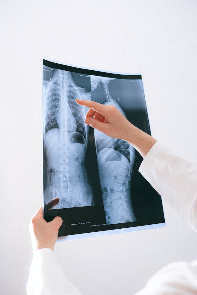
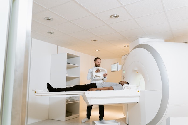
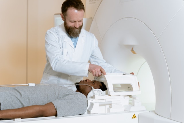
Each piece of technology encompassed in medical imaging focuses on a different area or system of the body. Take x-rays for example. X-rays are used to view the skeletal system – the bones – of the body. X-rays are used to identify different issues with a patient’s bones and joints. Other types of medical imaging includes ultrasounds, Magnetic Resonance Imaging (MRI), Computed Tomography (CT), and even mammography.

diagnostic imaging

Medical imaging, as a practice, has been around since the last 19th century. It began with the development of x-rays. The first x-ray was taken in 1895, and since then the industry has grown as new pieces of technology have been developed. And women have played a pivotal role in this development.

Women in Medical Imaging

There have been innumerable women who have had a hand in the development and advancement of medical imaging. Of those, there are a few that we’d like to highlight.

Marie Curie

The first woman of medical imaging was a Polish scientist named Marie Curie. While this name is often familiar to many people, it’s familiar because of her discovery of radium. Radium is a naturally occurring radioactive element that was instrumental to the development of medical imaging.

Without Marie Curie’s discoveries, medical imaging may not exist, or would at least have been seriously delayed. Medical imaging uses radiation to see within the human body, so Marie Curie and her developments were imperative.

Florence Ada Stoney

Florence Ada Stoney was a radiologist during World War I. While she may not have created a new technology or radiology treatment, she was the lynchpin of women’s participation in medical imaging and radiology.

Florence Ada Stoney
Florence Ada Stoney in the center pictured with her sister and father

Stoney was not allowed to serve in the x-ray department of a London hospital during the war effort because she was a woman. Rather than admit defeat, Stoney created a volunteer unit comprised entirely of women. In 1915, Stoney was hired as the head of the Fulham Military Hospital’s x-ray department – the first time a woman headed such a department.

Muyinatu Bell

Muyinatu Bell is a modern counterpart of the forerunners of medical imaging. In the mid-2010s, Bell developed a new ultrasound technique that produces higher-quality images and is most effective for use in obese patients.

Without these women, modern medical imaging wouldn’t be where it is today. But this list is not exhaustive. There are countless more women who have found their place in medical imaging and left their mark.

While the number of women actively working in medical imaging is rarely higher 30%, no matter what area or discipline, they are still making serious gains. Women are a small portion of the medical imaging world, but they are mighty. 

How Covid Impacted Vital Health Screenings

So many aspects of our lives have changed since the outbreak of COVID19; shopping, working, education, entertainment, and our everyday lives look pretty different, even from a few years ago. The pandemic has also impacted our health and the way we approach medicine. Advancements in technology are moving quickly in medicine. Telehealth and remote medical services are becoming more mainstream, however, convincing patients to get health screenings, particularly for cancer, has become a challenge. It’s important, now more than ever, to keep up good health practices and let patients know they shouldn’t delay getting screened.

Timely screenings are incredibly important, says Imaging Technology News. Since the pandemic began there have been massive delays that have continued into 2023. Many facilities closed temporarily during the lockdown and many other facilities had severe staff shortages, says National Cancer Institute. When facilities did open back up, people were fearful of going to hospitals and other places where COVID might be prevalent, says the article.

covid impact on health
The COVID pandemic has caused many to delay important screenings

According to a survey between June and November of 2020, 11% to 36% of the 7,115 participants delayed their screening. The screenings ranged from mammograms, stool blood tests, pap smears, colonoscopies, and HPV tests. In another study done by Tulane University, screening rates decreased to nearly zero in 2020 of the 45,000 women enrolled in Medicaid in Louisiana. Should this trend continue, it’s predicted that later-stage cancer diagnosis will be significant, says Imaging Technology News.


How Can Medical Facilities Encourage Patients to Get Vital Screenings?

What medical professionals can do is encourage their patients as much as possible to get screened. This may be tricky, with COVID still lingering around the country. Appealing to patients’ health, and letting them know that a screening can save their life can be the most effective, says Imaging Technology News, as well as encouraging patients through evidence-based interventions, says the National Cancer Institute.

campaigns to increase mammogram awareness
Cancer prevention begins with screening

When cost is a barrier, says the article, there are options. Through the Affordable Care Act there are affordable cancer screening tests available as well as low-cost screening options for those without insurance or physician referrals. Medical professionals and facilities may also consider expanding the locations and access to screenings through mobile screening programs, says the article. Many of those in low-income areas who do not have transportation to access screenings can take advantage of mobile programs, says the American College of Surgeons.

For people who aren’t visiting doctors or medical facilities, making the general public aware of how important screenings are can help, says the National Cancer Institute. Social media, campaigns, and patient education are a few ways to reach everyone. In a multipronged approach, some hospitals educated virtually through community focus groups, asking specific questions as to why the people in the area weren’t getting screened, said the article. Hospital staff also worked with parish nurses to encourage and educate congregations on screening. In addition, some hospitals encouraged their own staff to be screened.

In our strange, new world, amidst COVID, staff shortages, and so much fear, we still must take care of ourselves and encourage others to do so. As healthcare professionals, we have the power to empower others to take charge of their own health.


Combating Radiologist Shortages

If your hospital, emergency room, or private practice is struggling with radiologist shortages, partnering with a teleradiology company like Vesta can help.

Our US Board Certified radiologists offer night, weekend, and holiday coverage to fill those gaps. Ask us about our seamless integrations: contact us now to learn more.

Reduced Radiation Exposure in Imaging

Medical imaging is a powerful tool for diagnosing and treating medical conditions; however, it can also expose patients to excess radiation levels. Fortunately, recent technological advancements have resulted in the development of new ways to reduce radiation exposure during imaging.

Here are a few ways these advancements are helping patients receive the care they need while reducing their radiation exposure.

New Guidelines

Clinicians have always used guidelines for the use of CT scans. Still, they recently developed new guidelines for the benefit of CT scans on the head, neck, chest, and abdomen/pelvis areas of the body–especially in pediatric practice.

x-ray safety for kids
New guidelines for CT scans


Clinicians can reduce or eliminate the need for CT scans if the child is awake and alert, if the blood work is normal, and if the patient is not experiencing abdominal pain.

A study of 146 children not exposed to the excess CT scan radiation using the new guidelines showed no clinically significant missed injuries.

Smart Software

One of the most significant advancements in this area is using smart software to reduce radiation dose in computerized tomography (CT) scans. This software analyzes patient size, age, and other factors to determine the dosage necessary for an accurate scan that minimizes radiation exposure. In some cases, reducing radiation doses by up to 90 percent has been possible without any decrease in diagnostic accuracy.


Lower Radiation Levels in the New CT Machines

In addition to using innovative software to reduce doses, researchers are also looking at new technologies, such as low-dose CT machines, that can achieve similar results without any additional processing needed.


Technicians have designed these machines with unique components that limit X-ray emissions while still providing high-quality imaging. As a result, patients can receive the same level of care without worrying about unnecessary radiation exposure.


Reducing Radiation Exposure with Ultrasound

Finally, reducing radiation exposure through ultrasound technology is more progressive. For clinicians using sound waves instead of X-rays or other forms of energy, ultrasound technology can provide doctors with highly detailed imaging with no risk of exposing patients to dangerous radiation levels.

Ultrasound is an ideal option for pregnant women and children who may be more sensitive to the effects of radiation than adults.

vesta teleradiology for women's wellness centers
An ultrasound tech consults a patient

Recent advancements have made it easier for medical professionals to provide high-quality care while minimizing their patients’ exposure to harmful radiation.

With the medical profession redefining the guidelines, using low-dose scans, better x-ray machinery, and more ultrasounds, many options are now available for reducing radiation exposures during imaging procedures—all without sacrificing diagnostic accuracy or quality care.

That’s great news for anyone who needs an imaging scan but wants to ensure they’re taking steps to protect their health and safety!


How AI is Making an Impact on Radiology and Imaging

The fields of science and medicine are always progressing. This progression intends to help both patients and providers.

Today, artificial intelligence (AI) is becoming common as a way to diagnose patients. It provides a more efficient way to collect and store information. The software can even analyze imaging to a high level of accuracy. This helps providers catch a problem that they may have missed before.

AI is a field that is advancing quickly. What progress have we seen in the past couple of years? What programs have we begun to put in place?

What Is Artificial Intelligence?

Artificial intelligence refers to highly advanced computers or computer-controlled robots. These computers are capable of performing incredibly complex tasks. Before, we thought these tasks could only be done by intelligent beings.

AI in imaging
AI is making advancements in the medical field

These computers are often associated with human characteristics. They seem to be able to reason and learn from past experiences.

How Is Artificial Intelligence Used For Diagnostic Imaging & Radiation?

Using AI in radiology and imaging has been gaining traction in the medical world. We use it largely to store and analyze data, helping physicians to make a prognosis. AI can store and analyze all a patient’s records. It can then make a diagnosis based on those records. The analysis is often far more accurate than what a human counterpart can do.

The use of AI is also helpful because of its storage capability. AI can have large imaging biobanks to hold more images than standard computers.

It also makes the lives of physicians easier by filtering patients by need. It can recommend appropriate diagnostic imaging based on the patient’s current records. It can also sort patients by priority in the case of an emergency.

What Advancements Have Been Made?

AI means to eliminate problems associated with human limitations. Traditional imaging takes a team of technicians. They must take the imaging as well as interpret it. This can be time-consuming. Plus, AI is able to analyze images with far greater accuracy than the human eye.

Radiomics

Radiomics is a tool that performs a deep analysis of tumors down to the molecular level. AI can perform radiomics with far better accuracy than the human eye or brain.

AI can analyze a specific region and extract over 400 elements. It then takes these features and correlates them with other data to form a diagnosis. The AI can analyze features from radiographs, CT, MRI, or PET studies.

Rapid Brain-Imaging AI Software

Hyperfine is the manufacturer of portable MRI machines. They are now creating these machines with new AI intelligence software. They believe that this new software will be able to perform brain scans in under 3 minutes.

AI-Generated Drugs

In 2020, an AI-created drug went to human clinical trials. The drug intends to treat OCD, and was designed entirely by AI. Exscientia is the manufacturer of the drug. They say that it normally takes about 4.5 years to get a new drug to this stage of testing. With AI generation, the drug got to the human clinical trial stage in under 12 months.

Making A Diagnosis

We stated earlier that AI is being used as a way to more efficiently diagnose patients. Still, relying entirely on AI to do this can complicate things and may be unwise.

So, the researchers of MIT’s Computer Science and Artificial Intelligence Lab worked to combat this. They created a machine learning system that analyzes the data and decides whether to diagnose.

If it “feels” it’s unable to make an accurate prediction, it will defer to a medical professional. It even considers whether to defer to an expert based on who in the medical team is available. It will consider each team member’s availability, level of experience, and specialty.

Conclusion  

AI in diagnostic imaging shows promise to truly advance quality of care for patients. We are excited to see more advancements in this arena. In the meantime, we don’t believe any machine can currently replace a trained human eye when it comes to interpretations. At Vesta, we provide US Board Certified radiologists who work to provide accurate preliminary and final interpretations. Learn how we can support your radiology department– contact us today. 

Recent Breakthroughs in Radiology and Imaging

Since the 19th century, radiology and imaging have been making breakthroughs in clinical practice, allowing for safer and more effective treatment for millions of patients. Recent breakthroughs have made the field of radiology more dependable, cost-effective, and practical.

In the healthcare realm, radiology has become the standard for modern equipment used for preventive care, surgical treatment, diagnostics, and more. Here are some ways radiology and imaging have expanded healthcare technology:

Imaging Tests Replace Invasive Surgery

Imaging tests have gone a long way to replace exploratory diagnostic testing and unnecessary invasive surgeries. Before radiology and imaging tests, medical professionals may have had a more challenging time identifying fractures, broken bones, infections, cancers, tumors, strokes, MS, epilepsy, etc.

Imaging tests help doctors, nurses, surgeons, dentists, and more visualize what’s happening inside your body using imaging scans like X-rays, MR scans, CT scans, ultrasounds, etc., without performing invasive surgery. Seeing what’s happening inside your body is an incredible superpower, which is how imaging tests have helped save millions of lives.

If you have to undergo surgical or dental procedures, imaging tests can help guide medical professionals for accurate results.

Scan for Cancer with PET/CT

PET scans combined with CT scanning allow medical professionals to look at your biological functions, metabolic changes associated with cancer, and changes to your organs. While radiation exposure is related to this process, radiology technology is working to reduce that.

Currently, healthcare providers work on limiting exposure using specific medications, lead aprons, and more. The payoff is detecting cancer much earlier than with traditional imaging and scans. Not only can these scans look for cancer, but they can also even monitor your chemotherapy treatment.

ct scan

Breast Cancer Screening with Digital Mammography

Digital mammography is a highly effective screening method for breast cancer, especially compared to traditional methods. Digital mammograms have proven to be more accurate, effective, reliable, and easier to share with other medical providers. 

CT Angiography Blood Vessel Imaging

Until recently, angiography was performed by inserting a catheter into an artery to inject a substance visible in an X-ray. This allows medical professionals to look for internal bleeding, blockages, and other health problems. This process can take a long time, require sedatives, and have a slight chance of bleeding or blood clots. High-tech CT angiography scans provide the same information without being invasive. Plus, the new process takes less than 30 minutes.

As radiology and imaging technology keeps experiencing breakthroughs, the impact on healthcare practice is definite. We can look forward to quicker, minimally-invasive diagnostic exams, more precise and more accurate images, and the ability to share scans instantly between providers.

For patients and providers both, this represents quick visits and satisfying results. Recent breakthroughs in radiology and imaging mean the process is safer, faster, and more cost-effective.

 Teleradiology

Teleradiology itself has been an amazing advancement in the medical field, allowing radiologists from anywhere in the world, the ability to receive and interpret scans. The US Board Certified Radiologists at Vesta are able to work with any healthcare provider to provide premlin and final interpretations, and we work nights, holidays and weekends to support your staff and operations. Contact us to learn more.

Why Hire a Teleradiology Company? The Benefits of Teleradiology

Teleradiology is an industry on the rise. Reports from Emergen Research project that the industry’s market size will reach $40 billion globally by 2028.

But why are so many medical clinics adopting teleradiology? It’s because this type of imaging offers many significant benefits. Today, let’s discuss just a few ways that teleradiology can benefit your practice.

How Teleradiology Helps Patients

Medical professionals have long been advocating for teleradiology as a tool to make healthcare more accessible. In fact, medical journals were promoting radiology as long ago as the 1980s!


One of the main arguments for teleradiology is the potential to expand your patient network, particularly in rural areas. Teleradiologists don’t need to travel to a hospital or physician’s office to read an image, which means rural hospitals can diagnose patients faster and more effectively.

In addition to the opportunity to work with more patients, the reality is that telehealth — including teleradiology — is becoming widely accepted by patients. The COVID-19 pandemic forced many patients to try this method of care for the first time, and the reception was generally positive. Today, many Americans favor telemedicine — in fact, 35% say they would consider replacing their primary care doctor with a telehealth physician!


Hiring a teleradiology company gives your office the opportunity to impact more patients’ lives than ever before.

How Teleradiology Helps Hospitals

It’s no secret that the medical industry is in the midst of a labor shortage, and radiology is no exception. In the next decade, the United States is expected to have a shortage of 17,000-42,000 radiologists! Teleradiology can help lessen this burden. By working with a teleradiology company, you have access to diagnostic imaging even if your facility doesn’t have a radiologist on staff.

teleradiology company
Teleradiology reviews an x-ray image


Additionally teleradiology companies can help keep your costs low. When you work with a teleradiology company, your costs are drastically reduced. Teleradiologists can access images from anywhere, so there’s no need to pay for travel expenses or hire a full-time, in-house employee. You can get the same quality patient diagnostics with far less overhead.


It is clear that teleradiology is a benefit to patients and physicians alike — which means it is very likely the future of healthcare. Hiring a teleradiology company for your practice will allow you to serve patients in a faster, economical, and more accessible way.

 Vesta Teleradiology

Contact Vesta to learn more about how teleradiology can help your practice today.

Pillars in Radiology History, Past, and Present

Pillars in Radiology History, Past, and Present

Radiology is a phenomenal tool. Medical teams depend on it to guide diagnosis and treatments. As a product of the early twentieth century, it is hard to imagine how medicine functioned without it. Today, our world is a much better place thanks to the people who discovered and developed the powers of radiology.

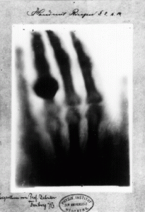
The father of radiology is Wilhelm Roentgen. Beginning with an interest in cathode rays in October of 1895, he is credited with having detected electromagnetic radiation in a specific type of wavelength on November 8, 1895. From that point, Roentgen submitted his first publication titled “On a New Kind of Rays” on December 28, 1895. When Roentgen was asked what he thought when he made his discovery, he responded, “I didn’t think, I investigated.”  Encouraged to name his discovery Roentgen Rays, he chose the letter “X” to name the rays due to their mysterious and unknown nature. The famed X-ray picture of his wife’s hand shows her bones and wedding ring, clearly indicating the rays would not penetrate bone or metal.  Few discoveries have gained notoriety as quickly as Roentgen’s did. Within a year, X-rays had changed the fields of physics and medicine exponentially. It earned him the inaugural Nobel Prize in Physics in 1901and opened doors for Roentgen personally and professionally, some of which he appreciated and some he did not. While he did not accept the title leading to German nobility offered to him, he did agree to accept the honorary degree of Doctor of Medicine his university extended to him.

first ever xray
First medical X-ray by Wilhelm Röntgen of his wife’s hand

In addition, he donated his Nobel Prize earnings to his university, Wuerzburg University in Germany. An indication of his impeccable character. He never took any patents on X-rays. He wanted the world to benefit from his work freely. Consequently, he died almost bankrupt during World War I, but his goal was achieved. The world has benefited greatly from his work.

After Roentgen’s discovery, other scientists soon followed with further exploration of the rays. The first scientists to extend Roentgen’s work were Sir William Henry Bragg and William Lawrence Bragg. They are a father and son who earned the Nobel Prize in Physics in 1915 together specifically, “for their services in the analysis of crystal structure by means of X-rays” soon to be known as Crystallography, the basis for analytic chemistry. “The diffraction of X-rays by crystals,” written in 1922 by William Henry Bragg, describes their beginning work. Credited with the creation of Bragg’s Law, learning the atomic structures of viruses, proteins, gemstones, and more has been possible.

Many scientific fields were developed from Roentgen’s discovery of the X-ray. Major names of the time, such as Albert Einstein, who established matter and energy to be equal in 1903, were contenders in the study of unseen forces associated with Roentgen’s work as well. Considering the most evocative pioneering scientists, perhaps the most influential one was Marie Curie.

Intently studying Roentgen’s work, Marie Curie took an interest in uranium, the weaker ray, also called Becquerel rays. Studying its compounds, she eventually identified that the electrical effects of uranium rays are constant, which led to a monumental shift in the understanding of the structure of the atom, which led to the development of radioactivity. With this information, she determined radiation is a powerful tool with a wide range of potential applications, including diagnostic and therapeutic procedures. Her continued work with her discovery of radium not only earned her a second Nobel Prize, but today we have vast benefits from her discoveries, including nuclear energy and radiotherapy (RT), a treatment for cancer.

Vesta Teleradiology

We truly applaud and honor these individuals for their immense contributions. As a teleradiology service provider, we understand that delivering accurate interpretations along with stellar support is how we can continue to contribute to the overall mission of helping others, ultimately the healthcare provider and their patients.