The Role of 3D Printing in Radiology

3D printing is an incredible technological tool both in and out of the medical world. Fortunately, this revolutionary technology has opened up an entirely new realm of possibilities for medical professionals to better their patients care. 

Why Is 3D Printing Important To Medical Professionals?

3D printing has enabled doctors to create models of patients’ organs, tissues, and bones that can show the precise image of the patient’s anatomy, enabling them to make more thorough and accurate diagnoses. This means that diseases can be caught faster, leading to a higher percentage rate of positive outcomes. 

These models are helpful to ensure quality by using test treatments on a physical model before trying them on a patient. This can help to reduce the risk of adverse reactions and ensure that the treatment is as effective as possible. In addition, 3D printing can be used to create prosthetic limbs, allowing patients to receive a personalized prosthesis that is custom fit to their body.
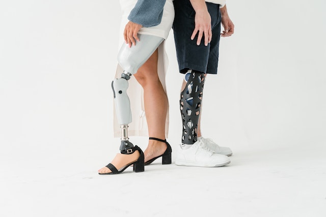
prosthetic limbs
Prosthetic limbs

Finally, 3D printing is being used to create custom medical implants. Medical implants are used to replace missing or damaged tissue. They are typically made of titanium or other materials, but 3D printing has enabled medical professionals to create custom implants that are specifically designed for each patient. This has improved patient outcomes and allowed doctors to create implants that are more effective than ever before.

3D Printing In Radiology

In an interview for the American College of Radiology, Kenneth C. Wang, MD, Ph.D. said, “the benefit of printing comes from the patient-specific nature of what is depicted. And that almost always starts with imaging. That’s why we, as radiologists, are so well positioned to be at the center of these workflows. We can use our deep understanding of imaging modalities, anatomy, and disease to create models, interpret findings and also to know the limitations of the images, and communicate with other providers.” 

3D printing has become an increasingly common and useful tool for radiologists. Because it can be used to create customized models of organs, bones, and other body parts, it gives radiologists a better visualization and understanding of medical conditions that couldn’t be seen before. The in-depth imaging 3D printing provides can also be used to create medical implants, such as scaffolds for bone regeneration and customized joint replacements. 
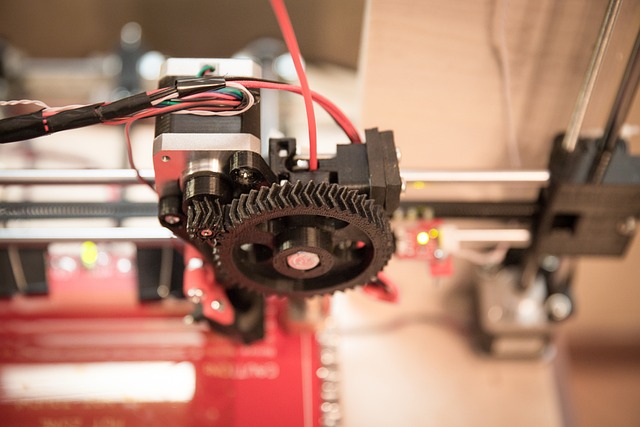
3d printing imaging
3D printer

3D-printed models of organs can be used to practice minimally invasive procedures and to create guides for surgeons to follow during operations. 

This will help with medical education, allowing students and radiologists to visualize the anatomy and better understand medical conditions. Finally, 3D printing can be used to create patient-specific medical devices, such as hearing aids and prosthetics, to customize them and make them more comfortable for patients.

Conclusion

Overall, 3D printing has revolutionized the world of radiology. It has allowed medical professionals to make more precise and accurate diagnoses, create custom medical devices, create models of organs, tissues, and bones, and create custom medical implants. This technology has enabled doctors to provide better patient care and has improved patient outcomes. With this kind of technology coupled with Artificial Intelligence, the sky is the limit.

Vesta Teleradiology is always looking to new technologies to improve our operations and further assist our clients in accurate and quick interpretations. Please reach out to us to learn about how we customize the process for your healthcare facility’s needs. 


Differences Between Preliminary and Final Radiology Interpretations  

A radiology report interprets images into words. The requesting physician who requests the radiology reports recommends treatment to their patient based on the findings a radiologist provides in these reports. A patient’s understanding of preliminary or final radiology interpretations is critical in treatment decisions.

An on-call radiology resident or technician may be issuing the preliminary radiology report at a hospital emergency room or an urgent care facility. A physician may need to act on the findings of this initial report before a final interpretation by the radiological physician overseeing the resident or technician.

Studies have shown a minimal discrepancy rate when physicians review the preliminary report with the final findings. A few factors, like the imaging technique and the technician’s experience level, account for most modifications in the final reports.

Why Have Preliminary Radiology Interpretation?

Hospital funding in rural areas and 24-hour urgent care facilities cannot always afford an on-staff radiology physician, and they rely on teleradiology interpretations. However, if a patient in pain arrives at the facility, the physician needs immediate information on how to proceed with the patient’s treatment.


preliminary reading
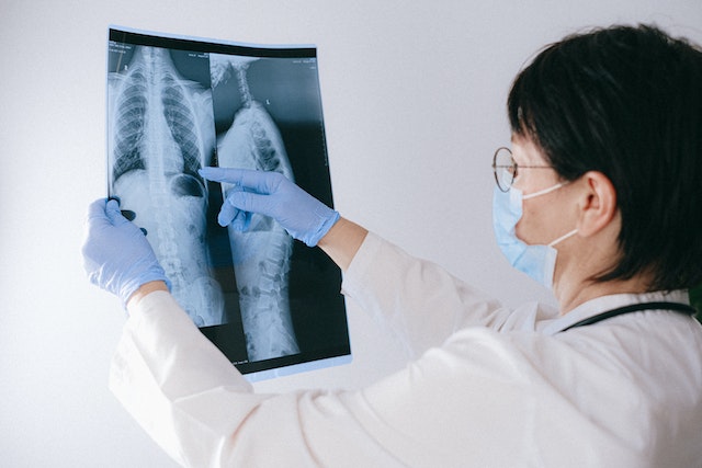
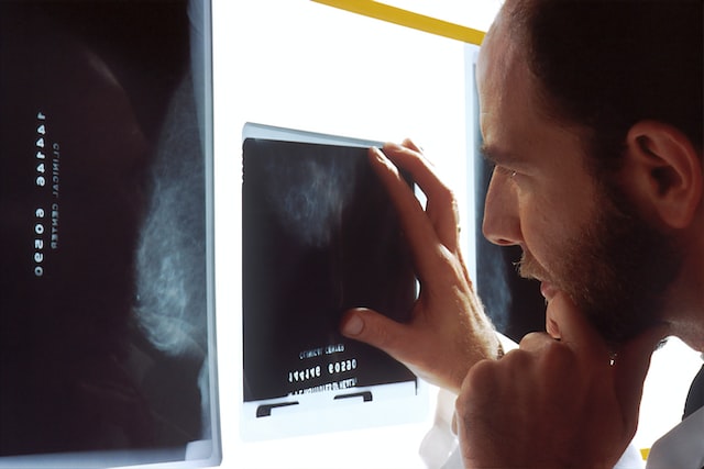
A radiologist examines an x-ray

An example may be if the patient complains about severe abdominal pain. The physician must rely on the technician’s preliminary report defining acute appendicitis and perform emergency surgery. Another example may be a possible stroke victim brought into the facility. The physician cannot wait for a final report to make a life-saving decision with the patients.

Radiologist Physician Expertise for a Final Report

Few people understand the extensive education and dedication a Radiologist Physician undergoes during their career. They graduate from medical school, complete their internships and residency requirements, and interpret thousands of exams under supervision. These are just a few of the provisions of licensing.

The extensive requirements of completing a radiologist physician program limit the number of physicians available for final radiology interpretations.

With the advancement of technology in the field, teleradiology has broadened the possibility of more accessible and punctual final reads from radiologists. In most cases, it can make the need for preliminary reporting almost obsolete.

Besides an emergency, another exception may be for very complicated cases where second reads of the imagery may be pertinent to a diagnosis or treatment plan. In these cases, it is most beneficial to have a preliminary report, a second read, then a final report issued before making any decisions.

The primary goal of physicians is to make a difference in the lives of their patients, and they can do this by having timely, accurate, and well-defined information. Continuing improvement of the technical aspects of the radiology field will allow the radiologist to expeditiously provide final radiology interpretations and assist in improving patient management.


final interpretation
Teleradiology offers hospitals and healthcare facilities an efficient way of completing interpretations

US Board Certified Teleradiologists from Vesta

At Vesta Teleradiology, our US Board Certified Radiologists can assist your facility with both preliminary and final interpretations, including subspecialty solutions like: nuclear medicine, body imaging, gastrointestinal/genitourinary diagnoses, cardiac angiography and thoracic radiology.


Are X-Rays Safe?

Over 125 years ago, Wilhelm Conrad Röntgen made a monumental discovery that changed medical practices forever. He was the first man to see the results of using radiological rays as a non-invasive way to investigate inside a human being’s body.

With the help of Röntgen’s discovery of what is today commonly known as X-rays, medical science has been able to advance at an accelerated rate. In the modern world, just about every person has experienced having an X-ray in one form or another.

X-ray technologies have advanced at a staggering rate in the past 125 years, and so has their uses. Professionals depend on X-rays in dental offices, doctor’s offices, chiropractic offices, hospitals, urgent care, and other medical service facilities.

With the medical community using X-rays so frequently, the public must understand the types of radiation we encounter and the risks involved with radiation exposure.

The air we breathe, soil and water, rocks, and plant life all have radiation. Radon is natural radiation found in soil and can be potentially harmful to humans. Cosmic radiation (which includes X-rays) constantly penetrates the earth’s atmosphere and is all around us. Cosmic radiation is unavoidable but is at low levels.

We have no choice but to live with the natural and cosmic radiation exposure, but X-rays are a choice we need to evaluate.

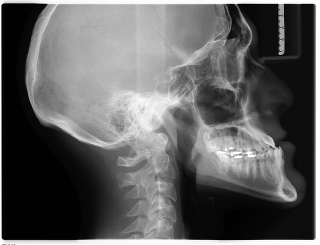
When professionals take a diagnostic X-ray, the image reflects on the X-ray negative plate depending on how much radiation is absorbed. The reason bones appear white on the negatives is because bones have a high calcium content that absorbs the radiation.


x-ray
An x-ray of a human head and neck


With all X-rays, ionizing radiation is used, potentially harming living tissue. Radiography is the most commonly used X-ray imaging and uses the smallest amount of radiation. Professionals use radiography to image bones, teeth, and chest X-rays.

Fluoroscopy also uses a small amount of radiation, but more than radiography. Professionals use fluoroscopy with barium drinks to watch how the body acts and reacts during digestion.

Computed Tomography, or CT, uses the highest amount of radiation. The higher radiation is because, during one procedure, the CT mechanism takes multiple X-rays.

If used appropriately by a professional, the benefits of having X-rays taken far outweigh any risk of radiation exposure. Using X-rays, medical staff can detect cancerous tumors, infections, and damaged blood vessels. The risk of developing cancer from the ionizing radiation of X-rays is small.

Even with the low risk of cancer from X-rays, patients receiving medical treatment involving radiation should ask questions and communicate their medical history to their provider. Also, when providers recommend X-rays for children, ask the technician to double-check that the X-ray machinery has been adjusted for a child. Risks are more significant for children than for adults.

Even though studies show the low dose of radiation from X-rays–when used appropriately—does not cause health problems, X-ray technology is improving every day. Researchers are discovering ways to reduce radiation dosage, improve imaging, and create better materials and methods of imaging.

Vesta Radiologists

We work with healthcare offices that provide x-rays as a diagnostic tool. We help them interpret their findings so they can continue to service their patients in a timely manner. Look to Vesta’s US Board Certified Radiologists to help take on any workload you need taken care of, nights, days and even weekends.

Vesta Teleradiology Presents Informative White Paper on Transitioning from ICD-9 to ICD-10

Vesta Teleradiology releases a white paper, entitled “How to Make Your Transition from ICD-9 to ICD-10 a Seamless One,” for the purposes of providing guidance to physician practices as they prepare to transition and “go live” with ICD-10 on October 1st.

icd 9 to icd 10

The white paper located at vestarad.com/mediablog/blog incorporates preparatory practices as well as answers to common questions for those practices who have found themselves less than adequately prepared for the transition. The white paper provides those all-important answers that will allow those practices whom are not quite prepared to continue to bill under old ICD-9 codes and be reimbursed, so long as specific criteria are met. Specific guidance is delineated in the white paper.

Vijay Vonguru, President, Vesta Teleradiology, knows how apprehensive many providers are feeling about going from some 13,000 diagnostic codes to 69,000 codes, but states that “in the end the changes are for the betterment of patients and providers alike, in that it will serve to streamline referrals and approvals on diagnostic testing, as well as gather epidemiological data” and “this information should hopefully alleviate some of the resistance surrounding the upcoming transition”.

For those practices who having done the hard work and started last year in preparation for the transition, with tutorials for their physician, billing and ancillary staff, coordination with their vendors of EMR software and technical support: Kudos to them and their teams as they are one step ahead of the game. There is still some time for preparation, and utilizing the tips provided in the white paper will help the practices prepare for the October 1st deadline!


ICD-9
Are you ready for the transition?

About Vesta Teleradiology:

Vesta offers teleradiology solutions and services, providing 24x7x365 access to their highly qualified Board Certified Radiologists through a secure PACS.

Vesta has been a pioneer in supportive diagnostic workflow technology and quality diagnostic services. The team is steadfast in their passion to remain at the forefront of innovation in healthcare. Prospective clients are offered a free test drive of Vesta’s teleradiology service.