The ongoing imbalance between radiologist supply and medical imaging demand in the U.S. is projected to continue through 2055 without significant intervention, according to recent research by the Neiman Health Policy Institute, (NHPI), published in the Journal of the American College of Radiology on February 12. As the population grows and ages, and imaging utilization increases, the shortage of radiologists poses a significant challenge for healthcare systems nationwide.
Projected Growth in Radiologist Supply
The NHPI study anticipates a nearly 26% increase in the supply of radiologists over the next 30 years, assuming residency numbers remain unchanged. However, even this growth may not be sufficient to meet rising imaging demands. If residency positions increase, the radiologist workforce could see a 40% expansion by 2055. Yet, attrition rates—especially post-COVID—pose a threat to this growth, highlighting the need for initiatives aimed at improving workplace well-being and retaining experienced radiologists.
Increasing Demand for Imaging Services
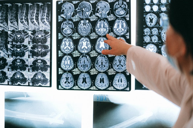
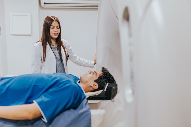
The demand for imaging services is expected to rise between 17% and 27% by 2055, driven largely by population growth and aging. Specific modalities like CT scans may see utilization increases as high as 59%, while others, such as nuclear medicine, may experience a decline. These projections underscore the urgency of balancing supply and demand to prevent prolonged patient wait times and compromised care.
 Current Impact on Patients and Healthcare Systems
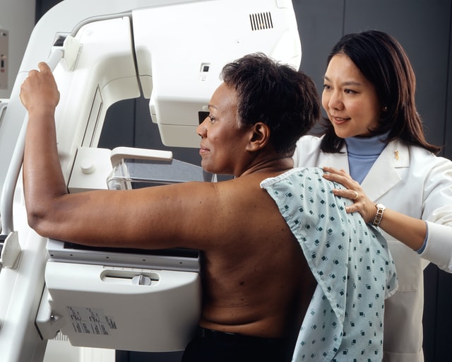
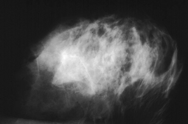
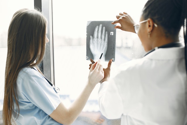
Patients across the U.S., including those in West Michigan, are already feeling the impact of the radiologist shortage. Delays in receiving imaging results have caused frustration, particularly for individuals with pressing health concerns such as fibroids and breast cancer risk. Healthcare providers, from radiologists to patient care technicians, are also facing mounting pressure to deliver timely care amidst workforce shortages.
Potential Solutions to Mitigate the Shortage
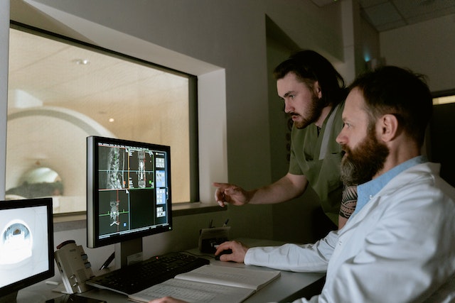
To address this crisis, experts emphasize the need to increase radiology residency slots and curb inappropriate imaging use. Monitoring attrition patterns and enhancing workplace conditions are also crucial. Technological advancements, such as AI for improving radiologist efficiency and clinical decision support systems, present promising avenues for alleviating some of the burden on the current workforce.
 Conclusion
The radiologist shortage in the U.S. is a complex issue that requires multifaceted solutions. Increasing residency positions, enhancing workplace well-being, and leveraging technology are essential steps to ensure patients receive timely and accurate imaging services.
Top Radiology Company: Onsite and Remote
At Vesta Teleradiology, we are committed to bridging the gap caused by radiologist shortages. Our team of U.S. board-certified radiologists offers both on-site and remote services, providing reliable imaging interpretations to meet your facility’s needs efficiently. Let us help you navigate the challenges of radiologist shortages with our expert solutions.

 Implications for Healthcare Providers
 What types of work environments are available for radiologists?
 What should I consider when evaluating a job offer?


 Sources: