As the demand for specialized medical imaging continues to rise, healthcare facilities face significant challenges in accessing qualified radiologists, particularly in subspecialties such as neuroradiology, musculoskeletal radiology, and pediatric imaging. This shortage is exacerbated in rural hospitals and underserved areas, where recruiting and retaining subspecialty radiologists is often difficult. Partnering with a teleradiology company that offers subspecialty expertise has become essential for ensuring timely and accurate diagnoses.
The Growing Demand for Subspecialty Teleradiology
Several factors contribute to the increasing need for subspecialty teleradiology services:
- Aging Population: The U.S. population aged 65 and older grew by 38.6% from 2010 to 2020, leading to a higher demand for imaging services. acr.org
- Radiologist Workforce Shortage: Approximately 56.4% of diagnostic radiologists are 55 or older, indicating a significant portion of the workforce is nearing retirement. medicushcs.com
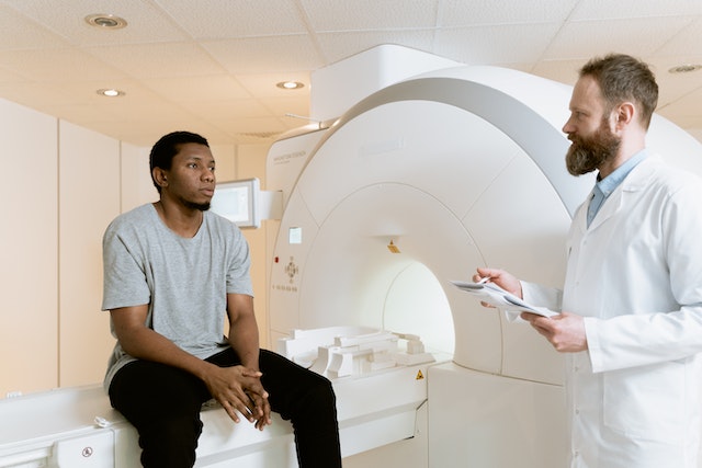
- Increased Imaging Utilization: Advancements in medical imaging technology have led to more frequent use of imaging studies, increasing the workload for radiologists. acr.org
These trends underscore the necessity for teleradiology services that provide access to subspecialty-trained radiologists, ensuring that healthcare providers can meet the growing demands of patient care.
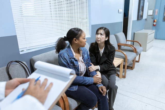
Supporting Rural Hospitals and Underserved Areas
Rural hospitals often face unique challenges in providing comprehensive radiology services due to limited access to subspecialty radiologists. Teleradiology bridges this gap by enabling remote interpretation of imaging studies, allowing rural healthcare providers to offer quality healthcare services locally and at lower costs. ruralhealthinfo.org
 How Vesta Teleradiology Provides Specialized Radiology Support
Vesta Teleradiology addresses these challenges by offering comprehensive teleradiology services nationwide, including:
- Access to Subspecialty-Trained Radiologists: Vesta provides access to a wide range of highly specialized, U.S.-trained, and American Board of Radiology-certified radiologists proficient in various modalities.
- Customizable Reporting and PACS Solutions: Our reporting module allows customization of reports to include the facility’s logo and adjust layouts to match existing reports. Our comprehensive PACS enables the creation of master accounts with sub-accounts, facilitating seamless integration into existing workflows.
- 24/7 STAT and Routine Reads: We interpret both STAT and routine cases, delivering detailed interpretations with quick turnaround times (Ohio, Illinois, Arizona, Georgia, Florida and more). Our flexible workflow supports various facility needs, from portable imaging units to stand-alone imaging centers and hospitals handling high-end cases.
- Efficient Communication with Referring Physicians: Our case managers facilitate communication between our radiologists and the facility’s referring physicians to answer questions and relay positive findings promptly. We customize the notification of significant findings to different recipients based on the time of day.
Why Subspecialty Teleradiology Matters for Patient Care
Utilizing subspecialty-trained radiologists through teleradiology services like Vesta ensures:
- Accurate Diagnoses: Specialized radiologists are adept at identifying subtle findings specific to their area of expertise, leading to precise diagnoses.
- Timely Treatment: Quick access to expert interpretations facilitates prompt decision-making and initiation of appropriate treatments.
- Cost Efficiency: Accurate and timely diagnoses can reduce unnecessary tests and procedures, optimizing healthcare resources.
The Right Teleradiology Partner Makes All the Difference
Choosing a teleradiology provider with subspecialty expertise is crucial for delivering high-quality patient care. Vesta Teleradiology offers:
- U.S.-trained, board-certified subspecialists
- Fast, detailed interpretations with high accuracy
- Seamless PACS and reporting system integration
- Dedicated support and case management
- Reliable coverage for rural and critical access hospitals
By partnering with Vesta Teleradiology, healthcare facilities can enhance their diagnostic capabilities, improve patient outcomes, and efficiently manage increasing imaging demands.


 Sources: