FDA Cautions Consumers Against Use of RoyalVibe Ultrasound Devices

The U.S. Food and Drug Administration issued a warning to consumers on July 17 about avoiding the use of ultrasound devices from a Texas-based manufacturer and distributor called RoyalVibe Health.

The company sells direct-to-consumer ultrasound scanners, marketing them to be used for various medical purposes, including diagnosing and treating serious conditions like cancer, diabetes, heart disease, Parkinson’s, and depression. However, the FDA has not reviewed these devices, and they lack the necessary 510(k) clearance or premarket approval.


What is 510(k) Clearance?

The 510(k) clearance is a regulatory pathway used by the U.S. FDA to evaluate and approve medical devices. If a new medical device is deemed substantially equivalent to a legally marketed device (known as a “predicate device”), the manufacturer can submit a 510(k) premarket notification to the FDA. The FDA reviews the submission to ensure the new device shares the same intended use and technology as the predicate device. If approved, the new device receives 510(k) clearance, allowing it to be marketed in the U.S. The 510(k) process is commonly used for moderate-risk medical devices and relies on the safety record of similar, already-approved devices to expedite approvals while ensuring safety and effectiveness.


Ultrasound Therapy

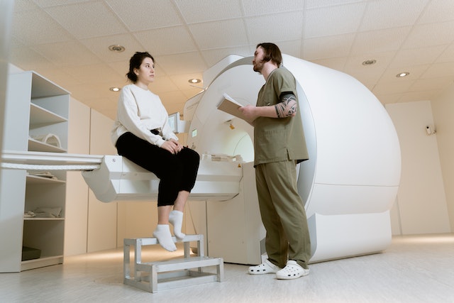
Ultrasound therapy is a non-invasive medical treatment that utilizes high-frequency sound waves to target specific tissues in the body. During the procedure, a handheld device emits these sound waves, which penetrate the skin and cause vibrations at a cellular level. This treatment process is believed to increase blood flow, relax muscles, and promote tissue healing. Ultrasound therapy is commonly used by physical therapists to alleviate pain, reduce inflammation, and improve tissue mobility in conditions like muscle strains, joint injuries, and certain musculoskeletal disorders. It is a safe and effective treatment that can provide relief and aid in the recovery process for various injuries and conditions.


Open Communication with Patients

Unfortunately, some consumers find products on the market that they can purchase directly in hopes to use for at-home self-care. Please advise your patients to be wary of these types of devices, including the products the FDA has not approved such as RoyalVibe Health. Patient education can go a very long way, so it is important to address their questions about their health, pain and concerns and emphasize that treatment should be administered by their healthcare provider.


Sources:

Fda.gov
verywellhealth.com
radiologybusiness.com
openai.com


New Studies in Ultrasound – An Exciting Frontier in Medical Research

Medical science makes incredible strides every year, finding new ways to heal the sick and injured. One of the most exciting of these new frontiers is ultrasound technology.

With its ability to safely and painlessly penetrate the human body, scientists and doctors are discovering new possibilities in diagnosing and treating conditions that previously required more invasive procedures.

Ultrasound in Cancer Treatment

In cancer treatment, chemotherapy often comes with various unpleasant side effects. But new research has shown that ultrasound technology can enhance chemotherapy effectiveness, sometimes even reducing the dosage of the drugs required.

Ultrasound can also help increase the uptake of cancer drugs into tumors, making their delivery more targeted and efficient.

Ultrasound and Alzheimer’s Disease

Alzheimer’s disease is one of the most heartbreaking conditions facing our aging population, and while there is no cure for it, there is hope for better treatment.

Alzheimer's

Recent research has discovered the possibility of using ultrasound to break up the plaques that form in the brains of Alzheimer’s patients, reducing the inflammation and damage that cause the disease. Research in this field is still in its early stages, but the possibility of a breakthrough treatment in the years to come is an exciting prospect.


Ultrasound Diagnostics

Of course, one of the primary uses of ultrasound in medicine is as a diagnostic tool. However, new studies are refining and expanding the possibilities of what ultrasound can detect.


For example, technicians now use ultrasound to locate and diagnose skin cancers and traditional mammography for breast cancer screening. Ultrasound also offers a non-invasive way to examine the heart and blood vessels in incredible detail, giving doctors a better understanding of the mechanisms of cardiovascular disease.


Ultrasound and Pain Management

In addition to being an incredible diagnostic tool, patients can use ultrasound to manage pain.


Ultrasound-guided nerve block injections can relieve patients suffering from chronic pain without the side effects of prolonged medication use. Studies are ongoing to refine this technique and expand its benefits beyond chronic pain.

pain

Other Applications of Ultrasound

The research and innovation happening in ultrasound are constantly expanding their possibilities.

For example, researchers are exploring ultrasound technology to treat kidney stones, sometimes allowing for less invasive procedures than traditional surgery.

They are also investigating ultrasound for use in regenerative medicine and even to monitor blood glucose levels in diabetic patients non-invasively.

The possibilities of ultrasound technology seem almost limitless, with new studies constantly uncovering ways to use this powerful tool for medical treatment and research.

From cancer treatment to pain management, diagnostics to disease prevention, ultrasound is a field that promises to revolutionize the face of medicine in the years to come. We can only look forward with enthusiasm and hope for what discoveries may come on this exciting frontier.


Teleradiology in Conjunction with Ultrasound


When you think about the role a teleradiology company like Vesta plays with healthcare providers, you might envision that the radiologists deal only with x-rays and MRI scans. We also work closely with hospitals for ultrasound readings, too. If you’re seeking an extra hand for preliminary and final ultrasound interpretations, please reach out to us today. We can handle any volume, large or small.


Diagnostic Imaging Trends: Point of Care Ultrasound – POCUS

Point of care ultrasound (POCUS) is revolutionizing the healthcare industry and changing how doctors prescribe treatment for patients. POCUS is a diagnostic tool that utilizes ultrasound imaging to diagnose, monitor, and guide treatments for medical conditions.

This technology has been around for decades but is presently utilized more broadly throughout the healthcare system. Let’s take a closer look at POCUS and how it transforms patient care.


What Is Point of Care Ultrasound (POCUS)?

Point of Care Ultrasound (POCUS) is an ultrasound-based diagnostic tool used in clinical settings that uses sound waves to create images allowing doctors to see inside the body without having to do surgery or other invasive procedures.

POCUS can detect various medical conditions, such as heart defects, abdominal diseases, vascular diseases, musculoskeletal problems, and gynecological issues. POCUS can also be used in emergency settings to assess a patient’s condition quickly and determine if further intervention or testing is needed.


How Is Point of Care Ultrasound Transforming Healthcare?

One benefit of POCUS is its cost-effectiveness compared with other imaging tests, such as MRI or CT scans, and POCUS does not require expensive equipment as those tests do.


POCUS
Point of care ultrasound


Additionally, it can be done quickly and easily at the point of care, which reduces wait times for patients and increases accuracy in diagnosis, as well as reduces unnecessary treatments or hospital admissions. Furthermore, since it does not use radiation as other imaging tests do, there are no additional health risks associated with this technology, making it safer overall for patients.

Another advantage of POCUS is its ability to provide real-time data about a patient’s condition, which helps doctors make more informed decisions about treatment plans for their patients.

Additionally, because POCUS used in most circumstances does not require special training or expensive equipment, these systems are becoming increasingly available in low-resource areas where access to traditional diagnostic imaging may be limited. This benefit means more people have access to high-quality healthcare regardless of where they live or available resources.

The Gates Foundation recently provided financing to bring 1,000 handheld ultrasound devices to Africa. When low to mid-income nations can improve the accuracy of an efficient diagnosis–local doctors can save more lives.

Since 2012, however, emergency medicine program accreditation requires competency in POCUS. Competency assessment in this field includes demonstrations of technical skill and how it relates to the specific clinical practice.

Point of Care Ultrasound (POCUS) offers numerous benefits over traditional imaging tests. Its cost-effectiveness allows physicians to provide accurate diagnoses without breaking the bank. In contrast, its portability allows it to reach underserved populations who may not otherwise have access to quality healthcare services.

With further advances in technology coming soon, we could see even more widespread use of this powerful diagnostic tool across all areas of medicine in the near future.


Vesta Teleradiology: At the Forefront of Medical Technology

Vesta believes it is crucial to stay on top of technological trends that can benefit our hospital and healthcare facility partners. Vesta is always at the forefront of technological advances in order to help bring more efficiency and accuracy to imaging and radiological interpretations. For more information about Vesta’s teleradiology services, please contact us today.


Advancements in Imaging: Colorectal Cancer

In the past, colorectal cancer was familiar as a disease to affect the older generation. However, advancements in treating and detecting colorectal cancer have shown the risk is prevalent in those younger, too. Now more than ever, screening tests are important so treatment can start sooner for better outcomes.  

What Is Colorectal Cancer?

The Centers for Disease Control and Prevention defines Colon cancer as “a disease in which cells in the colon or rectum grow out of control.” The rectum can develop polyps, with some growing in size. Those larger polyps can turn into cancerous ones. Smaller polyps rarely form a malignant growth. 

How Is Rectal Cancer Treated?

Doctors stress the importance of recognizing the early symptoms of colorectal cancer and getting regular screenings to check it before it spreads. “If the patient is diagnosed with a stage one or localized cancer, where it’s just in the colon and hasn’t moved elsewhere in the body, it’s about 90% curative just with surgery,” Dr. Paula Denoya, director of the Colorectal Surgery Residency Program at Stony Brook Medicine told NBC news. 

The first step for doctors to create a treatment plan for colon cancer is by finding what stage the cancer is in. Medical advancements in imaging have allowed this process to be more thorough and quicker to detect the area and size of cancer. The recent death of Kirstie Alley brought more attention to colorectal cancer since doctors shared this was the cause of her death. Unfortunately, Alley’s family said she had only found cancer shortly before her untimely death Monday. She died at 71 years of age. 

How Imaging Helps With Colorectal Cancer Detection

Diagnostic imaging allows doctors to see what the insides of the colon are doing, which will point out if any tumors are more than benign. Newer imaging procedures give doctors an even closer look at the rectal and surrounding areas in the body. 

colorectal cancer

Endoscopic ultrasounds are more invasive than routine colonoscopies. While both are meant to detect and/or provide a more thorough diagnosis, endoscopic ultrasounds go even further, using sound waves that go back and forth off of the inner body. This provides a detailed picture that doctors can study to see how rectal cancer has progressed or lessened if additional treatments were already in the queue. 

Endoscopic ultrasounds have two types; upper and lower endoscopic ultrasounds. Lower endoscopic ultrasounds deal with the parts of the body in the rectum to examine tumors closely. Doctors will use an endoscopic ultrasound after other imaging screenings show potential for malignant tumors. It gives doctors the ability to treat cancer with a more reliable decision. 

CT scans are another way doctors find and diagnose what stage the cancer in the colon is at. However, endoscopic ultrasounds have a higher percentage of precisely showing what stage the cancer is in.

Additional Imaging Diagnostics For Colon Cancer

A newer technique of diagnostic imaging is molecular imaging. Molecular imaging projects a picture of the cells in the body and their activity. This means doctors can see more information about a mass before it gets worse. If a tumor is present, many times before symptoms begin to aggravate a patient, this technology is able to see that cancer in the beginning stages.

Molecular imaging uses positron emission tomography or (PET) technology. A mixture, called a radioactive, is injected into the blood, and omits an image of the patient’s molecules and/or cells. If cells are numerous, this may mean colorectal cancer has spread.

Staying on top of sudden changes in the bowels, appetite, and illness is key in fighting to stop colorectal cancers before they grow larger. Regular screenings can catch otherwise undetected cancer. The advancements that technology has granted the medical world has saved more lives. The important part is ensuring these technologies are used.


Vesta Teleradiology: At the Forefront of Scientific Advancements

Vesta Teleradiology is a tech-forward teleradiology company, assisting various healthcare facilities with preliminary and final interpretations. If you’re seeking someone to fill your gaps, like nights and weekends, please contact us:  877-558-3782


Healthcare Quality Week: Thank you Radiology and Imaging

This week, your mind might be on the upcoming Halloween holiday and fall festivities that come around this time of year. But did you know that October 17-23 is actually Healthcare Quality Week?  This week of observance is “a dedicated time to celebrate the profession and raise awareness of the positive impact healthcare quality professionals have in their organizations and communities.”

Since Vesta has a team of dedicated radiologists as well as clients in healthcare facilities across the nation, we want to take time to acknowledge these amazing people and the processes they expertly and caringly carry out in order to provide quality healthcare.

Radiology and Imaging: A Game Changer

Radiology and imaging is one of the greatest inventions of the late nineteenth century. This powerful tool has been studied and developed, enabling millions of lives to be saved and an array of scientific discoveries in the fields of physics and biology.


xray radiology
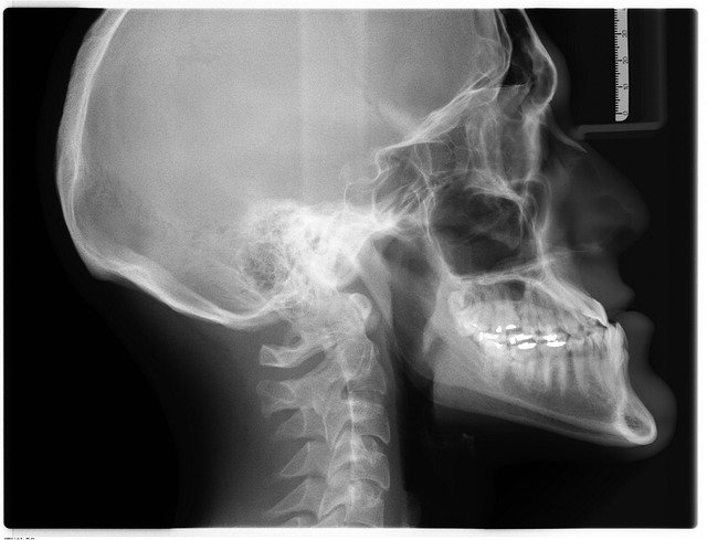
Going back to the beginning, the first X-ray picture in 1895 by Wilhelm Rontgen led to many scientific discoveries, ultimately earning Rontgenthe Nobel Prize in physics in 1901. Since then, the field of Imaging and Radiology has grown exponentially. The mystery of how matter converts energy became the focus of study as Albert Einstein furthered the exploration in 1903, allowing the world to begin to understand how these powerful rays demonstrated what our senses could not detect. Subsequently, fossils, art masterpieces, the earth, solar systems, and the universe were radioactively dated, and in 1953 the double helix of DNA was captured. From these and many other discoveries, multiple fields of study have evolved in Radiology and Imaging.

x-ray 1895

Cancer

Using radiation instead of chemotherapy to treat cancer is advantageous because radiation can be directed to the infected cells and avoid surrounding cells to a large degree. Benefits to radiation and the way it works is described in the following quote:

Radiation works by making small breaks in the DNA inside cells. These breaks keep cancer cells from growing and dividing and cause them to die.”

Utilizing radiation to treat cancer minimizes symptoms and preserves healthy cells, as opposed to chemotherapy. In some cases, chemotherapy drugs can weaken cells making them more sensitive to radiation. Ultimately, the ability to direct radiation can effectively kill specific cancer cells. This is a powerful phenomenon, especially when faced with a deadly disease.

Working as a medical imaging technologist combines a wide array of skills. Patient care, technological aptitude, data analysis, and people skills allow for a challenging and rewarding multilayered career, one that is only possible due to the discovery of electromagnetic radiation in a wavelength range over one hundred years ago. Saving lives and improving quality of life due to what we all commonly call the “X-ray”, it is only appropriate to marvel at the work of Wilhelm Rontgen and thank him.

More Notable Imaging Methods that Changed the World

Fluoroscopy is a medical imaging test that uses an x-ray beam. It passes continuously through the body to create a projected image on a monitor which allows doctors to see the movement of internal organs in real-time. This is extremely helpful during surgery. Other ways fluoroscopy benefits patients are with barium X-ray enemas to examine the gastrointestinal tract, catheter insertion to direct a catheter through blood vessels, placing devices in the body such as stents, and in orthopedic surgery with joint replacement.

Ultrasound, also called sonography, uses sound waves to create internal images of the body. It is commonly used to confirm and date a pregnancy. It is also used in diagnosing a wide variety of other conditions. Diseases affecting the organs and soft tissues can be diagnosed with the help of ultrasound.  This includes the heart, blood vessels, liver, gallbladder, spleen, pancreas, kidneys, bladder, uterus, ovaries, eyes, thyroid, and testicles.

3D ultrasound
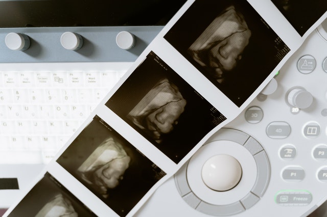
Fun Fact: the 3-D ultrasound actually takes thousands of photos at once to create a 3d image that is extremely clear.

Magnetic resonance imaging (MRI), or ultrasound imaging, has aided many women in avoiding having a hysterectomy. Many times, the cause of bleeding is a fibroid and a uterine fibroid embolization (UFE) is needed instead. An interventional radiologist can perform this minimally invasive treatment for women with symptomatic fibroids which can be assessed through an (MRI) and then also offer further assessment and counseling.

Transvaginal ultrasound lets most women with malignant gestational trophoblastic disease be cured. Without this procedure, the cancerous and potentially cancerous cells of the pregnancy would continue to thrive putting the mother’s life at risk. Through early detection, partial and complete molar pregnancy can be detected and treated by removing it from the uterus and reproductive function can be preserved.

Medical Ultrasound Awareness Month: Advancements Through the Ages

Ultrasounds—to many not in the medical field, the image of a woman getting her pregnant belly examined is often something that comes to mind first. However, as medical industry professionals, we understand that ultrasounds go way beyond that.

ultrasounds

It’s really amazing to actually look at the history and advancement of such an important diagnostic tool especially during October–Medical Ultrasound Awareness Month.

In simple terms, the ultrasound scan isn’t just a medical device that can help track the development of a baby during pregnancy. The ultrasound scan can also help with the following:

  •       Observing the ovaries and uterus
  •       Evaluating blood flow
  •       Diagnosing gallbladder disease
  •       Examining a lump in the breast
  •       Checking thyroid glands
  •       Guiding a needle
  •       Diagnosing prostate issues

This machine is non-invasive, safe, and does not use ionizing radiation.

History of Ultrasound

The first recorded use of the ultrasound was in 1794 when Italian physiologist Lazzaro Spallanzani used ultrasound to study how bats navigated at night. This became the foundation for modern ultrasound physics. 

Bats produce ultrasounds in order to catch prey.

Ultrasound was initially used to detect flaws in metal casings. It was not used clinically until 1956 by Dr. Ian Donald and Tom Brown. There are no known side effects for ultrasound, making it an incredibly safe and efficient imaging device.

Advancements in Ultrasound

Ultrasound machines have become progressively mobile, easy to carry, and smaller while providing high-quality imaging. Since they are non-invasive and cost-effective, doctors can use them to track patient development without the risk of radiation.

Ultrasound technology can allow us to have an excellent view of the body’s vascular system by tracking microbubbles. 3-D imaging is also possible for more precise diagnostics.

As technology continues to expand, our medical devices tend to shrink inversely. Now, medical professionals can increase their diagnostic capabilities from anywhere in the world. For example, some handheld ultrasound devices weigh under a pound and can hook up to your smartphone, help healthcare providers quickly scan their patients—and possibly save lives.

These economical, easy-to-handle devices can change lives globally, predominantly in rural areas. It’s exciting to think that we may only be seeing the beginning of the modern ultrasound age.

How Teleradiology Can Help

Teleradiology, also referred to as remote radiology, provides medical professionals instant imaging result views remotely.  This means that patients have access to better, more immediate health care.

teleradiology servicesStudies suggest that teleradiology offers benefits, including the availability of second opinions and the possibility for remote viewing services. We at Vesta can certainly attest to this! Our clients – hospitals, urgent car centers, physician offices, mobile imaging companies, diagnostic imaging centers, and government institutions—enjoy cost-effective and unrivaled Teleradiology and Telemedicine services any time of day or night.

5 Tips for Women’s Wellness Healthcare Facility

The fitness and wellness industry has become one of the biggest markets in the world. If you are the owner of a women’s health or wellness center, there is great potential for growth in this $3.4 trillion industry. This competitive industry is constantly creating new approaches to attract patients. If you are searching for new ideas to enhance your wellness center and the care for your patients, here are 5 tips for increasing patient flow.

  1. teleradiology service for women's clinic
    A woman discusses the treatment with her patient

    Offer educational workshops and speakers. Being able to have a cup of coffee and listen to an informed speaker on a subject of interest can bring in more patients. Those who want to help themselves or improve their life will like this approach. Stress-free opportunities to listen and learn about money management, cooking, gardening, yoga, fashion, or more could entice women with enjoying a nonthreatening space to absorb information. These could be mini-breakout sessions of an upcoming wellness conference which may interest participants to attend.

  2. Bundle services. Having one facility that offers multiple services is attractive. Women are busy. Their time is valuable. If there was an opportunity to make one stop to take care of a mammogram, pedicure, and haircut (for example) many women would like this option. In addition, having a nonjudgmental environment to talk to a therapist on a variety of topics such as mental health issues, menopause, and sexual health could bring in more patients.
  3. Act as a safehouse. Employing experienced professionals who have the awareness and abilities to support women in crisis is needed in every city in the world. Doctors, psychologists, and lawyers on site who are in place to readily protect and prevent violence against women and their children will attract patients. Partnering with local authorities may bring wellness center funding opportunities.
  4. Incorporate wellness retreats. Offering purposeful, affordable, well designed retreats for specific groups allows wellness opportunities beyond the wellness center facility. Programs organized with the intentional components of mental, emotional, physical, and social can provide transformational experiences for patients. These deep connections can be life changing and create interest in future retreats.
     
  5. Outsource your radiology imaging needs to a reliable teleradiology service. With this service, a patient can have her mammogram, ultrasound, or other imaging service performed, screened, and sent digitally to all parties involved. The image and its information is compressed and encrypted and stored in a database server accessed by the radiologist, provider, transcriber, and other workers across geographically diverse facilities. The final report is automatically sent to the doctor and the original facility. This saves time and allows you to focus more on attracting new patients.
ultrasound
An ultrasound underway

Vesta Teleradiology has been assisting Women’s Wellness centers, hospitals, mobile imaging, assisted living and other diagnostic imaging centers for nearly 15 years. It’s not just our speedy service and accurate readings from US Board Certified radiologists, it’s our ability to help you customize the service to tailor it to your facility and patients. Look to Vesta if you want accuracy, speed, affordability, support and customization.