This week, your mind might be on the upcoming Halloween holiday and fall festivities that come around this time of year. But did you know that October 17-23 is actually Healthcare Quality Week? This week of observance is “a dedicated time to celebrate the profession and raise awareness of the positive impact healthcare quality professionals have in their organizations and communities.”
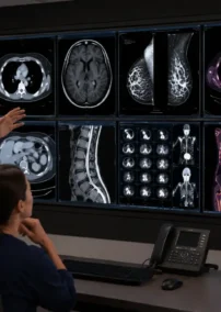
Since Vesta has a team of dedicated radiologists as well as clients in healthcare facilities across the nation, we want to take time to acknowledge these amazing people and the processes they expertly and caringly carry out in order to provide quality healthcare.
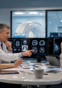
Radiology and Imaging: A Game Changer
Radiology and imaging is one of the greatest inventions of the late nineteenth century. This powerful tool has been studied and developed, enabling millions of lives to be saved and an array of scientific discoveries in the fields of physics and biology.
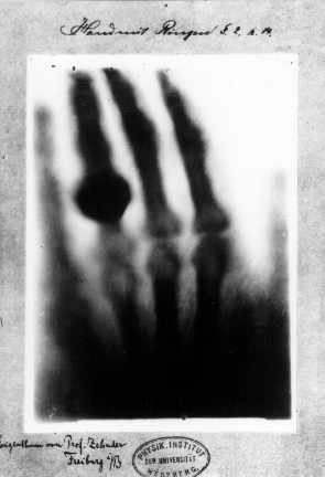
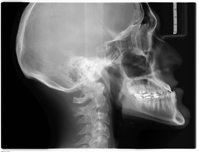 Going back to the beginning, the first X-ray picture in 1895 by Wilhelm Rontgen led to many scientific discoveries, ultimately earning Rontgenthe Nobel Prize in physics in 1901. Since then, the field of Imaging and Radiology has grown exponentially. The mystery of how matter converts energy became the focus of study as Albert Einstein furthered the exploration in 1903, allowing the world to begin to understand how these powerful rays demonstrated what our senses could not detect. Subsequently, fossils, art masterpieces, the earth, solar systems, and the universe were radioactively dated, and in 1953 the double helix of DNA was captured. From these and many other discoveries, multiple fields of study have evolved in Radiology and Imaging.
Cancer
Using radiation instead of chemotherapy to treat cancer is advantageous because radiation can be directed to the infected cells and avoid surrounding cells to a large degree. Benefits to radiation and the way it works is described in the following quote:
“Radiation works by making small breaks in the DNA inside cells. These breaks keep cancer cells from growing and dividing and cause them to die.”
Utilizing radiation to treat cancer minimizes symptoms and preserves healthy cells, as opposed to chemotherapy. In some cases, chemotherapy drugs can weaken cells making them more sensitive to radiation. Ultimately, the ability to direct radiation can effectively kill specific cancer cells. This is a powerful phenomenon, especially when faced with a deadly disease.
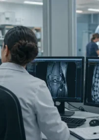
Working as a medical imaging technologist combines a wide array of skills. Patient care, technological aptitude, data analysis, and people skills allow for a challenging and rewarding multilayered career, one that is only possible due to the discovery of electromagnetic radiation in a wavelength range over one hundred years ago. Saving lives and improving quality of life due to what we all commonly call the “X-ray”, it is only appropriate to marvel at the work of Wilhelm Rontgen and thank him.
More Notable Imaging Methods that Changed the World
Fluoroscopy is a medical imaging test that uses an x-ray beam. It passes continuously through the body to create a projected image on a monitor which allows doctors to see the movement of internal organs in real-time. This is extremely helpful during surgery. Other ways fluoroscopy benefits patients are with barium X-ray enemas to examine the gastrointestinal tract, catheter insertion to direct a catheter through blood vessels, placing devices in the body such as stents, and in orthopedic surgery with joint replacement.
Ultrasound, also called sonography, uses sound waves to create internal images of the body. It is commonly used to confirm and date a pregnancy. It is also used in diagnosing a wide variety of other conditions. Diseases affecting the organs and soft tissues can be diagnosed with the help of ultrasound. This includes the heart, blood vessels, liver, gallbladder, spleen, pancreas, kidneys, bladder, uterus, ovaries, eyes, thyroid, and testicles.
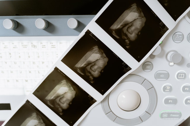 Fun Fact: the 3-D ultrasound actually takes thousands of photos at once to create a 3d image that is extremely clear.
Magnetic resonance imaging (MRI), or ultrasound imaging, has aided many women in avoiding having a hysterectomy. Many times, the cause of bleeding is a fibroid and a uterine fibroid embolization (UFE) is needed instead. An interventional radiologist can perform this minimally invasive treatment for women with symptomatic fibroids which can be assessed through an (MRI) and then also offer further assessment and counseling.
Transvaginal ultrasound lets most women with malignant gestational trophoblastic disease be cured. Without this procedure, the cancerous and potentially cancerous cells of the pregnancy would continue to thrive putting the mother’s life at risk. Through early detection, partial and complete molar pregnancy can be detected and treated by removing it from the uterus and reproductive function can be preserved.