The Latest in Brain Imaging News

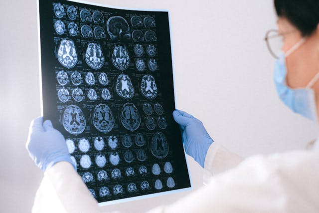
In recent years, awareness surrounding brain injuries has steadily risen, prompting significant strides in diagnostic technologies and treatment modalities. As we delve into the latest developments in this critical area of healthcare, it becomes increasingly apparent that advancements in medical imaging, particularly in the realm of neurological disorders, are poised to revolutionize the landscape of brain injury diagnosis and management.


AI-based Quantitative Brain Imaging System

Philips and Synthetic MR have joined forces to advance the diagnosis of neurological disorders through cutting-edge quantitative brain imaging tools. Their collaboration introduces the Smart Quant Neuro 3D MRI software suite, combining Philips’ SmartSpeed image-reconstruction technology, the 3D SyntAc clinical application, and SyntheticMR’s SyMRI NEURO 3D software. This innovation employs AI to analyze brain tissues, enhancing the detection and analysis of conditions like multiple sclerosis, traumatic brain injuries, and dementia.

The rise of AI in diagnostic imaging, projected to reach $1.2bn by 2027, signifies a transformative shift in improving accuracy and patient outcomes. With the diagnostic imaging market expected to grow to $9.1bn by 2030, fueled by demand for early disease diagnosis and personalized medicine, this partnership underscores the crucial role of AI in enhancing medical imaging.

Read the press release here.


A New Way of Diagnosing Mild TBIs

Researchers have developed a novel brain imaging method to diagnose mild traumatic brain injuries (mTBIs), which are often missed by standard techniques like MRI. This method involves loading gadolinium, a common MRI contrast agent, into micropatches attached to immune cells called macrophages. These cells migrate to areas of brain inflammation caused by mTBIs, enabling MRI detection. The technique, called M-GLAMs, was successfully tested in mice and pigs, showing promise for accurately diagnosing mTBIs. It also allows imaging at lower gadolinium doses, potentially benefiting patients with kidney issues. While unable to pinpoint injury locations, M-GLAMs could aid in identifying and treating brain inflammation. The researchers aim to bring this technology to clinical trials, with support from grants and intellectual property protection.

Read the study here.

tbi

New Imaging Tech that Captures Neuronal Activity Across the Brain During Recovery

Researchers at Tufts University School of Medicine have developed a novel imaging technology to monitor neuronal activity throughout the entire brain during the initial weeks of recovery from traumatic brain injury (TBI). Their study, published in Cerebral Cortex, reveals that TBI can induce changes in brain function beyond the injury site. Using a combination of fluorescent sensors and electrodes, they observed altered connectivity patterns in mice post-injury, even in regions distant from the impact. Despite the mice’s ability to perform physical tasks normally, their brain activity during both exercise and rest differed significantly from healthy brains. This impaired ability to switch between states suggests underlying brain state dysfunction post-injury. The findings highlight the brain’s plasticity in response to injury and have potential clinical implications for understanding TBI impacts and tailoring treatments. The researchers aim to further investigate long-term neural activity changes post-recovery and explore the technology’s potential in predicting specific dysfunctions or long-term outcomes of TBI. 

Read the study here.


Sources:

Medicaldevice-network.com
Otd.harvard.edu
Scitechdaily.com
Openai.com


Diagnostic Imaging Trends: Point of Care Ultrasound – POCUS

Point of care ultrasound (POCUS) is revolutionizing the healthcare industry and changing how doctors prescribe treatment for patients. POCUS is a diagnostic tool that utilizes ultrasound imaging to diagnose, monitor, and guide treatments for medical conditions.

This technology has been around for decades but is presently utilized more broadly throughout the healthcare system. Let’s take a closer look at POCUS and how it transforms patient care.


What Is Point of Care Ultrasound (POCUS)?

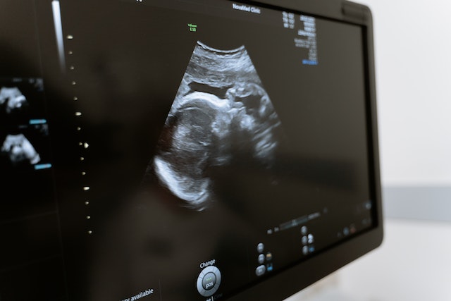
Point of Care Ultrasound (POCUS) is an ultrasound-based diagnostic tool used in clinical settings that uses sound waves to create images allowing doctors to see inside the body without having to do surgery or other invasive procedures.

POCUS can detect various medical conditions, such as heart defects, abdominal diseases, vascular diseases, musculoskeletal problems, and gynecological issues. POCUS can also be used in emergency settings to assess a patient’s condition quickly and determine if further intervention or testing is needed.


How Is Point of Care Ultrasound Transforming Healthcare?

One benefit of POCUS is its cost-effectiveness compared with other imaging tests, such as MRI or CT scans, and POCUS does not require expensive equipment as those tests do.


POCUS
Point of care ultrasound


Additionally, it can be done quickly and easily at the point of care, which reduces wait times for patients and increases accuracy in diagnosis, as well as reduces unnecessary treatments or hospital admissions. Furthermore, since it does not use radiation as other imaging tests do, there are no additional health risks associated with this technology, making it safer overall for patients.

Another advantage of POCUS is its ability to provide real-time data about a patient’s condition, which helps doctors make more informed decisions about treatment plans for their patients.

Additionally, because POCUS used in most circumstances does not require special training or expensive equipment, these systems are becoming increasingly available in low-resource areas where access to traditional diagnostic imaging may be limited. This benefit means more people have access to high-quality healthcare regardless of where they live or available resources.

The Gates Foundation recently provided financing to bring 1,000 handheld ultrasound devices to Africa. When low to mid-income nations can improve the accuracy of an efficient diagnosis–local doctors can save more lives.

Since 2012, however, emergency medicine program accreditation requires competency in POCUS. Competency assessment in this field includes demonstrations of technical skill and how it relates to the specific clinical practice.

Point of Care Ultrasound (POCUS) offers numerous benefits over traditional imaging tests. Its cost-effectiveness allows physicians to provide accurate diagnoses without breaking the bank. In contrast, its portability allows it to reach underserved populations who may not otherwise have access to quality healthcare services.

With further advances in technology coming soon, we could see even more widespread use of this powerful diagnostic tool across all areas of medicine in the near future.


Vesta Teleradiology: At the Forefront of Medical Technology

Vesta believes it is crucial to stay on top of technological trends that can benefit our hospital and healthcare facility partners. Vesta is always at the forefront of technological advances in order to help bring more efficiency and accuracy to imaging and radiological interpretations. For more information about Vesta’s teleradiology services, please contact us today.


Advancements in Mammography

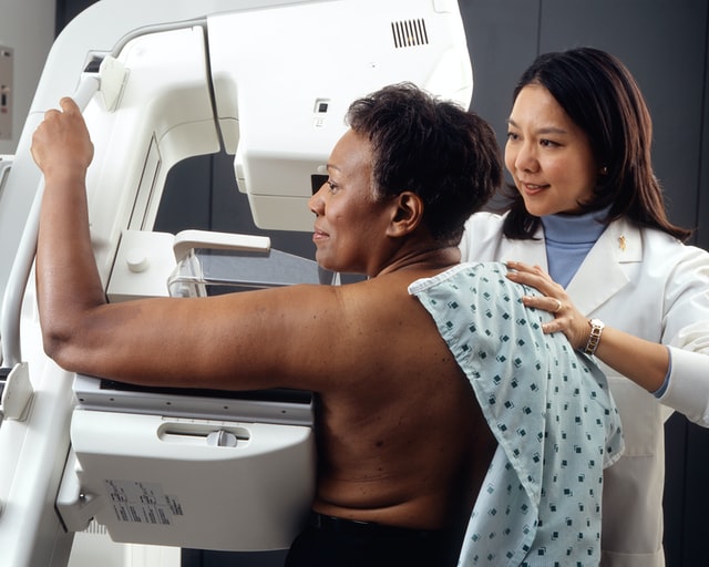
Mammography is one of the necessary tests physicians use to detect the early stages of breast cancer and other breast diseases. Fortunately, mammogram technology has advanced rapidly within the last few years and has positively impacted women’s health and wellness.

Radiological mammography has been in use through most of the 1900s, but the FDA didn’t approve digital mammography until 2000. The digital technology advancement opened up a whole new world for physicians to diagnose breast cancer earlier. Digital mammography accesses computer technology to enhance the X-ray images of the breast.

After digital mammography came into use, 3D breast imaging technology emerged in 2011. The 3D digital mammography (also known as 3D tomosynthesis) is where a technician takes multiple breast images from different angles. The technician then processes these images using computer software to create a three-dimensional reproduction of the breast.

With a three-dimensional reproduction of the breast, a radiologist can analyze the imaging slice-by-slice in great detail. This process has reduced many of the physician’s false-positive diagnoses given to women and reduced the stress of call-back appointments.

Since the 3D technology, companies have developed more advanced mammography equipment, tests, and computer-aided diagnosis systems (CAD). Researchers also have advanced imaging tools like whole breast ultrasound (WBUS) and magnetic resonance imaging  (MRI) to aid the mammography process.

Physicians may recommend patients perform regular year-to-year screening mammograms so any changes in the patient’s breast that may cause concern can be detected. A physician orders a diagnostic mammogram when the screening mammogram shows an abnormality or if the patient notes other extraordinary symptoms.


A diagnostic mammogram is similar to a screening mammogram, except the technician will take more images using more positions to get more explicit photos of the area. A diagnostic mammogram can define if a biopsy is needed.

Throughout mammogram use, the human eye has been depended on to detect abnormalities in a patient’s breast X-rays, leading to false positives and false negative exams. With the advancements in equipment, technology, and software, radiologists can detect any abnormality in breast tissue with more certainty.

Increased research and equipment advancements in mammograms have also decreased patients’ exposure to radiation. Studies have concluded that the benefits of mammograms nearly always outweigh the potential harm from radiation exposure. However, patients should always disclose to the X-ray technicians if they are pregnant or have other health issues at risk by using any level of radiation.

Newer mammography imaging tests help physicians diagnose the smallest of tumors and most minimal cell defects. These tests include positron emission mammography (PEM), optical imaging, electrical impedance tomography (EIT), and molecular breast imaging (MBI).

Positron emission mammography (PEM) is a scan that uses sugar attached to a radioactive particle to look for cancer cells. This test is sometimes a replacement for an MRI.


detecting breast cancer

Optical imaging is a test where technicians monitor the light passed into the woman’s breast and compare it to the measurement of light passing through the breast tissue. An altered reading of light will detect an area of the breast that warrants further exploration. Researchers are using this test with MRIs or 3D mammograms.


Since breast cancer cells conduct electricity differently than normal cells, physicians sometimes use electrical impedance tomography (EIT) as a diagnostic tool. During the test, a technician passes a bit of current through the patient’s breast and looks for changes with small electrodes applied to the skin.

Another test that researchers have developed is molecular breast imaging (MBI). This test is used with mammograms for women who have dense breasts. Doctors inject a radioactive drug into a patient’s vein, and the drug attaches to cancer cells, and a special camera can locate those cancer cells through the imaging process.

Researchers are continuing their efforts to improve mammogram results. Safe and effective screening and diagnostic mammograms will continue to improve survival statistics for women no matter what their genetic makeup, family history, or any other risk factor may indicate.

Vesta Teleradiology

At Vesta, our US Board Certified Radiologists are trained to read mammography scans as well as an entire host of other types of diagnostic imaging results. Look to us to support your team. Learn more about our teleradiology services here.