National Cancer Prevention Month: The Role of Imaging in Early Detection

February is National Cancer Prevention Month, a time to focus on the impact of early cancer detection and prevention. As healthcare providers, you know that catching cancer in its earliest stages significantly improves patient outcomes. Advanced imaging technologies continue to play a crucial role in detecting cancers before they progress, giving patients the best chance for successful treatment. This is especially important for facilities in rural or underserved areas, where access to specialized medical services is more limited.

The Importance of Early Detection Through Imaging

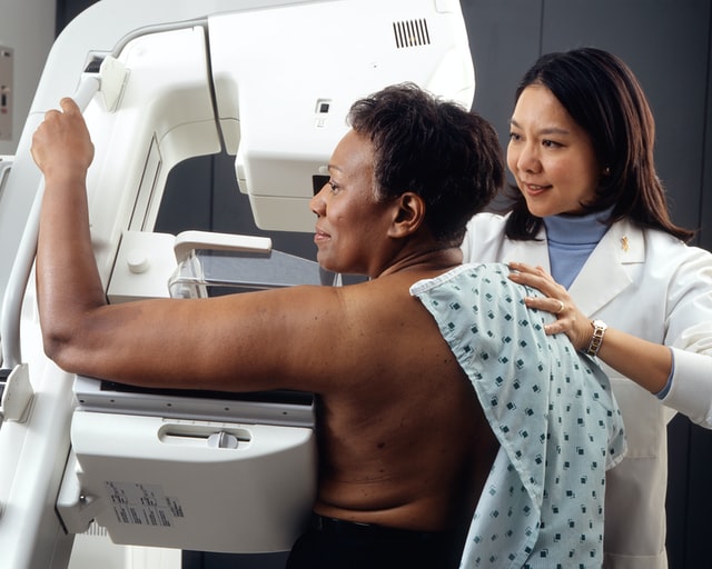
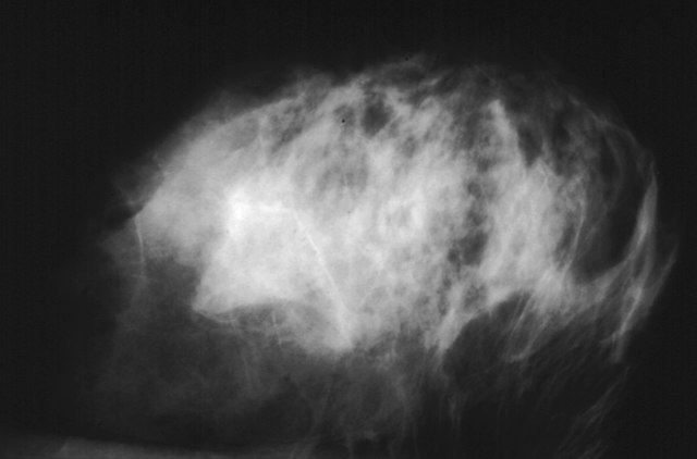
Early detection of cancer through imaging allows for interventions at stages when treatment is most effective. Techniques such as mammography, low-dose computed tomography (LDCT), and magnetic resonance imaging (MRI) are instrumental in identifying cancers like breast, lung, and prostate in their nascent stages. Regular screenings can lead to early diagnosis, which is associated with higher survival rates and a broader range of treatment options.

Teleradiology: Bridging the Gap in Underserved Areas

Teleradiology plays a pivotal role in expediting cancer diagnoses, particularly in rural or underserved regions. By transmitting radiological images from one location to another for interpretation by specialists, teleradiology ensures that patients receive timely and accurate diagnoses regardless of their geographical location. This technology mitigates the challenges posed by a shortage of on-site radiologists and enhances the quality of care in remote areas.

radiologistsRecent developments underscore the significant benefits of teleradiology for healthcare providers. A report from Healthcare IT News highlights how teleradiology enables radiologists to interpret scans remotely, increasing flexibility in work schedules and expanding access to specialized expertise. This approach addresses staffing challenges and enhances patient care by providing timely, expert interpretations.

Furthermore, a comprehensive guide by Philips emphasizes that teleradiology ensures 24/7 availability of radiology experts, allowing patients to receive timely diagnoses regardless of the time of day. This continuous access to radiological services helps alleviate the workload on in-house radiologists, leading to more efficient and focused patient care (Philips).

Additionally, RealTime Medical outlines several advantages of teleradiology, including cost savings, improved patient outcomes, increased efficiency, and better quality control. These benefits collectively enhance the overall performance of healthcare facilities by streamlining operations and ensuring high-quality diagnostic services.

Latest Screening Guidelines

Staying informed about current cancer screening guidelines is essential for both healthcare providers and patients. Here are the latest recommendations for breast, lung, and prostate cancer screenings:

  • Breast Cancer: The American Cancer Society recommends that women aged 45 to 54 undergo annual mammograms. Women aged 40 to 44 should have the option to start annual screening if they wish, and those 55 and older can transition to biennial screening or continue yearly exams. Screening should continue as long as a woman is in good health and expected to live at least 10 more years (American Cancer Society).
  • Lung Cancer: The U.S. Preventive Services Task Force advises annual screening for lung cancer with LDCT in adults aged 50 to 80 years who have a 20 pack-year smoking history and currently smoke or have quit within the past 15 years. Screening should be discontinued once a person has not smoked for 15 years or develops a health problem that substantially limits life expectancy (USPSTF).
  • Prostate Cancer: The American Urological Association suggests that men aged 55 to 69 engage in shared decision-making with their healthcare provider about the benefits and risks of prostate-specific antigen (PSA)–based screening for prostate cancer. For men younger than 55 at higher risk, such as those with a family history of prostate cancer or African American men, discussions about screening may be appropriate (AUA).

Optimizing Radiology Practices

Healthcare facilities can enhance their radiology services by:

  • Implementing Teleradiology: Adopting teleradiology services can provide access to subspecialty radiologists, reduce turnaround times for image interpretations, and offer continuous coverage, which is particularly beneficial for facilities in underserved areas.
  • Investing in Advanced Imaging Technologies: Upgrading to the latest imaging equipment ensures higher resolution images, leading to more accurate diagnoses.
  • Continuing Education for Radiologists: Encouraging radiology staff to engage in ongoing education keeps them abreast of the latest advancements and best practices in imaging and interpretation.
  • Community Outreach and Education: Educating the community about the importance of regular screenings and early detection can lead to increased participation in screening programs, ultimately improving public health outcomes.
  •   Recent developments underscore the significant benefits of teleradiology for healthcare providers. A report from Healthcare IT News highlights how teleradiology enables radiologists to interpret scans remotely, increasing flexibility in work schedules and expanding access to specialized expertise. This approach addresses staffing challenges and enhances patient care by providing timely, expert interpretations.
  •   Furthermore, a comprehensive guide by Philips emphasizes that teleradiology ensures 24/7 availability of radiology experts, allowing patients to receive timely diagnoses regardless of the time of day. This continuous access to radiological services helps alleviate the workload on in-house radiologists, leading to more efficient and focused patient care.

By focusing on early detection through advanced imaging and leveraging teleradiology, healthcare providers can significantly impact cancer outcomes, offering patients the best chance for successful treatment. National Cancer Prevention Month serves as a reminder of the strides we can make in cancer care through prevention and early detection.

At Vesta Radiology, we are committed to leveraging the advantages of teleradiology to provide exceptional diagnostic services. Our team of experienced radiologists is available both on-site and remotely, ensuring that healthcare providers receive timely and accurate interpretations tailored to their specific needs. By partnering with us, facilities can enhance their radiology services, improve patient outcomes, and optimize operational efficiency.

For more information about our services and how we can support your healthcare facility, please visit our website at Vesta Radiology.


What to Expect During a Breast Cancer Screening at an Imaging Center

The best way to detect breast cancer in its early stages is by mammograms–merely an X-ray of the breast. There is no financial strain because health insurance plans are required to cover the costs of mammograms every one or two years.

When you reach your forties, you should begin conversing with your medical providers about when to start and how often you should get a mammogram.

breast cancer awareness
Take care to get your screenings


Specialists usually recommend the procedure for women between 50 to 74 years of age at average risk of breast cancer and performed every one or two years depending on the woman’s risk factors.

How to Prepare for a Mammogram

When your healthcare provider suggests scheduling a mammogram for the first time, there are some considerations.

If possible, you should consider making your appointments at a nearby facility that specializes in mammograms. In doing this, you can have technicians who are very familiar with the process and accurately compare your mammograms yearly.

mammogram technology
Mammogram screening

If you need to change facilities, it’s best to arrange to have all your records sent to the new clinic for X-ray comparison. It is also an excellent idea to bring all the dates and locations of any previous breast procedures done.

Specialists advise that women avoid making mammogram appointments the week before their periods. Breasts are more swollen and sensitive during this time which can cause the mammogram procedure to be uncomfortable for them and may interfere with taking clear X-rays.

Technicians will advise that women wear pants or a skirt with a top that they can easily remove along with their bra. They also recommend that women do not use any deodorant, lotions, or powders that may show up as spots on the X-ray.

When You Arrive at the Imaging Center

Most technicians are very considerate and sensitive to what women experience during mammograms. They will ask you to undress above the waist and will give you a wrap to wear.

Many clinics have private dressing areas where you undress and wait for your technician to escort you into the X-ray room. There will only be the two of you in this area which is helpful to reduce any anxieties you may have.

You will stand in front of the mammogram machine, and the technician will position your body and breast for the most accurate X-rays. Your breast will rest on the device, and a plastic upper plate will lower and compress your breast.

The process takes 10 to 15 seconds for each body position, and there are usually only two or three X-rays taken per breast. The whole procedure only takes about 20 minutes.

Your Mammography Results

Mammography produces black-and-white digital images of your breast tissue that will be sent to a physician specializing in radiology to interpret. An imaging center could also refer to a teleradiology company to have a radiologist do the interpretation. Most clinics offer 2D mammograms for breast screening, but many facilities now offer 3D mammograms.

The radiologist will then report their findings to your healthcare provider. This process will take approximately ten days but could take longer. The radiology report will determine if you will need further X-rays, possible MRIs, or treatments. Physicians call back about 10 to 13% of women for abnormal findings, but most of these findings are not breast cancer.

Regular mammograms are an easy, safe way to screen for breast cancer. The most challenging part about the process may be remembering to make your annual appointment, and many women make their birthdays a reminder to make their yearly appointment.

What can be more celebratory than scheduling what may be a life-saving procedure for your health on the most important day of the year for you, your friends, and your loved ones? 

Advancements in Mammography

Mammography is one of the necessary tests physicians use to detect the early stages of breast cancer and other breast diseases. Fortunately, mammogram technology has advanced rapidly within the last few years and has positively impacted women’s health and wellness.

Radiological mammography has been in use through most of the 1900s, but the FDA didn’t approve digital mammography until 2000. The digital technology advancement opened up a whole new world for physicians to diagnose breast cancer earlier. Digital mammography accesses computer technology to enhance the X-ray images of the breast.

After digital mammography came into use, 3D breast imaging technology emerged in 2011. The 3D digital mammography (also known as 3D tomosynthesis) is where a technician takes multiple breast images from different angles. The technician then processes these images using computer software to create a three-dimensional reproduction of the breast.

With a three-dimensional reproduction of the breast, a radiologist can analyze the imaging slice-by-slice in great detail. This process has reduced many of the physician’s false-positive diagnoses given to women and reduced the stress of call-back appointments.

Since the 3D technology, companies have developed more advanced mammography equipment, tests, and computer-aided diagnosis systems (CAD). Researchers also have advanced imaging tools like whole breast ultrasound (WBUS) and magnetic resonance imaging  (MRI) to aid the mammography process.

Physicians may recommend patients perform regular year-to-year screening mammograms so any changes in the patient’s breast that may cause concern can be detected. A physician orders a diagnostic mammogram when the screening mammogram shows an abnormality or if the patient notes other extraordinary symptoms.


A diagnostic mammogram is similar to a screening mammogram, except the technician will take more images using more positions to get more explicit photos of the area. A diagnostic mammogram can define if a biopsy is needed.

Throughout mammogram use, the human eye has been depended on to detect abnormalities in a patient’s breast X-rays, leading to false positives and false negative exams. With the advancements in equipment, technology, and software, radiologists can detect any abnormality in breast tissue with more certainty.

Increased research and equipment advancements in mammograms have also decreased patients’ exposure to radiation. Studies have concluded that the benefits of mammograms nearly always outweigh the potential harm from radiation exposure. However, patients should always disclose to the X-ray technicians if they are pregnant or have other health issues at risk by using any level of radiation.

Newer mammography imaging tests help physicians diagnose the smallest of tumors and most minimal cell defects. These tests include positron emission mammography (PEM), optical imaging, electrical impedance tomography (EIT), and molecular breast imaging (MBI).

Positron emission mammography (PEM) is a scan that uses sugar attached to a radioactive particle to look for cancer cells. This test is sometimes a replacement for an MRI.


detecting breast cancer

Optical imaging is a test where technicians monitor the light passed into the woman’s breast and compare it to the measurement of light passing through the breast tissue. An altered reading of light will detect an area of the breast that warrants further exploration. Researchers are using this test with MRIs or 3D mammograms.


Since breast cancer cells conduct electricity differently than normal cells, physicians sometimes use electrical impedance tomography (EIT) as a diagnostic tool. During the test, a technician passes a bit of current through the patient’s breast and looks for changes with small electrodes applied to the skin.

Another test that researchers have developed is molecular breast imaging (MBI). This test is used with mammograms for women who have dense breasts. Doctors inject a radioactive drug into a patient’s vein, and the drug attaches to cancer cells, and a special camera can locate those cancer cells through the imaging process.

Researchers are continuing their efforts to improve mammogram results. Safe and effective screening and diagnostic mammograms will continue to improve survival statistics for women no matter what their genetic makeup, family history, or any other risk factor may indicate.

Vesta Teleradiology

At Vesta, our US Board Certified Radiologists are trained to read mammography scans as well as an entire host of other types of diagnostic imaging results. Look to us to support your team. Learn more about our teleradiology services here.