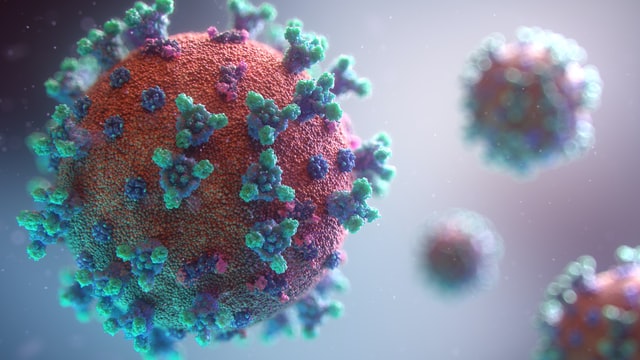
The COVID-19 pandemic may seem never ending. While the exposure and infection numbers may be shrinking, the long-lasting effects of this illness are revealing themselves. One of the biggest and most concerning shortages is the labor shortage.
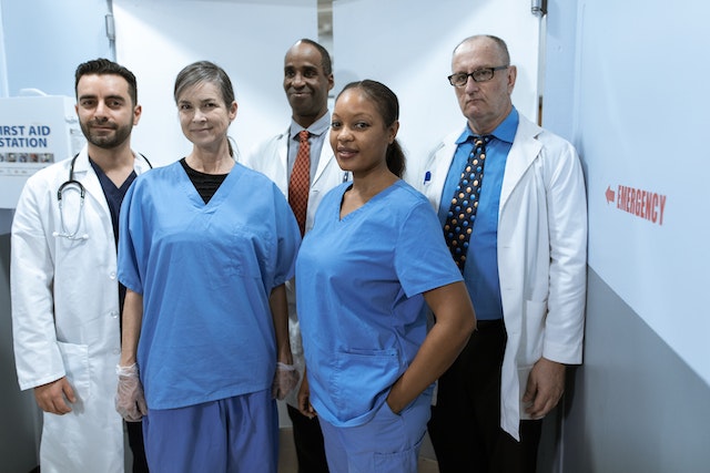
Of course, labor shortage can be vague. What industries are seeing these shortages, and how do those shortages affect customers? Many industries are seeing labor shortages, but one of the most concerning is the healthcare labor shortage.

The Healthcare Labor Shortage
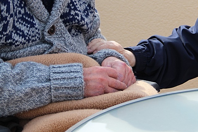
The COVID-19 pandemic proved to be tough on several fronts, especially for the first responders providing care and assistance to those suffering. Doctors and nurses were on the front lines treating patients, finding answers, and working long hours. Many healthcare workers were forced out for different physical and mental reasons like burnout. The pressures were so great that nearly 1.5 million healthcare workers left the profession in the first two months of the pandemic.
As the pandemic continues to wane, healthcare workers are returning to hospitals and doctors’ offices. The return is great, but the numbers are still down, and jobs are still left vacant. In fact, healthcare employment has still not returned to pre-pandemic numbers. Even in those who have returned, anecdotal evidence suggests many are thinking of leaving soon. This means sick people, some of the most vulnerable in society, will feel the consequences.
Technology and the Healthcare Labor shortage
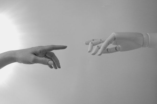
How do healthcare providers keep their invaluable workers and staff? How do they combat the pressures and stressors created and highlighted by the worldwide pandemic? The short answer is technology.
Healthcare providers can automate different tasks to allow healthcare providers – doctors and nurses – the freedom and space to care for patients. The best news is we live in the age of technology. There are dozens of different technological applications that can be used in these areas.
Inbound Calls
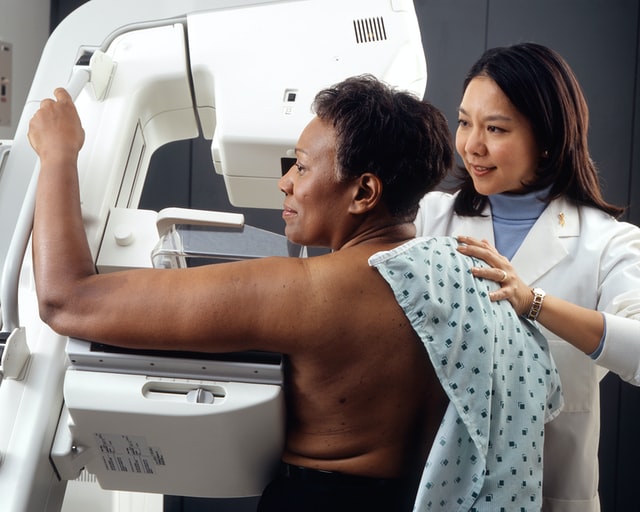
Hospitals and doctors’ offices are often overloaded by inbound calls, even when they are fully staffed. When these providers are understaffed, however, it can be time-consuming to field these ceaseless inbound calls. Patients can and should be encouraged to schedule their own appointments through web-based applications and portals. Not only will this open up more time and space for healthcare providers, but these tasks help empower patients to be more involved in their healthcare journey.

Intake Process
The amount of paperwork in the healthcare industry is daunting. Technology, however, can limit the paperwork and streamline the intake process altogether. Mobile check-in and registration can make it easier for patients to check in, but it also limits the person-to-person contact that so easily spreads diseases.
Access and Availability
Perhaps the best advantage of medicine is the access and availability afforded through telemedicine. Telemedicine isn’t necessarily “new,” but it has been brought to the forefront. Telemedicine is the ability to meet with medical professionals and healthcare workers to get information and establish treatment plans.
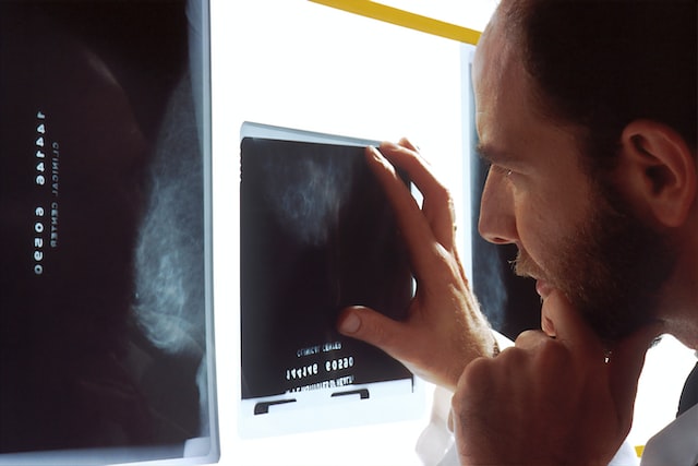
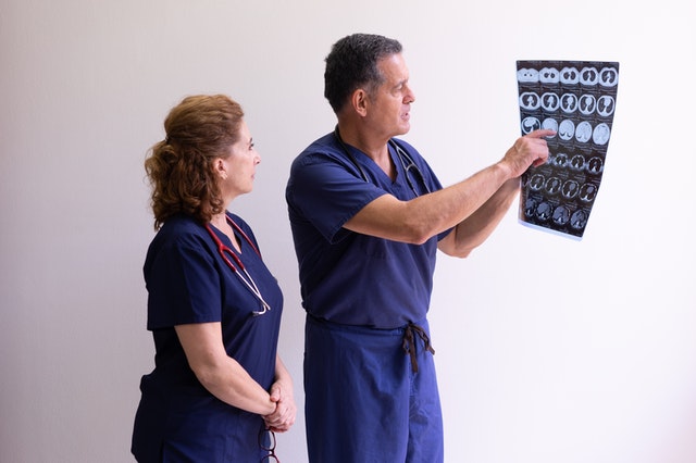
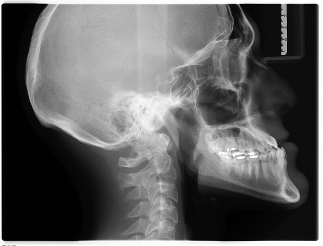
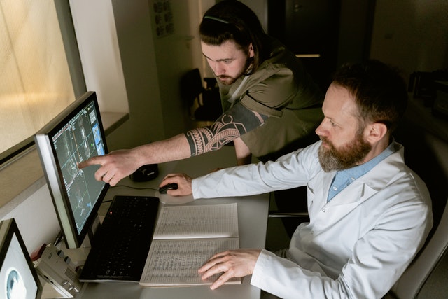
It’s especially beneficial when it comes to specialized medicine, like radiology. Teleradiology, the term widely used for this specific section, is a much more recent development. It helps patients get information and necessary access to radiology professionals.
Teleradiology allows a radiologist to get, review, and interpret CT or MRI images. Radiologists are able to communicate important information to patients who are desperate for that information. It means fewer radiologists can meet with more patients and get those patients the information and treatment they need.
Virtual Monitoring Systems
Telesitter programs help reduce the workload and potential burnout for nurses. With these systems, cameras are setup so that virtual monitoring can take place and track patient activity. Any time there are concerns or emergencies, staff would be notified.

The world is changing. It’s the one true constant. But technology offers us the chance to adapt and modify the ways we move about in the world. Technology can make things easier and fill in the gaps that form.