Vizamyl’s New PET Label: Quantify & Monitor Amyloid—What It Means for Imaging Teams


What changed—and why it matters

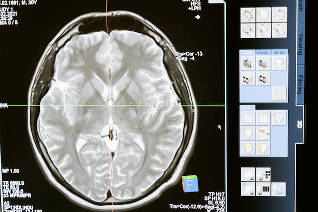
The FDA has expanded the label for flutemetamol F 18 (Vizamyl), enabling quantification of amyloid plaque burden and long-term therapy monitoring in Alzheimer’s disease. This shift moves amyloid PET beyond a qualitative “positive/negative” decision toward objective, longitudinal assessment that can inform treatment choice, dose intervals, and discontinuation decisions. Business Wire

Professional groups report the update aligns amyloid PET with the clinical era of disease-modifying anti-amyloid therapies (e.g., lecanemab, donanemab), clarifying roles for baseline confirmation, on-treatment monitoring, and response tracking in routine care. Notably, SNMMI stated the FDA granted supplemental indications—including quantitative measurement and use for therapy monitoring—to three amyloid PET agents (flutemetamol F-18/Vizamyl, florbetapir F-18, and florbetaben F-18). SNMMI

Operational updates for radiology leaders

  • Protocols & quant pipelines: Build or validate a quant workflow (SUVr or comparable metrics) that’s scanner-calibrated and reproducible across sites. If you operate multi-vendor fleets, document harmonization steps in your SOPs.
  • Structured reports: Add fields for quantified burden at baseline, change from baseline, and interpretive guidance tied to therapeutic decisions (initiation, continuation, or discontinuation).
  • Scheduling & throughput: Expect rising referral volume from neurology and geriatrics as therapy monitoring enters routine practice; protect access with extended hours or overflow capacity.
  • Quality & governance: Define thresholds for biologically meaningful change, reader training for quant review, and reconciliation rules when quant and visual impressions diverge.

For additional context, trade coverage underscores that the updated label formally removes previous limitations around therapy monitoring and permits quant analysis in routine reporting. Empr

How Vesta Teleradiology helps

Vesta’s subspecialty neuro and nuclear medicine radiologists provide:

  • Amyloid PET expertise: Visual+quant reads with structured templates aligned to your therapy pathway.
  • Coverage when you need it: After-hours, weekends, or daytime overflow—without sacrificing turnaround time.
  • Interoperability: Seamless delivery to your PACS/RIS and EMR; clear flags for therapy decisions and recall intervals.
  • QA you can see: Peer review, consistency checks across readers, and optional double-reads during program ramp-up.

If you’re standing up or scaling amyloid PET services, we can supply immediate subspecialty coverage and templates tuned to your neurologists’ needs.

 

Recent Advancements in Nuclear Medicine

The medical community is always looking for new and better ways to serve patients and save lives. Science, medicine, and technology often intersect to break barriers and create innovative new treatments – and nowhere is that truer than in the field of nuclear medicine.

What is Nuclear Medicine?

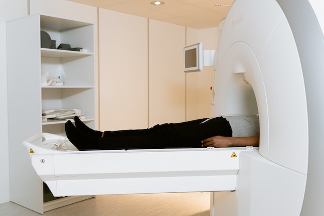
The National Institute of Biomedical Imaging and Bioengineering defines nuclear medicine as a specialty that uses radioactive tracers to diagnose and treat disease. Nuclear medicine is invaluable for patient care, as it can help detect disorders in the bones, gall bladder, heart, and much more.

advancements in nuclear medicine

This field has seen tremendous advancements in recent years, which offer the potential for incredible and life-saving benefits. Here are some of the latest developments in nuclear medicine.

Making AI More Effective

Artificial intelligence has been an integral part of medicine for decades, particularly in the realm of diagnostics. And now, new research suggests that nuclear medicine may make AI-based diagnostics even more effective.

radiology interpretations

For example, researchers in the Journal of Nuclear Medicine (JNM) suggest that nuclear imaging can help with machine learning and AI cancer diagnoses. This is because nuclear imaging creates a high contrast between tumors and normal tissue, making it much easier for the machine to identify abnormalities. Combining AI diagnostics with nuclear medicine can make the machines more accurate, which will ultimately result in better patient care over time.

Detecting Heart Disease

Radionuclide imaging has long been used to detect issues in patient heart function. However, researchers are beginning to explore new uses for this technology – including the examination of the heart’s very molecules.

Research from 2020 found that radionuclide imaging is successful at detecting cardiac amyloidosis, a rare condition in which a protein called amyloid is deposited in the cardiac muscle. Amyloid deposits can cause buildup over time and ultimately lead to heart failure, so it is very important to detect this condition as early as possible.

Discovering New Treatments

Nuclear medicine has many potential uses for imaging and diagnostics. However, it also offers many benefits for researchers.

For example, scientists at the Vanderbilt University Institute of Imaging Science recently used a radioligand (a radioactive substance used to study receptors in the body) to study whether an antioxidant called ERGO could penetrate the brain and protect against oxidative stress. The study successfully proved that ERGO can penetrate the brains of mice, which opens doors for further research on using this antioxidant to treat conditions like Alzheimer’s disease.

Nuclear medicine is always developing and advancing, and each advance makes it easier to give patients the care they deserve. 


Nuclear Radiology Readings


We are proud of our talented pool of teleradiologists who specialize in a variety of subspecialties, including nuclear radiology. If you’ would like to learn more about how we can integrate with your current workflow in order to provide preliminary and final interpretations, please contact us now at 1-877-55-VESTA