What’s New in Breast Density and Mammography: Fall 2025 Update

Why breast density remains a frontline issue

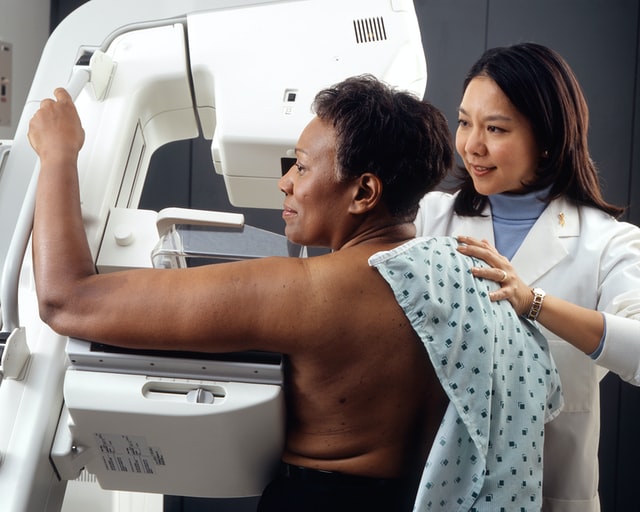
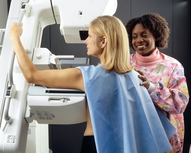
Breast density continues to be one of the most important—and complex—factors in breast cancer screening. Dense breast tissue not only raises cancer risk but also makes abnormalities harder to detect on mammograms. For hospitals and imaging centers, keeping up with evolving regulations, trial data, and technology is no longer optional. It’s central to compliance, patient communication, and imaging strategy.

FDA updates the national reporting standard

In July 2025, the FDA approved changes to the breast density reporting standard under the Mammography Quality Standards Act (MQSA). This builds on the September 2024 rule requiring that all mammography reports inform patients whether their breasts are “dense” or “not dense.”

Hospitals should review their reporting templates now. The updated language affects how results must be communicated to both patients and referring clinicians. Staying compliant avoids liability and ensures consistent, patient-friendly communication across facilities.

Doctors reviewing breast density mammogram results for Fall 2025 hospital updates.New trial evidence favors MRI and contrast-enhanced mammography

The interim results of the BRAID trial in the U.K. made headlines this summer. Among women with dense breasts and negative mammograms, supplemental abbreviated MRI and contrast-enhanced mammography (CEM) identified significantly more invasive cancers than ultrasound.

  • MRI and CEM: ~15–19 extra cancers detected per 1,000 women screened
  • Ultrasound: ~4 extra cancers detected per 1,000

These findings were reported in the OBG Project’s summary of the BRAID interim results.

While recalls and contrast risks remain a concern, the data strengthen the case for offering advanced supplemental imaging in high-density populations. Hospitals may want to begin planning how to integrate MRI or CEM into workflow, or establish referral pathways for patients with very dense breasts.

MBI joins the conversation

Molecular breast imaging (MBI), when paired with digital breast tomosynthesis, is showing early promise in improving invasive cancer detection in women with dense breasts. Findings from the Density MATTERS trial highlight MBI as a potential alternative for hospitals with limited MRI or CEM capacity.

AI-enabled density assessment and multimodal risk stratification

Artificial intelligence tools are advancing rapidly in breast imaging. A recent clinical study demonstrated that multimodal AI systems can reduce recall rates by over 30% while maintaining sensitivity. Other work shows promise in improving density quantification and developing 5-year breast cancer risk models from imaging features.

Hospitals considering AI adoption should focus on how these tools can streamline workflow, support compliance, and reduce unnecessary patient callbacks.

Shifting clinical culture: from notification to action

At the 2025 Society of Breast Imaging annual meeting, a clear theme emerged: simply notifying patients about dense breast status is not enough. The expectation is shifting toward offering supplemental imaging or providing clear, individualized next steps.

Hospitals that rely on tomosynthesis alone may increasingly be asked to justify why they do not offer MRI, CEM, or other supplemental options.

Key takeaways for hospitals and imaging centers

  • Compliance check: Ensure your reporting language matches the updated FDA standard.
  • Workflow planning: Prepare for increased demand for supplemental imaging in dense-breast populations.
  • Technology assessment: Evaluate the role of MRI, CEM, MBI, and AI tools in your facility.
  • Patient communication: Move beyond dense-breast notification toward structured shared decision-making.
  • Equity focus: Consider insurance coverage and access barriers that could affect your patient population.

Hospitals that adapt now will not only stay compliant but also lead in patient-centered breast cancer screening strategies.


FDA’s 2025 AI Draft Guidance: A Buyer’s Checklist for Imaging Leaders

In January 2025, the U.S. Food and Drug Administration released a draft guidance for AI-enabled medical devices that lays out expectations across the total product life cycle—design, validation, bias mitigation, transparency, documentation, and post-market performance monitoring. For imaging leaders, it’s a clear signal to tighten procurement criteria and operational guardrails before piloting AI in CT, MRI, mammo, ultrasound, or PET.

As teams lock in Q4 budgets and head into RSNA season, the FDA’s AI lifecycle draft (Jan 2025) and the now-final PCCP (Dec 2024) have reset what buyers should expect from AI in imaging—devices, software, and workflows. Vendors are updating claims and governance; this issue distills a practical buyer’s checklist—multisite validation with subgroup results, drift monitoring and version control, clear in-viewer transparency—and how pairing those tools with Vesta’s subspecialty coverage and QA turns promise into measurable gains across CT/MRI/US/mammography.

A practical buyer’s checklist

Use this when evaluating AI for your service lines:

  1. Intended use fit: Verify indications, inputs/outputs, and claims match your pathway and patient mix.
  2. Validation depth: Prefer multisite, diverse datasets; stratified results; pre-specified endpoints; documented data lineage and splits.
  3. Bias mitigation: Demand subgroup performance (sex, age, race/ethnicity when available), scanner/vendor variability analyses, and site-transfer testing.
  4. TPLC plan: Require drift monitoring, retraining triggers, versioning, and how updates are communicated.
  5. Human factors & transparency: Ensure limitations, failure modes, and interpretable outputs are presented in-viewer without slowing reads.
  6. Security & support: Patch cadence, vulnerability disclosure, SOC2/ISO posture, uptime SLAs, and rollback paths for version issues.
  7. Governance: Define metrics owners, review cadence, and thresholds to pause or roll back a model.

Implementation playbook: pilot → scale without disruption

Start with a 60–90 day pilot in one high-impact line (e.g., ED stroke CT or mammography triage) and lock in baselines: median TAT, positive/negative agreement, recall rate, PPV/NPV, and discrepancy rate. Set guardrails—when to auto-triage vs. force human review—and document escalation paths for model failures. Require case-level confidence and structured outputs your radiologists can verify quickly. Stand up a model governance huddle (modality lead, QA, IT security, and your teleradiology partner) that meets biweekly to review drift signals, subgroup performance, and near-misses. Bake in a rollback plan (version pinning) and a quiet-hours change window so updates don’t collide with peak volumes. As results stabilize, scale by cohort (e.g., expand to non-contrast head CT, then CTA) and keep training “micro-bursts” for techs/readers—short videos or checklists in-workflow. Tie vendor SLAs to uptime, support response, and clinical KPIs so the AI program stays accountable to operational value.

Where teleradiology fits

AI only delivers when it’s welded to coverage, quality, and speed. A teleradiology partner should provide:

  • 24/7 subspecialty + surge capacity: Vesta absorbs volume peaks so AI never becomes a bottleneck.
  • QA you can see: We benchmark pre/post-AI performance, add targeted second looks for edge cases, and feed variance data back to your team.
  • Standardized outputs: Structured reports that integrate model outputs with radiologist findings—no black-box surprises.
  • Smooth rollout: Pilot by service line (stroke CT, mammo triage, PE workups), then scale with tracked KPIs (TAT, PPV, recalls).
  • Interoperability & security: Seamless PACS/RIS/EMR integration with strict access controls, audit trails, and support for change-controlled updates.

Bottom line: Pairing AI with Vesta Teleradiology gives you round-the-clock subspecialty reads, measurable QA, and operational breathing room while you pilot and scale responsibly. If you’re mapping your AI roadmap under the FDA’s 2025 draft guidance, we’ll be your coverage and quality backbone—so your clinicians see faster answers and your patients see safer care. Visit vestarad.com to get started.


Why Second Opinions Matter: How Our Teleradiology Services Support Healthcare Facilities with Subspecialty Expertise

When it comes to accurate diagnoses and effective patient care, getting a second opinion on imaging results can make all the difference. Our teleradiology company is here to support healthcare facilities that need a reliable and expert review of radiology reports. Whether you’re looking for a second set of eyes for complex cases or want to ensure the highest level of diagnostic accuracy, our team of board-certified radiologists—with subspecialties in areas such as neuroradiology, musculoskeletal imaging, and oncology—are ready to assist.

Why Choose a Teleradiology Partner for Second Opinions?

Second opinions are often requested in cases where findings are ambiguous, complex, or where the stakes are high. By partnering with a teleradiology company like ours, healthcare providers can access subspecialized expertise quickly and cost-effectively, regardless of their location. This can be a game-changer for facilities with limited in-house radiology staff or those handling a high volume of specialized imaging studies.

In fact, a multidisciplinary tumor board at a National Comprehensive Cancer Network (NCCN) center found that 43% of patients referred for second opinions on breast cancer imaging had their initial diagnosis changed. This statistic highlights the significant impact that expert second opinions can have on patient outcomes​.


Types of Imaging Studies That Often Need a Second Opinion

Not all imaging studies are straightforward, and some require deeper insight to reach a definitive diagnosis. Here’s a look at the imaging tests that most frequently benefit from a second opinion:

MRI (Magnetic Resonance Imaging)

    • Neuroradiology (Brain and Spine Imaging): Brain and spinal MRIs are complex studies that may reveal subtle findings like micro-bleeds, white matter lesions, or small tumors. Our neuroradiologists can provide expert insight into these challenging cases.
    • Musculoskeletal MRI: Joint injuries, ligament tears, and early-stage bone lesions often require subspecialty evaluation to avoid misdiagnosis.

CT (Computed Tomography) Scans

      • Abdominal and Pelvic CT: Complex abdominal findings, such as small tumors or liver masses, can be challenging to interpret. Our abdominal imaging specialists ensure accurate diagnoses for conditions like pancreatic cancer or complex GI issues.
      • Thoracic CT: Pulmonary nodules or lung masses require careful analysis to differentiate benign from malignant findings, especially in patients with a history of smoking or cancer.

Mammography and Breast Imaging

        • Given the sensitivity and potential impact of findings on patient care, a second opinion can confirm initial readings and prevent over- or under-treatment. Our radiologists with breast imaging expertise can re-evaluate mammograms, breast MRIs, and ultrasounds.

Providing second opinions on breast imaging cases not only ensures diagnostic accuracy but also places significant demands on radiologists’ workloads. A study found in the Journal of the American College of Radiology revealed that second opinions for breast cancer cases required an estimated 3,135 to 3,804 work relative value units (wRVUs) over a three-year period, but the reimbursement received did not match the effort involved​.

Ultrasound

    • Obstetric and Gynecologic Ultrasounds: Subtle findings such as ovarian cysts or fetal abnormalities often require expert review to confirm a diagnosis.
    • Vascular Ultrasound: Evaluating blood flow and vessel abnormalities can benefit from a second opinion to ensure treatment decisions are accurate.

Oncologic Imaging

    • PET-CT and Whole Body MRIs: Cancer staging is critical for treatment planning, and our subspecialized oncologic radiologists can help refine staging or detect subtle metastases that may have been missed.

Who Benefits from Our Teleradiology Second Opinion Services?

Our second opinion teleradiology services are a valuable resource for a wide variety of healthcare facilities, each with unique needs:

Hospitals and Health Systems

    • From large urban hospitals to smaller rural facilities, hospitals often have to handle complex imaging cases where a second opinion is invaluable. This is especially true for trauma cases, oncology patients, and complex neurological conditions.

Outpatient Imaging Centers

    • Standalone imaging centers that offer diagnostic imaging services such as MRI, CT, and X-ray can enhance the quality of their reports by providing second opinions from subspecialized radiologists. This builds trust with referring physicians and patients.

Urgent Care Centers

    • Urgent care facilities frequently deal with musculoskeletal injuries and abdominal pain that require precise interpretations. A teleradiology partner with musculoskeletal and abdominal imaging expertise can help confirm initial findings and ensure appropriate follow-up care.

Primary Care and Family Practice Clinics

    • When general practitioners encounter unexpected or unusual imaging findings, a second opinion from a subspecialist can guide the next steps in patient management.

Specialty Clinics (Neurology, Orthopedics, Oncology)

    • Specialty clinics can use our second opinion services to validate complex imaging studies, such as intricate spinal MRIs or oncology staging scans, ensuring the highest standard of care for their patients.

Our Subspecialized Expertise Sets Us Apart

Our team includes radiologists who are not only board-certified but also have subspecialty training in fields such as:

  • Neuroradiology
  • Musculoskeletal Imaging
  • Breast Imaging
  • Abdominal Imaging
  • Cardiothoracic Imaging
  • Pediatric Radiology

This means that when you request a second opinion, your cases are reviewed by experts who focus on these specific areas every day, providing a level of precision that general radiology might not offer.

How Our Teleradiology Process Works

  1. Submit the Case Securely: Our HIPAA-compliant platform allows you to upload imaging studies quickly and securely.
  2. Subspecialized Review: We assign your case to a radiologist with the relevant subspecialty training to review the images and provide an expert second opinion.
  3. Receive a Detailed Report: You receive a comprehensive report with detailed findings, recommendations, and any necessary follow-up steps, ensuring that your patients get the best possible care.choosing a teleradiologist
Ready to Partner for Second Opinions? Choose Vesta Teleradiology

If your healthcare facility needs expert support for complex or high-stakes imaging cases, contact us today. Our team is dedicated to helping you provide accurate, timely, and comprehensive care for your patients through our teleradiology second opinion services.

Optimize patient outcomes and build trust in your imaging results by partnering with us for your second opinion needs!

Contact Vesta now to learn more about our subspecialty teleradiology services.


Sources:

jacr.org
pubmed.ncbi.nlm.nih.gov
openai.com


Mammography: Is AI Better than Humans?

In recent years, artificial intelligence (AI) has made remarkable strides in revolutionizing the landscape of the medical field, offering unprecedented opportunities for enhanced patient care, diagnosis, and treatment. From accelerating the analysis of medical imagery to predicting disease outcomes with unparalleled accuracy, AI-powered technologies have swiftly established themselves as indispensable tools for healthcare professionals. Beyond diagnostics, AI has played a pivotal role in drug discovery, streamlining clinical trials, and personalizing patient interventions. As AI continues to evolve, its potential to transform healthcare systems globally is becoming increasingly evident, promising not only improved medical outcomes but also cost-effective solutions and optimized resource allocation. The fusion of AI’s computational prowess with medical expertise heralds a new era of medical advancements that hold the potential to alleviate the burden on healthcare systems, save lives, and redefine the standards of patient well-being.

In the United States alone, it is estimated that around 40 million mammograms were performed each year. Mammograms are crucial as they are the primary method for early detection of breast cancer, enabling timely intervention and improving survival rates. By detecting small abnormalities and tumors that may not be palpable, mammograms help identify potential breast cancer cases in their earliest stages, allowing for more effective and less invasive treatment options.

Abnormal mammogram

Radiologists often find themselves overwhelmed due to the increasing volume of medical images requiring analysis, coupled with a shortage of radiology specialists. The demand for accurate and timely diagnoses, especially in fields like mammography, can lead to extended work hours and heightened stress levels among radiologists. Introducing AI technologies can alleviate this burden by assisting in image analysis, enabling radiologists to focus on complex cases and ensuring more efficient patient care.

How AI Helps in Mammography

A recent study published in The Lancet Oncology suggests that artificial intelligence (AI) may outperform trained doctors in detecting breast cancer from mammogram images. Mammograms face challenges due to factors like breast density, leading to missed cancer cases. The study analyzed 80,000 mammograms from Swedish women, finding that AI-assisted readings detected 20% more cancers compared to human radiologists. While not a standalone solution, AI could alleviate doctors’ workloads, enhancing accuracy without increasing false negatives. Despite FDA-approved AI technologies, integration with conventional methods is likely, aiding radiologists in managing a growing workload. The balance between AI and human expertise remains essential, ensuring optimal patient care and early cancer detection.

Healthcare experts, including the NHS and the Royal College of Radiologists, acknowledge AI’s promise in enhancing efficiency, decision-making, and prioritizing critical cases.

mammograms

Vesta Teleradiology

AI applied to diagnostic imaging holds the potential to significantly enhance the level of patient care. We eagerly anticipate further progress in this field. However, we maintain the viewpoint that presently, no machine can effectively substitute for the expertise of a skilled human observer for interpretations. At Vesta, we offer the services of radiologists who are US Board Certified, dedicated to delivering precise preliminary and final analyses. Discover how we can bolster your radiology department by reaching out to us today.


Sources:

Criver.com
health.com
theguardian.com
openai.com


Advancements in Mammography

Mammography is one of the necessary tests physicians use to detect the early stages of breast cancer and other breast diseases. Fortunately, mammogram technology has advanced rapidly within the last few years and has positively impacted women’s health and wellness.

Radiological mammography has been in use through most of the 1900s, but the FDA didn’t approve digital mammography until 2000. The digital technology advancement opened up a whole new world for physicians to diagnose breast cancer earlier. Digital mammography accesses computer technology to enhance the X-ray images of the breast.

After digital mammography came into use, 3D breast imaging technology emerged in 2011. The 3D digital mammography (also known as 3D tomosynthesis) is where a technician takes multiple breast images from different angles. The technician then processes these images using computer software to create a three-dimensional reproduction of the breast.

With a three-dimensional reproduction of the breast, a radiologist can analyze the imaging slice-by-slice in great detail. This process has reduced many of the physician’s false-positive diagnoses given to women and reduced the stress of call-back appointments.

Since the 3D technology, companies have developed more advanced mammography equipment, tests, and computer-aided diagnosis systems (CAD). Researchers also have advanced imaging tools like whole breast ultrasound (WBUS) and magnetic resonance imaging  (MRI) to aid the mammography process.

Physicians may recommend patients perform regular year-to-year screening mammograms so any changes in the patient’s breast that may cause concern can be detected. A physician orders a diagnostic mammogram when the screening mammogram shows an abnormality or if the patient notes other extraordinary symptoms.


A diagnostic mammogram is similar to a screening mammogram, except the technician will take more images using more positions to get more explicit photos of the area. A diagnostic mammogram can define if a biopsy is needed.

Throughout mammogram use, the human eye has been depended on to detect abnormalities in a patient’s breast X-rays, leading to false positives and false negative exams. With the advancements in equipment, technology, and software, radiologists can detect any abnormality in breast tissue with more certainty.

Increased research and equipment advancements in mammograms have also decreased patients’ exposure to radiation. Studies have concluded that the benefits of mammograms nearly always outweigh the potential harm from radiation exposure. However, patients should always disclose to the X-ray technicians if they are pregnant or have other health issues at risk by using any level of radiation.

Newer mammography imaging tests help physicians diagnose the smallest of tumors and most minimal cell defects. These tests include positron emission mammography (PEM), optical imaging, electrical impedance tomography (EIT), and molecular breast imaging (MBI).

Positron emission mammography (PEM) is a scan that uses sugar attached to a radioactive particle to look for cancer cells. This test is sometimes a replacement for an MRI.


detecting breast cancer

Optical imaging is a test where technicians monitor the light passed into the woman’s breast and compare it to the measurement of light passing through the breast tissue. An altered reading of light will detect an area of the breast that warrants further exploration. Researchers are using this test with MRIs or 3D mammograms.


Since breast cancer cells conduct electricity differently than normal cells, physicians sometimes use electrical impedance tomography (EIT) as a diagnostic tool. During the test, a technician passes a bit of current through the patient’s breast and looks for changes with small electrodes applied to the skin.

Another test that researchers have developed is molecular breast imaging (MBI). This test is used with mammograms for women who have dense breasts. Doctors inject a radioactive drug into a patient’s vein, and the drug attaches to cancer cells, and a special camera can locate those cancer cells through the imaging process.

Researchers are continuing their efforts to improve mammogram results. Safe and effective screening and diagnostic mammograms will continue to improve survival statistics for women no matter what their genetic makeup, family history, or any other risk factor may indicate.

Vesta Teleradiology

At Vesta, our US Board Certified Radiologists are trained to read mammography scans as well as an entire host of other types of diagnostic imaging results. Look to us to support your team. Learn more about our teleradiology services here.