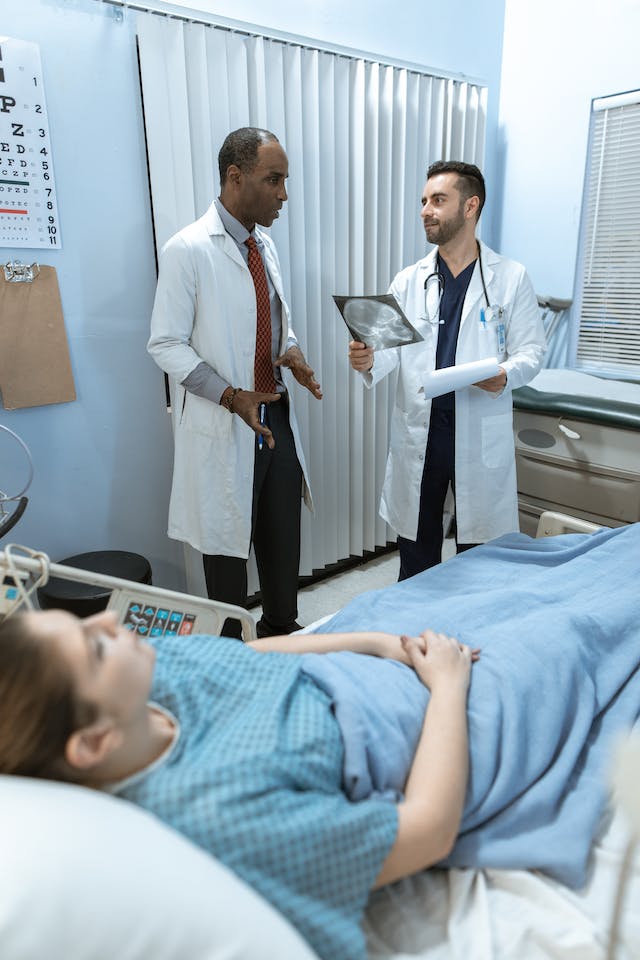
Creative Mammogram Awareness Campaigns  

Just before October 2022–Breast Cancer Awareness Month–Katie Couric publicly revealed that her mammogram a few months earlier had alerted her doctors that she needed further medical tests, and this testing led to her diagnosis of breast cancer. Fortunately, she and her doctors had caught her cancer early on and had time to act on the diagnosis.

Katie’s career as a successful news anchor brought attention to the reality that breast cancer can happen to anyone. She felt her bold announcement of the disease might help reduce new cases of invasive breast cancer. That was her objective with many interviews and publicity on television, in magazines, and other media.

cancer awareness

Campaigns are More Important This Year

During the pandemic, many women went without seeing their physicians or getting mammograms. The Journal of American Medical Association estimates breast cancer screenings have reduced by 6% in the past two years. With breast cancer causing worldwide deaths of over 685,000 in 2020, early detection with mammograms is essential.

Mammograms are just an X-ray to detect abnormalities in a woman’s breast tissue; however, these images have found cancers up to 3 years before a patient can feel any lumps in the breast tissue.

Just about everyone is aware of the importance of mammograms in the fight against breast cancer, but many do not follow through with the process of making and showing up for mammogram appointments. Creative campaigns to motivate women to follow through with their health care can save many lives.

Campaign Strategies

The Centers for Disease Control and Prevention has issued a digital media toolkit to focus constructive messages to the public about Breast Cancer Awareness. The toolkit is structured to empower women to take control of their health and do what they must do for an early diagnosis of breast cancer.

breast cancer awareness campaign

Every campaign initiated by organizations, workplaces, and individuals should emphasize routine mammograms because they can be the key to saving a woman’s life. The mammogram is an easy exam, but it’s equally easy to forget or delay.

During October, many clinics or private practitioners offer free mammograms to low-income or uninsured women between the ages of 40 and 64. Individuals can help these organizations get the word out to the public by volunteering to help spread the word. The word free is a very effective campaign strategy.

The Susan G. Komen Foundation has multiple races across the country to bring awareness and financial support for breast cancer awareness. Their strategy is to create an event for the participants to go out and raise money for their cause. Their strategy is power in numbers, and the walks have been very successful for the foundation.

Employers can get involved with campaigns for their employees as well. Workplace wellness plans have partnered with their insurance companies to offer personal assessments for their employees, bring in mobile mammography vans, and promote educational information for the women in their workplace.

Anyone can have a successful campaign to bring awareness to breast cancer prevention and early detection. Finding strategies that work for each individual, community, and culture can sometimes be challenging but worth the effort. After all, it is about getting the word out and, more importantly, saving lives.

Read More Mammograms with Teleradiology

By partnering with a teleradiology company like Vesta, you will be able to get more mammogram screenings done since we can handle the interpretations for you, days, nights and weekends. Want to learn more about how we integrate with your workflow? Contact us now for a free quote and overview: 1-877-55-VESTA.

Why is There a Radiologist Shortage and What Can Be Done?

Association of American Medical Colleges states that radiologist burnout and the demographics of the workforce have been critical for years. For example, 82% of the 20,950 currently active radiologists are over 45 and over, while 53%  are 55 or over. According to a recent Mayo Clinic study, radiologists ranked fifth out of 23 specialties that reported burnout rate, particularly among those screening for breast cancer. So not only are radiologists aging out, the newcomers aren’t lasting that long.

Why Are Radiologists Being Burned Out?

Radiology Business says radiologists mention things like “lack of respect” from administrators or colleagues as one of the leading causes of burnout 60%, along with lack of control or autonomy over one’s life, long work hours, too many non-essential tasks, and more. That tempered with COVID-related burnout and we are now facing a global shortage of radiologists and radiology trainees.

shortage of radiologists
Burnout has impacted healthcare staffing


According to a Diagnostic Imaging podcast, numerous factors result in radiologist burnout, from outdated and clunky software, sinking reimbursements, continuing education requirements, stressful imaging reports turnout expectations, to reduced interaction with colleagues related to the pandemic, all resulting in a slow, laborious system death.

Should You Consider Teleradiology/Outsourced Radiology Company?

If you have never heard of teleradiology or outsourcing radiology, you may be interested in learning that many clinics, offices, and hospitals are turning to medical outsourcing. Hospitals will likely start to engage more and more outsourced services that can include long-distance radiology and teleradiology. While there are pros and cons for hospitals and clinics using teleradiology, the fact is that this may quickly become a fact of life. Patient care can still be high-quality, immediate, and efficient, for example.

There are incredible benefits that come with radiology outsourcing companies and teleradiology. Many teleradiology practices employ board-certified and trained radiologists for neurological and Body Imaging needs. These radiologists are available for patient consultations to go over results, answer any questions, and much more. Plus, these services are often quick and done promptly. Many of these companies and services are coupled with a quality assurance program to maintain their ACR accreditation. Finally, these services tend to be more cost-effective than in-house services. Teleradiology and outsourced services are often used in the following practices:

  •     Private practices
  •     Urgent care clinics
  •     Imaging practices
  •     Mobile services
  •     Physician groups
  •     Independent diagnostic testing facilities
teleradiology
Teleradiology can assist with staffing shortages

What Does the Future of Radiology Look Like?

One popular solution that addresses the shortage of radiologists includes using a teleradiology/outsourced radiology company as mentioned above.

Another solution is using trained radiographers to double-read screening mammograms, extending their expertise and skill set to perform as radiologists when needed. Double reading has been used in Europe and has been incredibly efficient at detecting cancer. Training non-radiologists may end up becoming a necessity for hospitals and breast screening facilities without having to lose patient standards.

Radiology remains a solid and stable career, especially as medical professionals and facilities adapt to shortages, and potential pandemics, especially when considering teleradiology and outsourced radiology companies.

Vesta Teleradiology: For Full or Supportive Radiology Staffing Needs

Whether your healthcare facility need full-time support, or just coverage for nights and weekend radiology interpretations, Vesta is here for you! Vesta has dedicated 15 years to serving diagnostic imaging centers, physician’s offices, hospitals and other healthcare facilities with their radiology needs. Contact us for a free quote: 1-877-55-VESTA.


Ovarian Cancer: Encouraging Patients to Get Screened

It’s a topic not many people enjoy talking about, or even thinking about. Cancer, of any kind, is complicated, and ovarian cancer is, arguably, one of the most complicated and aggressive cancers there is. About 20% of women receive an early diagnosis, and of those detected early, 94% live longer than 5 years after their diagnosis, says the American Cancer Society. Encouraging patients to get screened for early detection, paying attention to the body’s signals, and regular exams are the biggest defenses we have against this deadly disease.


Ovarian cancer is difficult to detect, which is why it is paramount for patients to have regular pelvic exams. To help encourage patients to get the proper care for early detection, or with a new diagnosis, empower them with communication so they feel in charge, says Cancer Care. Suggesting the patient takes notes of the session will help, says the article, including dates, names, and discussion points. This will provide physical evidence of what went on during the appointment and a reference point for the possibly overwhelmed patient. Bringing a trusted friend or family member to the appointment can also ease the possible isolation or fear the patient may have. Another set of eyes and ears never hurts and the extra person may provide different questions and concerns the patient hadn’t thought of. Encouraging patients to write down questions or worries they may have before, during or after the appointment also gives the control back to the patient, says the article.


Persistent symptoms, even seemingly dismissible, should be examined. The fact is that ovarian cancer moves quickly, so before symptoms become worrisome, it’s important the patient knows her family history, says the American Cancer Society. If the patient has a strong family history of breast or ovarian cancer, has an inherited genetic syndrome, like Lynch syndrome, or a gene mutation such as BRCA, her high risk status must be presented and she must be heavily encouraged to get regular exams and to pay close attention to any changes within her body.


cancer awareness


The two most common screening tests for ovarian cancer are the transvaginal ultrasound (TVUS) and the CA-125 blood test, says American Cancer Society. The sound waves used during a TVUS detects abnormal shapes or measurements, says Healthline, and is about 75% effective, which is why you may order at CT scan, MRI, or a biopsy for further testing, says the article. The CA-125 blood test is not as reliable for ovarian cancer detection because high levels of the protein may not be an indicator of ovarian cancer, per se, but of inflammatory disease or endometriosis. Unfortunately, there aren’t many sure and simple tests to detect early ovarian cancer or recommend for your patient, says the CDC. Keeping your patients aware of these options may seem alarming, but they’ll know what to expect should the concern arise.


Telling your patients about genetic counseling is also a good option, says WebMd. For your high risk patients, genetic counseling will give them more concrete knowledge about their own bodies and family history. Should the patient’s test come back positive of a gene mutation, you will be aware of her risks and what to look out for during exams.


encourage your patients to get screened


Whether your patient is at high risk of ovarian cancer or not, urge them to consider regular exams and screening. There is research being done currently, says American Cancer Society, although the best and most proven way is through ultrasound and the CA-125 blood test. Reminding them you are always available if something in their body feels off, if they are in pain, or have been worried, is never a bad idea. Empower them to take charge of their health.

Teleradiology Interpretations for TVUS

Managing a healthcare practice means providing optimal care for your patients, and that includes providing the proper education and addressing patient concerns. We understand it is not always easy to balance running tests, interpretations and patient communication. That’s why Vesta has a team of US Board Certified radiologists who work with your team for preliminary and final interpretations – 24×7, nights, weekends and even holidays. Please contact us to learn more about our outsourced radiology services: 1-877-55-VESTA. 

Differences Between Preliminary and Final Radiology Interpretations  

A radiology report interprets images into words. The requesting physician who requests the radiology reports recommends treatment to their patient based on the findings a radiologist provides in these reports. A patient’s understanding of preliminary or final radiology interpretations is critical in treatment decisions.

An on-call radiology resident or technician may be issuing the preliminary radiology report at a hospital emergency room or an urgent care facility. A physician may need to act on the findings of this initial report before a final interpretation by the radiological physician overseeing the resident or technician.

Studies have shown a minimal discrepancy rate when physicians review the preliminary report with the final findings. A few factors, like the imaging technique and the technician’s experience level, account for most modifications in the final reports.

Why Have Preliminary Radiology Interpretation?

Hospital funding in rural areas and 24-hour urgent care facilities cannot always afford an on-staff radiology physician, and they rely on teleradiology interpretations. However, if a patient in pain arrives at the facility, the physician needs immediate information on how to proceed with the patient’s treatment.


preliminary reading
A radiologist examines an x-ray

An example may be if the patient complains about severe abdominal pain. The physician must rely on the technician’s preliminary report defining acute appendicitis and perform emergency surgery. Another example may be a possible stroke victim brought into the facility. The physician cannot wait for a final report to make a life-saving decision with the patients.

Radiologist Physician Expertise for a Final Report

Few people understand the extensive education and dedication a Radiologist Physician undergoes during their career. They graduate from medical school, complete their internships and residency requirements, and interpret thousands of exams under supervision. These are just a few of the provisions of licensing.

The extensive requirements of completing a radiologist physician program limit the number of physicians available for final radiology interpretations.

With the advancement of technology in the field, teleradiology has broadened the possibility of more accessible and punctual final reads from radiologists. In most cases, it can make the need for preliminary reporting almost obsolete.

Besides an emergency, another exception may be for very complicated cases where second reads of the imagery may be pertinent to a diagnosis or treatment plan. In these cases, it is most beneficial to have a preliminary report, a second read, then a final report issued before making any decisions.

The primary goal of physicians is to make a difference in the lives of their patients, and they can do this by having timely, accurate, and well-defined information. Continuing improvement of the technical aspects of the radiology field will allow the radiologist to expeditiously provide final radiology interpretations and assist in improving patient management.


final interpretation
Teleradiology offers hospitals and healthcare facilities an efficient way of completing interpretations

US Board Certified Teleradiologists from Vesta

At Vesta Teleradiology, our US Board Certified Radiologists can assist your facility with both preliminary and final interpretations, including subspecialty solutions like: nuclear medicine, body imaging, gastrointestinal/genitourinary diagnoses, cardiac angiography and thoracic radiology.


Recent Advancements in Nuclear Medicine

The medical community is always looking for new and better ways to serve patients and save lives. Science, medicine, and technology often intersect to break barriers and create innovative new treatments – and nowhere is that truer than in the field of nuclear medicine.

What is Nuclear Medicine?

The National Institute of Biomedical Imaging and Bioengineering defines nuclear medicine as a specialty that uses radioactive tracers to diagnose and treat disease. Nuclear medicine is invaluable for patient care, as it can help detect disorders in the bones, gall bladder, heart, and much more.

advancements in nuclear medicine

This field has seen tremendous advancements in recent years, which offer the potential for incredible and life-saving benefits. Here are some of the latest developments in nuclear medicine.

Making AI More Effective

Artificial intelligence has been an integral part of medicine for decades, particularly in the realm of diagnostics. And now, new research suggests that nuclear medicine may make AI-based diagnostics even more effective.

radiology interpretations

For example, researchers in the Journal of Nuclear Medicine (JNM) suggest that nuclear imaging can help with machine learning and AI cancer diagnoses. This is because nuclear imaging creates a high contrast between tumors and normal tissue, making it much easier for the machine to identify abnormalities. Combining AI diagnostics with nuclear medicine can make the machines more accurate, which will ultimately result in better patient care over time.

Detecting Heart Disease

Radionuclide imaging has long been used to detect issues in patient heart function. However, researchers are beginning to explore new uses for this technology – including the examination of the heart’s very molecules.

Research from 2020 found that radionuclide imaging is successful at detecting cardiac amyloidosis, a rare condition in which a protein called amyloid is deposited in the cardiac muscle. Amyloid deposits can cause buildup over time and ultimately lead to heart failure, so it is very important to detect this condition as early as possible.

Discovering New Treatments

Nuclear medicine has many potential uses for imaging and diagnostics. However, it also offers many benefits for researchers.

For example, scientists at the Vanderbilt University Institute of Imaging Science recently used a radioligand (a radioactive substance used to study receptors in the body) to study whether an antioxidant called ERGO could penetrate the brain and protect against oxidative stress. The study successfully proved that ERGO can penetrate the brains of mice, which opens doors for further research on using this antioxidant to treat conditions like Alzheimer’s disease.

Nuclear medicine is always developing and advancing, and each advance makes it easier to give patients the care they deserve. 


Nuclear Radiology Readings


We are proud of our talented pool of teleradiologists who specialize in a variety of subspecialties, including nuclear radiology. If you’ would like to learn more about how we can integrate with your current workflow in order to provide preliminary and final interpretations, please contact us now at 1-877-55-VESTA

Bone Health with BMD and DEXA Scans

Bone density is the ratio of skeletal weight (mass) to the volume or area of the bones. The heavier the bones, the stronger they will be. It affects physical activity levels, menopause, nerve signals, and more. A bone mineral density (BMD) scan compares your bone mass to an established norm and produces a score unique to you. This is different than a bone scan that looks for infections or cancer, or the presence of a fracture. A BMD scan helps determine the presence of osteopenia, osteoporosis, and the probability of future falls and fractures. A BMD score, combined with personal and family medical history, can help doctors get a complete picture of bone health.


bone density x-ray
bone scan


The types of diagnostic imaging used to measure bone density have included ultrasonography, CT and MRI images, and central dual-energy x-ray absorptiometry (DXA or DEXA) tests. In 1988, the dual-energy x-ray absorptiometry (DEXA) scan was approved by the Food and Drug Administration (FDA) for clinical use. Since then, DEXA has become the gold standard for measuring bone mineral density. Its scan of the large bones at the lumbar spine and hips is most used. Shorter scan times and minimal radiation exposure makes it safe. DEXA transmits photons at two energy levels for soft tissue and cortical bone and aids in the diagnosing of osteopenia, osteoporosis, and fracture risk assessment. It is inexpensive and the most accurate imaging modality for assessing bone mass density and health.

Doctors and radiologists use the BMD score to comprise a T-score or Z-score, which is a comparison to a reference group on a standard deviation scale. T-scores are given to adults and are determined by comparison to a young gender-matched group with peak bone mass. Z-scores are given to children and are determined by comparison to an age-matched group. These scores are used in risk fracture assessment, low bone mass or osteoporosis diagnosis, patient criteria for clinical trials, and management guidelines for osteoporosis. It is crucial that BMD measurements are correct, as well as differences in T-score and Z-score population groups. Accurate documentation is necessary for dependable results. Any variation used in this process can affect the actual T-score and Z-score. Improvements in calculation methods are currently ongoing.

Maintaining strong bones is essential. Daily calcium, vitamin D supplements, and weight-bearing exercises can help slow bone loss. In addition, patients should have their BMD checked regularly. Patients should also be counseled on safety measures like fall prevention.

patient and doctor
Doctors should counsel their patients on bone health

Top Teleradiology Company: Vesta

At Vesta Teleradiology, our U.S. Board Certified Radiologists are able to read and interpret DEXA scans. If you need supporting staff to cover nights, weekends and holidays, please reach out to us today: 1-877-55-VESTA.

Benefits of Mobile Imaging for Outpatient Healthcare

Most of the healthcare provided by physicians involves some method of imaging. In the past, technicians have performed diagnostic services in the imaging departments of hospitals. Today, imaging services are brought to the patient with the help of mobile imaging.


Since there is such a high demand for x-rays, ultrasounds, MRI, CT scans, and EKGs, in-house hospital departments tend to be overwhelmed, and patients can have long wait times in crowded waiting rooms. Even getting an appointment for the imaging service can take a long time which delays treatment for the patient’s care.


With the emergence of mobile imaging, patients have more accessible, more efficient access to imaging services. With faster access to the patient, healthcare professionals can diagnose their patients more quickly and begin necessary treatments.

mobile radiology for assisted living
A man takes care of a resident in an assisted living facility

Senior Population and Imaging

The best example of the benefits of mobile imaging is with the senior population–many of whom reside in assisted living facilities and nursing homes. Persons over the age of 65 are a fragile population who need imaging services frequently.

The older age group has an increased fall rate, a higher rate of pneumonia and cancers, and compromised immune systems.  They need prompt diagnoses, but transportation to receive medical care is complicated.

The benefits are vast when mobile imaging can accommodate this population’s needs:

  • Overall, costs are reduced for the service because the facility doesn’t need to transport the patient to a hospital imaging department.
  • Understaffed facilities don’t need to assign an employee to accompany the patient for diagnostic imaging in a hospital.
  • There are fewer transfers between facility and hospital.
  • Mobile imaging services reduce anxiety in the elderly because transporting and waiting for imaging services accentuates fear in the undiagnosed.
  • There is a reduced need for hospitalizations and outpatient treatments of the patients because the assisted living facilities and nursing homes can provide prescribed care.
  • Mobile imaging can provide needed diagnostic information to the patient’s attending physician faster than an imaging department can, which expedites a treatment plan for the patient.
  • The patient can remain in familiar surroundings (and with people they know) while receiving diagnostic imaging services. This benefit is significant when the patient has difficulty understanding or processing information.

When people of any age are hurting, fearful, anxious, or lack understanding, their comfort is the most crucial factor in beneficial treatments. Mobile imaging comes to the patient and provides fast, efficient, accessible, and cost-effective diagnostics for the most fragile patients.

 Working with a Teleradiology Company

With Vesta Teleradiology, we work with mobile imaging centers and any health facility that provides this technology to their patients. We work with your workflow and integrate to your technology so sending and receiving scans is a breeze. Learn more about how we can help you now: call us at 1-877-55-VESTA.

teleradiology services
Vesta Teleradiology services include nighthawk, weekend and holiday interpretations

Pediatric Radiology Trends

Pediatric radiology covers a wide range of uses. From broken bones to dental exams to chronic conditions, it’s arguably one of the most important advancements in medical history. Even more impressive, the improvement hasn’t stopped there. Pediatric radiology has made multiple advancements over time, many of them in the past few years, including lower exposure techniques and non-invasive imaging.

One of the most influential advancements in pediatric radiology is the use of ionizing radiation. According to a 2021 article by Imaging Technology News, radiation is a big factor in medical imaging for children. Because their organs are still developing, they are more sensitive to radiation, and can develop illnesses, including leukemia or brain and thyroid cancer, if exposed to too much of it. To combat exposure, medical professionals use computed tomography, fluoroscopy, and the x-ray. All three imaging procedures use a form of ionizing technology which allows doctors to diagnose patients non-invasively. These life-saving advancements are incredibly useful, however, over time and with cumulative exposure, radiation is still a concern, according to the article.

In 2019, Business Wire wrote about a recently approved FDA technology that reduces the dose of radiation to pediatric patients while still producing a clear image. The S-Vue, produced by Samsung, “reduced x-ray dose up to 45% for pediatric abdomen exams, 15.5% for pediatric chest exams, and up to 27% for pediatric skull exams.” S-Vue uses noise-reducing technology to produce these clear images. As if this wasn’t extraordinary enough, Samsung also released an updated version of the S-Vue for adults earlier this year, said Design and Development Today.

For the smallest patients, a new MRI system recently became approved just last year and is being used in Yale New Haven Children’s Hospital, and hospitals around the world, according to Forbes. The Embrace Neonatal MRI System accommodates newborns and infants for clear images while not moving the tiny patient, who may be in critical condition, to different parts of the hospital. According to their website, the Embrace is the first FDA approved neonatal MRI system for exclusive use inside NICUs for newborns.

In addition to technologies used for newborns, ultrasounds have become advanced as well. Usually, an ultrasound is the first record of a new life, producing that fuzzy, albeit beautiful image of a parents’ child. However, ultrasounds are used for many more reasons. According to Forbes, the ultrasound has gotten a bit of an upgrade, including 3-D and 4-D technologies, and an ultra-doppler advancement technique, among other innovations. According to the article, ultrasound elastography is a technique used to detect different stages of liver fibrosis. This technology reduces the need for young patients to undergo a biopsy where sedation and anesthesia may be required.


teleradiology pediatric

While the reason for these technologies may not be our favorite thoughts, it is a comfort and an uplifting notion that the innovators, scientists, and medical professionals behind these machines are working hard to help the youngest among us and to reinforce the idea that we are truly here to help one another.

How AI is Making an Impact on Radiology and Imaging

The fields of science and medicine are always progressing. This progression intends to help both patients and providers.

Today, artificial intelligence (AI) is becoming common as a way to diagnose patients. It provides a more efficient way to collect and store information. The software can even analyze imaging to a high level of accuracy. This helps providers catch a problem that they may have missed before.

AI is a field that is advancing quickly. What progress have we seen in the past couple of years? What programs have we begun to put in place?

What Is Artificial Intelligence?

Artificial intelligence refers to highly advanced computers or computer-controlled robots. These computers are capable of performing incredibly complex tasks. Before, we thought these tasks could only be done by intelligent beings.

AI in imaging
AI is making advancements in the medical field

These computers are often associated with human characteristics. They seem to be able to reason and learn from past experiences.

How Is Artificial Intelligence Used For Diagnostic Imaging & Radiation?

Using AI in radiology and imaging has been gaining traction in the medical world. We use it largely to store and analyze data, helping physicians to make a prognosis. AI can store and analyze all a patient’s records. It can then make a diagnosis based on those records. The analysis is often far more accurate than what a human counterpart can do.

The use of AI is also helpful because of its storage capability. AI can have large imaging biobanks to hold more images than standard computers.

It also makes the lives of physicians easier by filtering patients by need. It can recommend appropriate diagnostic imaging based on the patient’s current records. It can also sort patients by priority in the case of an emergency.

What Advancements Have Been Made?

AI means to eliminate problems associated with human limitations. Traditional imaging takes a team of technicians. They must take the imaging as well as interpret it. This can be time-consuming. Plus, AI is able to analyze images with far greater accuracy than the human eye.

Radiomics

Radiomics is a tool that performs a deep analysis of tumors down to the molecular level. AI can perform radiomics with far better accuracy than the human eye or brain.

AI can analyze a specific region and extract over 400 elements. It then takes these features and correlates them with other data to form a diagnosis. The AI can analyze features from radiographs, CT, MRI, or PET studies.

Rapid Brain-Imaging AI Software

Hyperfine is the manufacturer of portable MRI machines. They are now creating these machines with new AI intelligence software. They believe that this new software will be able to perform brain scans in under 3 minutes.

AI-Generated Drugs

In 2020, an AI-created drug went to human clinical trials. The drug intends to treat OCD, and was designed entirely by AI. Exscientia is the manufacturer of the drug. They say that it normally takes about 4.5 years to get a new drug to this stage of testing. With AI generation, the drug got to the human clinical trial stage in under 12 months.

Making A Diagnosis

We stated earlier that AI is being used as a way to more efficiently diagnose patients. Still, relying entirely on AI to do this can complicate things and may be unwise.

So, the researchers of MIT’s Computer Science and Artificial Intelligence Lab worked to combat this. They created a machine learning system that analyzes the data and decides whether to diagnose.

If it “feels” it’s unable to make an accurate prediction, it will defer to a medical professional. It even considers whether to defer to an expert based on who in the medical team is available. It will consider each team member’s availability, level of experience, and specialty.

Conclusion  

AI in diagnostic imaging shows promise to truly advance quality of care for patients. We are excited to see more advancements in this arena. In the meantime, we don’t believe any machine can currently replace a trained human eye when it comes to interpretations. At Vesta, we provide US Board Certified radiologists who work to provide accurate preliminary and final interpretations. Learn how we can support your radiology department– contact us today. 

The State of Telehealth Today

The pandemic changed many ways we function in today’s society, but the most significant changes came in the healthcare industry. Many of our doctor visits and consults were via the internet on one of many telehealth websites developed during the pandemic time frame.

The telehealth websites allowed physicians and other medical professionals to manage their patient’s health care services during a time when social distancing was necessary for the public’s health and safety. The internet brought medical care to patients who could remain in the comfort of their homes.

Advantages of Telehealth

There were many advantages to providing healthcare through telehealth technology. These advantages include:

  • Easier access to healthcare for rural communities
  • Easier access to healthcare for patients with limited mobility
  • Safer and easier access to healthcare for patients with compromised immune systems
  • Easier access to professional medical specialists
  • Easier access for patients for medical consultations or advice on self-management of healthcare
  • More immediate and easier access for mental health patients with mental health professionals
  • A physician’s ability to monitor “at-risk” patients more closely using devices that monitor blood pressure, heart rates, oxygen levels, blood sugar levels, and certain medications

The primary difficulty with the changeover to telehealth technology has been the acceptance by insurance companies to include exceptions for payments. In the past, Medicare has dictated trends for other insurance companies to follow.

telemedicine

To take advantage of telehealth technology, the U.S Department of Health and Human Services needed to adopt many Medicare healthcare policy changes during the pandemic. Most of the changes were temporary and scheduled to end when the pandemic was over.

Because of the significant benefits telehealth technology has brought to the healthcare industry, Congress has approved additions and extensions to  Medicare. These are a few of the extensions Congress supported:

  • Medicare will pay health care providers for telehealth services to patients who are at their homes, or any other location
  • Medicare pays health care providers such as physicians and nurses and will include paying qualified occupational therapists, physical therapists, audiologists, and speech pathologists.
  • Medicare will continue to pay for audio-only telehealth technology
  • Medicare will continue to compensate and provide support for rural communities dependent on telehealth technology

telemedicine

Even though the public has been made aware of how vital telehealth technology has become in filling gaps in the health care system, there are still concerns about payment and fraud abuse. Medicare and other insurance systems will be more accepting if programmers expand on advanced reporting and payment safeguards designed into the programs.


Even with the concerns insurance companies may have, the future looks promising to include telehealth technology in everyday healthcare. Patients and healthcare providers will continue to enjoy the convenience, the swift access to professional help, and the ability to remain safe from exposure to other illnesses.


In summary, telehealth technology will probably not go away, but demand will insist on expansion. In the future, we might see:

  • Patient care plans including some form of virtual service
  • Changes from a fee-for-service model to a value-based care model of healthcare
  • Expansion of specialized physical health and mental health care options available using telehealth technology
  • Longer one-on-one healthcare professional-to-patient time talking and listening
  • It may become a solution to the projected shortage of healthcare professionals in the future


Presently, there is no continuity in the regulation of telehealth technology resulting in every state having different policies. It’s imperative to check with your health care insurance to evaluate coverage by your insurance policy.

Telehealth technology came to us out of necessity, but the benefits have given it staying power. The future for expansion in the field is limitless, allowing more freedom and productivity for providers and their patients.

tele-radiologist

Vesta Teleradiology

It goes without saying that Telemedicine is our specialty. In fact, healthcare facilities look to Vesta to support in full or part, their radiology departments. Our U.S. Board Certified radiologists work nights, weekends, and even holidays so you can continue to provide quality care for your patients. Even more, Vesta works with you in terms of the format and style of the interpretation report adapting to your interpretive mode.