
The healthcare industry continues to experience supply chain challenges brought to the surface during the recent global pandemic. There are still medical supplies, equipment, devices, and labor shortages.
The shortages of supplies have had a domino effect on the stability of the healthcare system. The consequences of hospital supply problems have caused shortages in personnel, financial instability, and weakened the safety and quality of patient care.
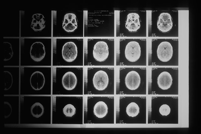
Imaging
Imaging scans like x-rays, MRIs and CTs are vital to any healthcare facility. Unfortunately, there is currently a shortage of a crucial item needed for CT scans—the liquid called IV contrast. This contrast dye that assist doctors in diagnosing conditions from a scan has been in shortage since a Shanghai plant went into lockdown and as a result, hospitals are rationing imaging tests.
Hospital Personnel
Hospital supply chain disruptions have created shortages of personal protective equipment for the hospital staff. The situation forced teams of health care professionals to reuse single-use gowns, gloves, and protective masks during the pandemic because of the supply shortages.
The lack of protection added another layer of stress to the hospital staff’s jobs and eventually led to experienced health care workers leaving their hospital employment.
The expense of supplying the hospitals without the use of technology or other updated distribution methods has also caused a reduction of funds available to staff the hospitals adequately.
One example is the increased demand for medical imaging, with fewer active radiologists available for active patient care. To keep up with the market, many hospitals have turned to non-physician radiology providers despite many concerns about the quality of service. One solution to this is something Vesta provides: teleradiology. Our US Board certified radiologists are here to help fill in those gaps.
Hospital Financial Stability
The pandemic-induced demand for medical supplies created an imbalance of medical supplies in the supply chain and increased the costs. Hospital inventory procedures needed to change drastically out of necessity.
Instead of maintaining just enough reserves on hand to meet the hospital’s immediate needs, hospital administrators needed to address a change in supply priorities.
The administrators were required to manage specific supply-type demands and the expense of eliminating an increase in waste from expired stock that did not serve the present needs.
Patient Care–Safety and Quality
The delay of cargo ships transporting much-needed supplies, the lack of truck drivers to transport the supplies to the hospitals, and the rapidly decreasing personnel in the hospitals have greatly affected the care patients receive. Many hospitals have had to turn patients away because of their inability to provide adequate care.

Possible Solutions
Hospitals are expecting that supply chain disruptions are not a temporary issue. There is a need to find solutions to the chain supply challenges through restructuring and designing more resilient systems for stable health care delivery.
One solution some hospital systems are implementing is to have more control over the delivery of supplies by making direct contact with manufacturers. These hospitals also use storage and distribution channels under their hospital control.
Multiple hospitals are also considering more system consolidation to increase the volume of supply purchasing to allow greater price bargaining. Some hospitals are working together to create a cooperative “just-in-time” model using a single distributor for many hospitals.
When the distributor delivers supplies daily to many hospitals, the hospitals are more efficient by reducing inventory to only the necessary items. Each hospital also eliminates much of the waste of outdated items.
The hospital’s financial gains overcome the financial risks of these changes by giving them more bargaining leverage with suppliers. The cost savings by system consolidation will also enable more available funds for additional personnel and direct patient care.
These system changes are possible through cooperative negotiations and the improvement of system technology. The hospital systems can take the challenges of supply chains experienced during the pandemic and improve their systems to avoid future problems and improve today’s health care system.