Medical Ultrasound Awareness Month: Advancements Through the Ages

Ultrasounds—to many not in the medical field, the image of a woman getting her pregnant belly examined is often something that comes to mind first. However, as medical industry professionals, we understand that ultrasounds go way beyond that.
ultrasounds

It’s really amazing to actually look at the history and advancement of such an important diagnostic tool especially during October–Medical Ultrasound Awareness Month.

In simple terms, the ultrasound scan isn’t just a medical device that can help track the development of a baby during pregnancy. The ultrasound scan can also help with the following:

  •       Observing the ovaries and uterus
  •       Evaluating blood flow
  •       Diagnosing gallbladder disease
  •       Examining a lump in the breast
  •       Checking thyroid glands
  •       Guiding a needle
  •       Diagnosing prostate issues

This machine is non-invasive, safe, and does not use ionizing radiation.

History of Ultrasound

The first recorded use of the ultrasound was in 1794 when Italian physiologist Lazzaro Spallanzani used ultrasound to study how bats navigated at night. This became the foundation for modern ultrasound physics. 

Bats produce ultrasounds in order to catch prey.

Ultrasound was initially used to detect flaws in metal casings. It was not used clinically until 1956 by Dr. Ian Donald and Tom Brown. There are no known side effects for ultrasound, making it an incredibly safe and efficient imaging device.

Advancements in Ultrasound

Ultrasound machines have become progressively mobile, easy to carry, and smaller while providing high-quality imaging. Since they are non-invasive and cost-effective, doctors can use them to track patient development without the risk of radiation.

Ultrasound technology can allow us to have an excellent view of the body’s vascular system by tracking microbubbles. 3-D imaging is also possible for more precise diagnostics.

As technology continues to expand, our medical devices tend to shrink inversely. Now, medical professionals can increase their diagnostic capabilities from anywhere in the world. For example, some handheld ultrasound devices weigh under a pound and can hook up to your smartphone, help healthcare providers quickly scan their patients—and possibly save lives.

These economical, easy-to-handle devices can change lives globally, predominantly in rural areas. It’s exciting to think that we may only be seeing the beginning of the modern ultrasound age.

How Teleradiology Can Help

Teleradiology, also referred to as remote radiology, provides medical professionals instant imaging result views remotely.  This means that patients have access to better, more immediate health care.
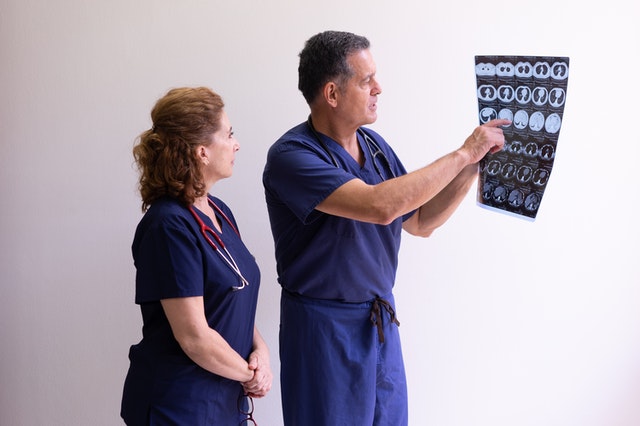
teleradiology servicesStudies suggest that teleradiology offers benefits, including the availability of second opinions and the possibility for remote viewing services. We at Vesta can certainly attest to this! Our clients – hospitals, urgent car centers, physician offices, mobile imaging companies, diagnostic imaging centers, and government institutions—enjoy cost-effective and unrivaled Teleradiology and Telemedicine services any time of day or night.
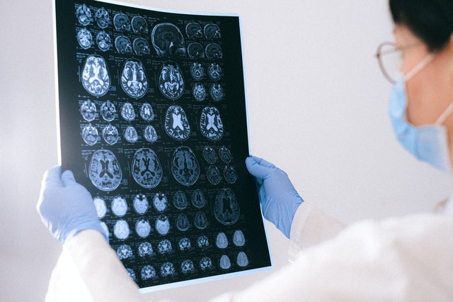
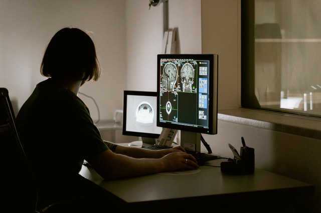
Failure to Properly Read Xrays, Mammograms, CAT Scans, and Other Diagnostic Tools: The Dangers

Every single day, healthcare facilities across America perform countless diagnostic procedures. These include x-rays, mammograms, CAT scans, and more, but they all have a common goal: to help healthcare workers gain a better understanding of their patients’ conditions.

failure to read
Imaging reading errors


Unfortunately, that isn’t always the case. While many hospitals and doctor’s offices are outfitted with top-of-the-line diagnostic tools, too often healthcare workers fail to read the results properly. These diagnostic errors can lead to delayed treatment or improper diagnoses — and that mistake can be extremely costly for the patient, the healthcare professional, and the facility where they work.

What is a Diagnostic Error?

Simply put, a diagnostic error is any failure to explain a patient’s health problem in a correct and timely manner. This can mean failing to spot a health issue (failing to notice a mass in a mammogram) or incorrectly diagnosing a problem (naming benign calcification present on a mammogram as cancerous masses).


Unfortunately, diagnostic errors are much more common — and more costly — than you might think. The Society to Improve Diagnosis in Medicine reports that diagnostic error is responsible for 40,000-80,000 American deaths every year!

The Risks of Misreading

What happens if one of your physicians misreads a diagnostic machine? If that mistake leads to delayed care, improper treatment, or serious harm to the patient, you might be in for a malpractice lawsuit.


The Society to Improve Diagnosis in Medicine also reports that diagnostic errors account for the largest fraction of malpractice claims in the country — and they are awarded the highest total of penalty payouts.


Patients can sue their healthcare providers for misdiagnosis, failure to diagnose, or delayed diagnosis if that diagnostic error caused them significant harm, and the average payout for a diagnostic error is around $494,000. No facility wants to cause their patients harm (or take on that financial penalty), so it is critical to avoid diagnostic error as much as possible.


How To Prevent Imaging Reading Errors

How can you prevent diagnostic errors in your healthcare facility? The best thing you can do is make sure your workers are not spread too thin. If your doctors, nurses, and other healthcare staff are overworked and stressed, they’re more likely to make mistakes.

night coverage radiology
Vesta offers fast turnaround times and night coverage.

One great way to avoid these errors is to outsource your radiology work to a teleradiology company. Teleradiology companies can alleviate some of the stress on your staff, and having a team of dedicated radiologists on your side can help ensure that every test is read appropriately.


You cannot afford to go with just any teleradiology company. Vesta not only has expert, US Board Certified Radiologists, we offer customized reporting, nighthawk coverage and fast tat (fast turnaround times).


Contact Vesta Teleradiology today to see how our teleradiology services can help your facility avoid errors and provide better patient care.

5 Tips for Women’s Wellness Healthcare Facilities

The fitness and wellness industry has become one of the biggest markets in the world. If you are the owner of a women’s health or wellness center, there is great potential for growth in this $3.4 trillion industry. This competitive industry is constantly creating new approaches to attract patients. If you are searching for new ideas to enhance your wellness center and the care for your patients, here are 5 tips for increasing patient flow.

  1. Offer educational workshops and speakers. Being able to have a cup of coffee and listen to an informed speaker on a subject of interest can bring in more patients. Those who want to help themselves or improve their life will like this approach. Stress-free opportunities to listen and learn about money management, cooking, gardening, yoga, fashion, or more could entice women with enjoying a nonthreatening space to absorb information. These could be mini-breakout sessions of an upcoming wellness conference which may interest participants to attend.
  2. Bundle services. Having one facility that offers multiple services is attractive. Women are busy. Their time is valuable. If there was an opportunity to make one stop to take care of a mammogram, pedicure, and haircut (for example) many women would like this option. In addition, having a nonjudgmental environment to talk to a therapist on a variety of topics such as mental health issues, menopause, and sexual health could bring in more patients.
  3. Act as a safehouse. Employing experienced professionals who have the awareness and abilities to support women in crisis is needed in every city in the world. Doctors, psychologists, and lawyers on site who are in place to readily protect and prevent violence against women and their children will attract patients. Partnering with local authorities may bring wellness center funding opportunities.
  4. Incorporate wellness retreats. Offering purposeful, affordable, well designed retreats for specific groups allows wellness opportunities beyond the wellness center facility. Programs organized with the intentional components of mental, emotional, physical, and social can provide transformational experiences for patients. These deep connections can be life changing and create interest in future retreats.
     
  5. Outsource your radiology imaging needs to a reliable teleradiology service. With this service, a patient can have her mammogram, ultrasound, or other imaging service performed, screened, and sent digitally to all parties involved. The image and its information is compressed and encrypted and stored in a database server accessed by the radiologist, provider, transcriber, and other workers across geographically diverse facilities. The final report is automatically sent to the doctor and the original facility. This saves time and allows you to focus more on attracting new patients.
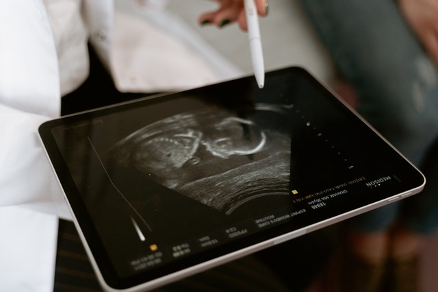
ultrasound
An ultrasound underway

Vesta Teleradiology has been assisting Women’s Wellness centers, hospitals, mobile imaging, assisted living and other diagnostic imaging centers for nearly 15 years. It’s not just our speedy service and accurate readings from US Board Certified radiologists, it’s our ability to help you customize the service to tailor it to your facility and patients. Look to Vesta if you want accuracy, speed, affordability, support and customization.

How Does Mobile Imaging Help Improve Patient Care at an Assisted Living Facility?

As radiography technology has evolved, so has the experience of the typical resident in a nursing home or assisted living facility. X-rays, CT scans, and ultrasounds are commonly needed to help diagnose residents of these communities with an appropriate treatment plan. The availability of mobile imaging improves patient care and quality of life in a multitude of ways.

assisted living teleradiology
Mobile imaging provides easier access for residents requiring these services.

1. Improved Access

With the immediate availability of mobile imaging onsite, more patients can access this type of diagnostic care. In a study conducted by BMC Health Services, researchers found that without mobile access, the use of diagnostic imaging for nursing home patients decreased by almost a third, from 14,500 to only 10,305. This data suggests that the ease of access to mobile radiography ensures that more patients receive the diagnostic care they need.

2. Fewer Transfers

Many risks are associated with transferring the elderly, especially those with mobility issues. By keeping radiology onsite, fewer transfers to hospitals and other centers will need to occur. A study in Norway found that transfers to a hospital’s radiology department were avoided in 71 percent of cases. This dramatic difference makes patient care simpler and more streamlined, and more importantly, it affords fewer opportunities for the elderly to sustain injuries, thereby increasing patient safety.

teleradiology for nursing home
There are risks associated with transferring the elderly, especially those who have mobility issues.

3. Reduced Spread of Contagious Diseases

The use of mobile imaging can help reduce the spread of contagious diseases like COVID-19. Rather than transferring patients out to imaging centers and increasing the number of people they come into contact with, a nursing home can have in-house imaging staff. A study out of Italy showed the potential for mobile imaging to reduce the spread of COVID-19 when certain safety protocols were followed.

4. Faster Imaging Results

A study conducted in Norway by BMC Health Services Research looked at the results of mobile imaging and found it to be of the same quality as hospital imaging. This research also concluded mobile radiography leads to a faster diagnosis. Offsite imaging can equate to days or weeks of wait time for interpreting results. If a scan is faulty and a patient needs to return for another scan, this wait time can become detrimentally long. With faster results, doctors can work quickly to begin treatment and improve outcomes. 

5. Better Treatment Plans

By having radiology staff on-site, doctors can better prescribe treatment plans specific to a patient’s needs. The aforementioned study in Norway also found that access to treatments improved with onsite mobile imaging in addition to patients receiving more specialized care.

Teleradiology Company that Fits Your Needs

teleradiology services
Vesta has been providing stellar teleradiology services for over 15 years.

For these reasons and more, it is imperative that nursing homes and assisted living communities work with an experienced teleradiology company to improve the quality of care for residents. Vesta Teleradiology provides nighthawk services with expert and customizable interpretations, thereby improving health outcomes for your entire community. Quality patient care is our top priority as well as giving assisted living facility or nursing home operators flexible and cost-effective, expert service.

Sarcoma & Bone Cancer Awareness Month: Newest Technology in Imaging

July is Bone Cancer and Sarcoma Awareness Month. According to the American Association for Cancer Research, in 2021, around 13,000 Americans will be diagnosed with soft tissue sarcoma, while 3,600 will be diagnosed with bone cancer. Luckily, the 5-year survival rate of both types of cancer is fairly high (65 for soft tissue and 66.8 for bone). In a comprehensive study from 1978 to 2004, the survival rate of these types of cancers has improved dramatically, thanks in part to improvements in imaging and early detection.

bone cancer
Sarcoma is a broad term for a group of cancers found in the bone.

Advances in Radiography

While x-ray technology has been used in a health context since 1899, many features surrounding it have changed. X-rays are now more defined with higher resolution, allowing doctors to zoom in to see smaller tumors and other issues. In fact, this past year, scientists at the University of Erlangen-Nuremberg have broken the record for x-ray resolution, down to under 10 nanometers. Furthermore, the process of obtaining can now be digitized, rather than having to use a darkroom to develop scans. These developments make radiology more accessible to patients and provide more immediate results, which helps bone cancer and sarcoma patients get diagnosed earlier when time is of the essence. There are multiple types of radiography used in diagnosing these types of cancer, and specific advances have emerged and evolved the diagnostic process for each type.


Bone Scans

During bone scans, a patient is injected with a tracer that allows oncologists to see abnormalities. Recent changes in bone scans allow doctors to quantify the metastasis of a bone from a scan. Bone scans can sometimes occur as a diagnostic tool before a CT scan, PET scan, or MRI.


CT Scans

CT scans can be ordered with “contrast,” where the patient swallows or takes an IV containing a dye. This can help doctors see certain organs more clearly. These scans can also help doctors see a cross-section of parts of the body and different perspectives of organs than they’d be able to see with a typical x-ray. The latest advancements in CT scans now offerlower radiation exposure by splitting the x-ray beams, making these procedures safer than ever.


PET Scans 

Positron emission tomography (PET) imaging is an emerging tool for bone cancer patients receiving radiation treatments. This tool is now being used increasingly by doctors to monitor tumors between treatments. These advanced scans allow more doctors to receive more information, including receptor expression and metabolism of a tumor. This information helps doctors to understand whether or not a particular type of treatment is working to shrink tumors and to help assess the appropriate dosage.


MRI Scans

Magnetic resonance imaging or MRI scans help oncologists to see the extent of bone and soft tissue cancers by taking cross-sections of affected organs and allowing them to be displayed at different angles. These are taken around a patient while a patient lies still inside a long tube. The most recent changes in MRI technologyhave occurred on the software side. One relevant change includes faster scan times, thereby reducing costs and discomfort for the patient. Furthermore, multiple layers of contrast can now be taken from a single scan, allowing doctors to highlight different issues easily.

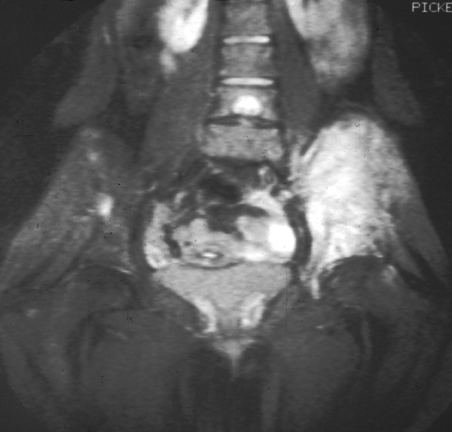
bone cancer imaging
Magnetic resonance imaging (MRI) image of human skeleton. MRI diagnoses Ewing’s sarcoma of the right hip. Source: National Cancer Institute

While bone and soft tissue cancers affect many Americans, advancement in imaging tools helps provide clarity and hope for these patients.

Tech Innovation in Teleradiology

Vesta Teleradiology prides itself in being a top innovator in technology and was even awarded this recognition in 2020 by Technology Innovators. We truly understand the importance of keeping up with medical and technological advancements in radiology, imaging and health.


Come to us for teleradiology services for your outpatient imaging centers, nursing homes, hospitals, mobile imaging, wellness centers and urgent care facilities. Expect flexible yet speedy service, with quality interpretations and customizable reports. Please contact us for a quote.