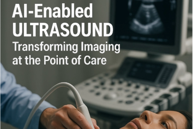
Latest News in Outpatient Radiology Centers

Outpatient radiology centers play a crucial role in the healthcare landscape by providing convenient, efficient, and cost-effective access to diagnostic imaging services for patients across a wide range of medical conditions. These services include X-rays, ultrasounds, MRIs, CT scans, mammography, and fluoroscopy, among others. Patients typically visit these centers for imaging tests prescribed by their healthcare providers to diagnose and monitor various medical conditions.

While these centers offer a convenient and efficient alternative to hospital-based imaging services, often providing faster appointments and reduced wait times, they do face challenges.

Issues with Outpatient Imaging Appointments

A recent study published in Academic Radiology reveals that nearly 24% of outpatient imaging appointments are missed, with the majority due to patient cancellations rather than no-shows. Factors such as younger age, being unwed, residing in disadvantaged neighborhoods, or lacking adequate insurance increase the likelihood of missing appointments. The study, conducted by researchers at the University of California, Irvine, analyzed data from their academic health center, finding that over 70% of cancellations were initiated by patients. Interventions are suggested to reduce missed appointments, such as self-scheduling, implementing checklists for necessary processes before imaging exams, and addressing health-related social risks like transportation access. Despite suggestions, limited research exists on reducing appointment cancellations in outpatient imaging.


Delays in MRI Orders

A recent study published in the Journal of the American College of Radiology reveals that nearly half of outpatient MRI orders experience significant delays, being performed more than 10 days from the date chosen by the referring provider. Led by Ronilda Lacson, MD, PhD, from Brigham and Women’s Hospital in Boston, the research emphasizes the critical importance of mitigating factors causing these delays, as they negatively impact patient care. Assessing over 97,000 outpatient MRI exams ordered between October 2021 and December 2022, the study identifies patient demographics, social determinants of health, and radiology practice- and community-level factors associated with delayed MR imaging. The study found that close to 50% of MRI orders had a prolonged order-to-performed interval, with factors such as higher Area Deprivation Index (ADI) scores contributing to delays. The authors stress the need for systemic approaches to address disparities in access to MRI examinations, including staff training, access to patient navigators, and programs tackling transportation barriers to outpatient imaging.


Other Challenges Outpatient Centers Face:

Technological Advancements: Keeping up with rapidly evolving imaging technologies requires significant investment and ongoing training for staff. Outpatient centers need to stay updated with the latest equipment and software to maintain competitiveness and provide accurate diagnostic services.

Regulatory Compliance: Compliance with healthcare regulations and standards, such as those related to patient privacy (HIPAA), radiation safety, and quality assurance, is essential but can be challenging to navigate. Failure to comply can result in fines, legal consequences, and damage to reputation.

Staffing and Workforce Management: Recruiting and retaining skilled radiologists, technicians, and support staff is crucial for maintaining quality and efficiency. Shortages in qualified personnel or high turnover rates can strain operations and affect patient care.

Integration with Healthcare Systems: Outpatient radiology centers need to effectively integrate with larger healthcare systems, including electronic health record (EHR) systems and referral networks. Seamless communication and coordination with referring physicians are essential for delivering comprehensive patient care.

Outpatient Centers Can Rely on Teleradiologists

In conclusion, outpatient radiology centers play a vital role in providing accessible, efficient, and high-quality diagnostic imaging services to patients. However, they face various challenges, including staffing shortages, which can impact their ability to deliver timely care. One solution to alleviate some of these challenges is the adoption of teleradiology services. Teleradiology services from reputable companies like Vesta, enables centers to access remote radiologists who can interpret images and provide diagnostic reports, helping to overcome staffing shortages and ensure continuous coverage. By embracing technology and innovative solutions like teleradiology, outpatient radiology centers can enhance their capabilities, improve patient care, and meet the evolving needs of healthcare delivery.


Sources:

Auntminnie.com
radiologybusiness.com
openai.com


February AI News in Radiology

Brain Tumor Spotted on PET Imaging

An AI algorithm named “JuST_BrainPET” identified a glioblastoma in a patient that had been missed by physicians. This finding, reported in the Journal of Nuclear Medicine, underscores the potential of AI-based decision support in diagnostic and treatment planning. The algorithm automatically segments metabolic tumor volume from healthy tissue on brain PET imaging. In a case study, it detected a lesion in the frontoparietal region, not identified by an expert, which progressed to a small tumor. The AI tool’s early detection could have influenced diagnostic and treatment decisions.


Using Eye-Tracking

Researchers in Lisbon, Portugal, have pioneered a method to enhance AI interpretability in radiology by integrating eye-tracking data into deep learning algorithms. This innovative approach, outlined in the European Journal of Radiology, aims to align AI systems more closely with human understanding, marking a significant leap towards more human-centered AI technologies in radiology. By leveraging eye-gaze data, the researchers sought to bridge the gap between human expertise and AI computational power, anticipating that AI models could learn from the nuanced patterns of image analysis observed by radiologists.


This integration promises AI models that prioritize image characteristics relevant for diagnosis, potentially reducing the disparity between AI decision-making processes and human radiologists’ diagnostic approaches. The potential benefits of this research are vast, potentially leading to AI systems that are not only more effective in identifying pathologies but also more understandable to radiologists, thus fostering trust in AI-assisted diagnostics and accelerating their adoption in healthcare.


Review Paper on AI and Cancer Detection

Professor Pegah Khosravi and her team of researchers explore how artificial intelligence (AI) can enhance anomaly detection in MRI scans to advance precision medicine. Their comprehensive review, published in the Journal of Magnetic Resonance Imaging, focuses on AI techniques like machine learning and deep learning, particularly in identifying tumors in the brain, lungs, breast, and prostate.

The authors discuss several AI strategies for improving tumor detection, including a holistic approach that integrates data from various imaging techniques such as MRI, CT scans, and PET scans, along with genomic information and patient histories. This approach not only enhances anomaly detection accuracy but also facilitates personalized treatments based on comprehensive patient profiles.

Furthermore, the paper explores the use of ensemble methods in AI, which combine different AI models’ strengths to improve anomaly detection. By leveraging these methods, a more thorough analysis of MRI data is ensured. The authors advocate for AI systems that are accurate and transparent in their decision-making processes, fostering trust among healthcare professionals. They also stress the importance of collaboration among researchers, clinicians, and policymakers to effectively implement AI in medical imaging, guiding future advancements in the field.


Sources:

Auntminnie.com
bnnbreaking.com
gc.cuny.edu
openai.com

New Studies in Ultrasound – An Exciting Frontier in Medical Research

Medical science makes incredible strides every year, finding new ways to heal the sick and injured. One of the most exciting of these new frontiers is ultrasound technology.

With its ability to safely and painlessly penetrate the human body, scientists and doctors are discovering new possibilities in diagnosing and treating conditions that previously required more invasive procedures.

Ultrasound in Cancer Treatment

In cancer treatment, chemotherapy often comes with various unpleasant side effects. But new research has shown that ultrasound technology can enhance chemotherapy effectiveness, sometimes even reducing the dosage of the drugs required.

Ultrasound can also help increase the uptake of cancer drugs into tumors, making their delivery more targeted and efficient.

Ultrasound and Alzheimer’s Disease

Alzheimer’s disease is one of the most heartbreaking conditions facing our aging population, and while there is no cure for it, there is hope for better treatment.

Alzheimer's

Recent research has discovered the possibility of using ultrasound to break up the plaques that form in the brains of Alzheimer’s patients, reducing the inflammation and damage that cause the disease. Research in this field is still in its early stages, but the possibility of a breakthrough treatment in the years to come is an exciting prospect.


Ultrasound Diagnostics

Of course, one of the primary uses of ultrasound in medicine is as a diagnostic tool. However, new studies are refining and expanding the possibilities of what ultrasound can detect.


For example, technicians now use ultrasound to locate and diagnose skin cancers and traditional mammography for breast cancer screening. Ultrasound also offers a non-invasive way to examine the heart and blood vessels in incredible detail, giving doctors a better understanding of the mechanisms of cardiovascular disease.


Ultrasound and Pain Management

In addition to being an incredible diagnostic tool, patients can use ultrasound to manage pain.


Ultrasound-guided nerve block injections can relieve patients suffering from chronic pain without the side effects of prolonged medication use. Studies are ongoing to refine this technique and expand its benefits beyond chronic pain.

pain

Other Applications of Ultrasound

The research and innovation happening in ultrasound are constantly expanding their possibilities.

For example, researchers are exploring ultrasound technology to treat kidney stones, sometimes allowing for less invasive procedures than traditional surgery.

They are also investigating ultrasound for use in regenerative medicine and even to monitor blood glucose levels in diabetic patients non-invasively.

The possibilities of ultrasound technology seem almost limitless, with new studies constantly uncovering ways to use this powerful tool for medical treatment and research.

From cancer treatment to pain management, diagnostics to disease prevention, ultrasound is a field that promises to revolutionize the face of medicine in the years to come. We can only look forward with enthusiasm and hope for what discoveries may come on this exciting frontier.


Teleradiology in Conjunction with Ultrasound


When you think about the role a teleradiology company like Vesta plays with healthcare providers, you might envision that the radiologists deal only with x-rays and MRI scans. We also work closely with hospitals for ultrasound readings, too. If you’re seeking an extra hand for preliminary and final ultrasound interpretations, please reach out to us today. We can handle any volume, large or small.


Vesta Teleradiology Presents Informative White Paper on Transitioning from ICD-9 to ICD-10

Vesta Teleradiology releases a white paper, entitled “How to Make Your Transition from ICD-9 to ICD-10 a Seamless One,” for the purposes of providing guidance to physician practices as they prepare to transition and “go live” with ICD-10 on October 1st.

icd 9 to icd 10

The white paper located at vestarad.com/mediablog/blog incorporates preparatory practices as well as answers to common questions for those practices who have found themselves less than adequately prepared for the transition. The white paper provides those all-important answers that will allow those practices whom are not quite prepared to continue to bill under old ICD-9 codes and be reimbursed, so long as specific criteria are met. Specific guidance is delineated in the white paper.

Vijay Vonguru, President, Vesta Teleradiology, knows how apprehensive many providers are feeling about going from some 13,000 diagnostic codes to 69,000 codes, but states that “in the end the changes are for the betterment of patients and providers alike, in that it will serve to streamline referrals and approvals on diagnostic testing, as well as gather epidemiological data” and “this information should hopefully alleviate some of the resistance surrounding the upcoming transition”.

For those practices who having done the hard work and started last year in preparation for the transition, with tutorials for their physician, billing and ancillary staff, coordination with their vendors of EMR software and technical support: Kudos to them and their teams as they are one step ahead of the game. There is still some time for preparation, and utilizing the tips provided in the white paper will help the practices prepare for the October 1st deadline!


ICD-9
Are you ready for the transition?

About Vesta Teleradiology:

Vesta offers teleradiology solutions and services, providing 24x7x365 access to their highly qualified Board Certified Radiologists through a secure PACS.

Vesta has been a pioneer in supportive diagnostic workflow technology and quality diagnostic services. The team is steadfast in their passion to remain at the forefront of innovation in healthcare. Prospective clients are offered a free test drive of Vesta’s teleradiology service.