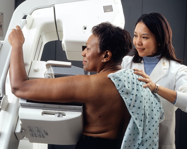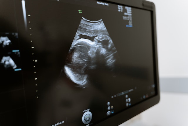
Point of care ultrasound (POCUS) is revolutionizing the healthcare industry and changing how doctors prescribe treatment for patients. POCUS is a diagnostic tool that utilizes ultrasound imaging to diagnose, monitor, and guide treatments for medical conditions.
This technology has been around for decades but is presently utilized more broadly throughout the healthcare system. Let’s take a closer look at POCUS and how it transforms patient care.
What Is Point of Care Ultrasound (POCUS)?
Point of Care Ultrasound (POCUS) is an ultrasound-based diagnostic tool used in clinical settings that uses sound waves to create images allowing doctors to see inside the body without having to do surgery or other invasive procedures.
POCUS can detect various medical conditions, such as heart defects, abdominal diseases, vascular diseases, musculoskeletal problems, and gynecological issues. POCUS can also be used in emergency settings to assess a patient’s condition quickly and determine if further intervention or testing is needed.
How Is Point of Care Ultrasound Transforming Healthcare?
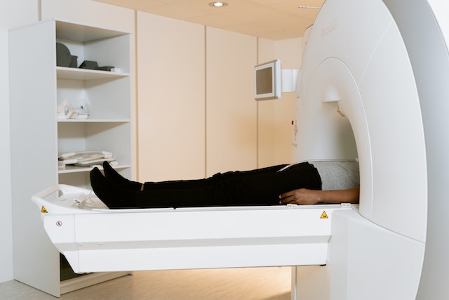
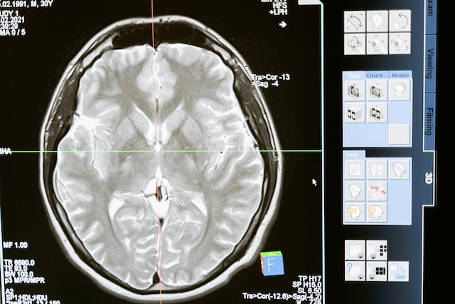
One benefit of POCUS is its cost-effectiveness compared with other imaging tests, such as MRI or CT scans, and POCUS does not require expensive equipment as those tests do.
Additionally, it can be done quickly and easily at the point of care, which reduces wait times for patients and increases accuracy in diagnosis, as well as reduces unnecessary treatments or hospital admissions. Furthermore, since it does not use radiation as other imaging tests do, there are no additional health risks associated with this technology, making it safer overall for patients.
Another advantage of POCUS is its ability to provide real-time data about a patient’s condition, which helps doctors make more informed decisions about treatment plans for their patients.
Additionally, because POCUS used in most circumstances does not require special training or expensive equipment, these systems are becoming increasingly available in low-resource areas where access to traditional diagnostic imaging may be limited. This benefit means more people have access to high-quality healthcare regardless of where they live or available resources.
The Gates Foundation recently provided financing to bring 1,000 handheld ultrasound devices to Africa. When low to mid-income nations can improve the accuracy of an efficient diagnosis–local doctors can save more lives.
Since 2012, however, emergency medicine program accreditation requires competency in POCUS. Competency assessment in this field includes demonstrations of technical skill and how it relates to the specific clinical practice.
Point of Care Ultrasound (POCUS) offers numerous benefits over traditional imaging tests. Its cost-effectiveness allows physicians to provide accurate diagnoses without breaking the bank. In contrast, its portability allows it to reach underserved populations who may not otherwise have access to quality healthcare services.
With further advances in technology coming soon, we could see even more widespread use of this powerful diagnostic tool across all areas of medicine in the near future.
Vesta Teleradiology: At the Forefront of Medical Technology
Vesta believes it is crucial to stay on top of technological trends that can benefit our hospital and healthcare facility partners. Vesta is always at the forefront of technological advances in order to help bring more efficiency and accuracy to imaging and radiological interpretations. For more information about Vesta’s teleradiology services, please contact us today.