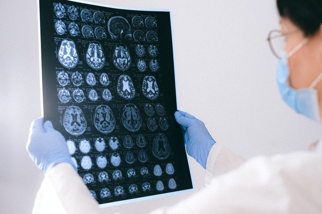
In 1895, Wilhelm Rontgen accidentally discovered X-rays. Since then, imaging technology has drastically advanced in all areas of healthcare, including cardiac imaging.
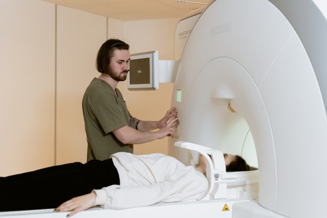
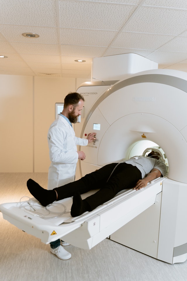
Medical professionals in the 1960s were using ultrasound imaging, and technicians developed Magnetic Resonance Imaging (MRI) and Computed Tomography in the 1970s. With these tools, the advancement of cardiac imaging escalated, creating real-time diagnosis of cardiovascular disease.
Advancing technology allowed cardiac physicians to perform diagnostic cardiac imaging like echocardiography and myocardial perfusion imaging in their medical offices. The trend for in-office imaging moved more to hospital settings because of the reduction of reimbursement rates in 2005 and the rapid technological changes in the industry.
Innovative technology advancement has needed to be a conglomeration of industrial, government, and academic research for cardiac imagery development to advance. Engineers, industry clinicians, and scientists have all participated in any advancements.
Two technology areas in cardiac imaging have had significant development over the past 15 years–nuclear cardiology and echocardiography.
Nuclear Cardiology
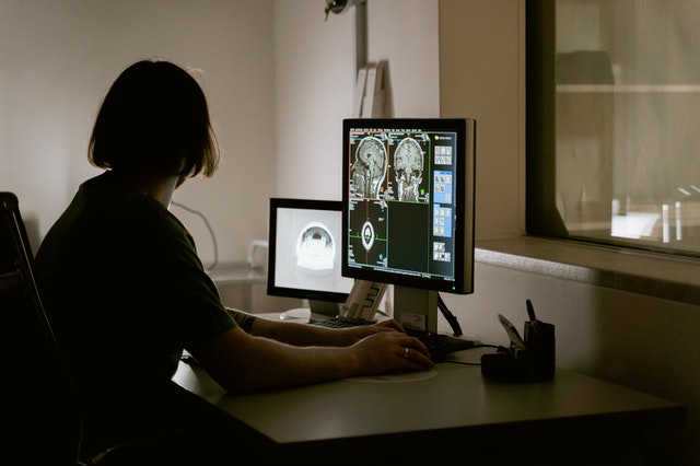
Nuclear Cardiology uses noninvasive techniques to assess and evaluate blood flow and define the internal location of a heart attack.
Commonly called positron emissions tomography (PET scans) or Computed X-ray tomography (CT scans), physicians can use this technology to discover the extent of heart muscle damage and the heart’s pumping function.
Nuclear Cardiology has proven to be a superior method for safe and cost-effective diagnosis. Technicians have upgraded cameras, giving physicians higher sensitivity and resolution views than the previous models used.
Echocardiography
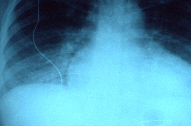
Echocardiography uses sound waves to display moving pictures of how heart chambers and valves work. This process can define areas where the heart muscles are not receiving adequate blood flow.
Physicians use echocardiography to locate possible blood clots within the heart and problems with the aorta. Physicians can also detect fluid buildup in the heart’s surrounding sac using echocardiography.
Echocardiography helps detect pediatric heart conditions because of the painless efficiency of the study.
The Future of Cardiac Imagery
Specialists have improved every technological aspect of cardiovascular practice within the past 50 years. Superior refinements in resolutions, measurement processes, and efficiency improve existing processes.
As with most technology-based sciences, the miniaturization of electrocardiography equipment will be the trend. Pocket-sized devices and combining diagnostic modalities through apps and computer applications will keep pace with changing health care practices.
Software development to capture real-time cardiac function and define the health of myocardial tissue is part of the newer developments in the field. Technicians are also refining more affordable methods of Cardiac MRI (Magnetic Resonance Imaging) through research.
A shift in diagnostic technology is advancing in identifying and decreasing cardiovascular risk in patients. Improved imagery will better equip physicians in preventing adverse outcomes. Physicians can more easily define patients’ risk of cardiac issues during exercise and stress-induced blood flow issues.
Another benefit to early detection and assessment of the cardiac disease process through imagery will be a better understanding of all the cardiac elements involved, leading to the development of new pharmaceutical drugs to prolong patient lives.
Cardiac Imagery will always be one of the best diagnostic tools available. Physicians and research teams assessing the cost-effectiveness and better patient outcomes will define the best approaches and safety in patient care management.
Outsourced Radiology
Vesta Radiologists are versed a cardiac imaging reading and a variety of other subspecialties. Further, we work nights and weekends so your staff can get the needed rest they require.
Please contact Vesta for outsourced radiology needs at 1-877-55-VESTA


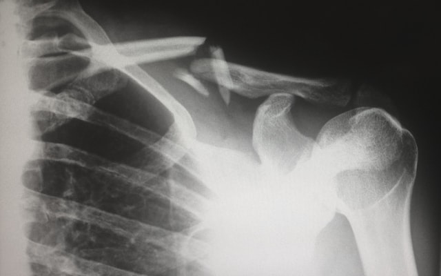
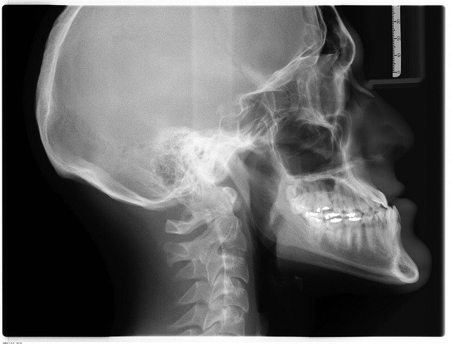 Going back to the beginning, the first X-ray picture in 1895 by
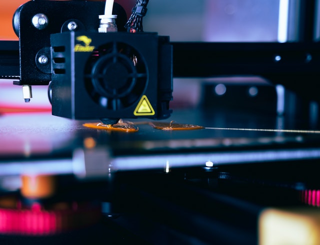
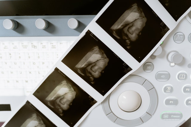 Fun Fact: the 3-D ultrasound actually takes thousands of photos at once to create a 3d image that is extremely clear.


 Studies suggest