Patient Comfort During Diagnostic Imaging

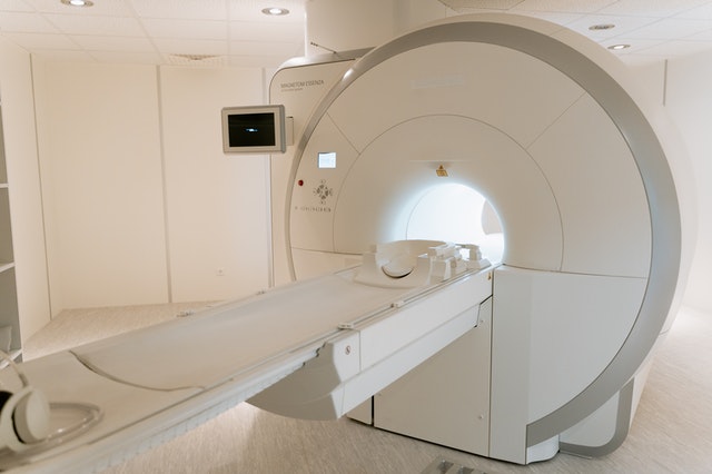
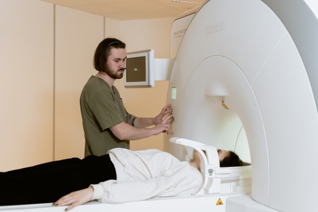
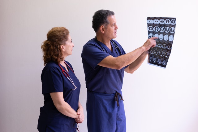
The role of the radiologist has changed over recent years. The health care world once thought of these professionals as sitting in a dark room interpreting imaging data and writing reports.

Today, diagnostic imaging is used by physicians more, and the radiologist’s role has become more centrally involved with patient care. There are opportunities to connect to the patients during the diagnostic imaging process that other health care providers don’t have.

Patient comfort during any healthcare procedure provides a series of activities that gain trust and provide emotional security in the process. Diagnosticians’ communication with their patients must convey that the staff is dependable, empathetic, and knowledgeable.

Environment

When patients first arrive for their appointment, they may be apprehensive and anxious about what their diagnostic imaging may reveal about their health and future.

The waiting room is where the patients not only wait for their appointment, but it is a place where they can talk with family members. Giving the seating lots of space allows areas with quiet corners for people to speak and express anxieties they may be experiencing.

A waiting room with plants, windows, and possibly waterfalls and nature scenes provides space for relaxation, gathering thoughts, and exploring possible questions about the procedure.

Physical Comfort

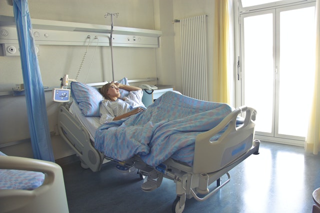
Diagnosticians performing a baseline assessment of the patient’s pain levels before, during, and after any invasive procedure is essential. It is also necessary to educate staff on any pain control protocols.

Patients appreciate simple office organization like short wait times in scheduling to minimize stress. Patients also appreciate comfortable procedure gowns, comfortable room temperatures, and warm blankets, along with sheets and pillows covering the procedure tables.

By reducing the technology sounds like beepers and overhead speakers, patients can have an easier time relaxing.

Communication

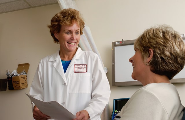
Effective communication can begin with the diagnostician partnering with the patient and their families to ensure that needs and preferences are being met. By involving the patient and their family, decision-making becomes more straightforward and more confident.

Patient-centered communication skills encourage the patient to talk about psychosocial issues that may affect their health or procedures. Through this communication process, the diagnostician can respect their patient’s requests and values and address or provide counseling and resources for patient support.

patient doctor communication

The top priority in the patient and health care provider relationship is maintaining an open and honest relationship. Diagnosticians can be sure the patient understands the procedures and the results of the processes by spending time, and using any tools that can assist is most valuable.

Tools like questionnaires, pamphlets, and visual aids can help the health care provider explain the benefits, risks, procedures, and processes or what is happening with the patient’s health.

Paying attention to the patient’s emotional concerns and using their own words when encouraging them to express themselves builds their trust with their health care providers.

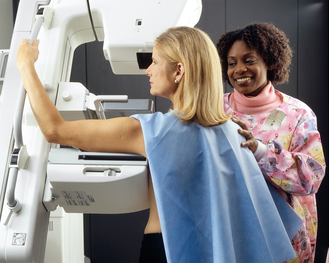
Imaging diagnosticians can use the time spent with the patient as teachable moments. For example, during mammography appointments, staff can inform and educate patients about the importance of colon screening and bone density tests.

It is also crucial for health care providers to practice restraint and allow quiet time for their patients. There is sometimes a tremendous amount of information to process, and it takes time for patients to deal with their feelings and formulate essential questions they may have.

enhanced patient experience


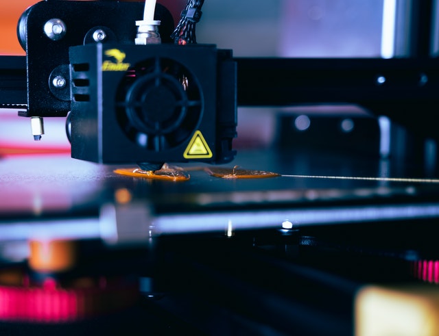
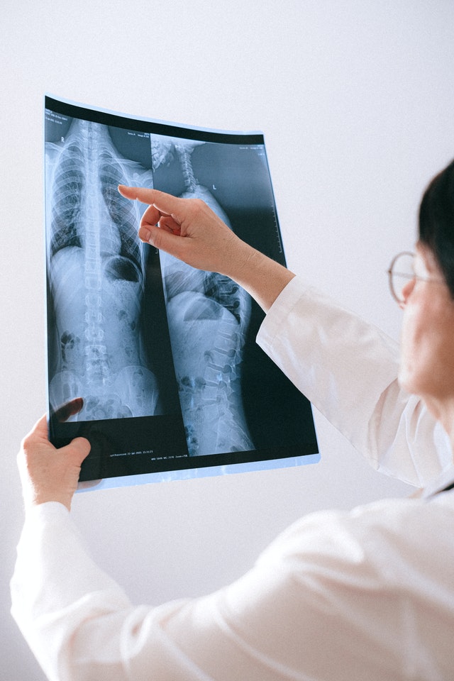
The importance of diagnostic imaging will continue to expand in the health care field. Advancements in cancer treatments, diagnoses, and disease prevention rely heavily on the professionalism of diagnostic imaging.

These health care professionals can experience positive relationships and trust with their patients by providing needed emotional support and conveying a genuine sense of caring.

Vesta Teleradiology wants to help you make your patients’ experience as streamlined as possible. That’s why we offer tools and customizable reports that work WITH your current workflow. We also offer 24x7x365 radiology reading services so your radiologists can have a better work life balance.

Solution and Ideas for Rural Hospital Challenges

Rural hospitals are the cornerstones of many rural communities. Not only are these facilities access points of care, but they are also significant financial support to rural areas by being primary employers.

These facilities are essential to the rural communities and make them vulnerable to financial difficulties. There are limited resources to comply with the increasing regulations on these facilities. Also, revenues are minimal due to low patient volume, shortage of physicians and other health care professionals, and a higher number of elderly, poor, and underinsured residents, along with a higher chronic illness rate.

small regional hospitals

The North Carolina Rural Health Research Program reported 95 rural hospitals closed between 2010 and 2018, resulting in a total loss of local care in some communities. Rural hospitals serve 20% of the population in the United States, making the survival of rural hospitals a priority in the health care system.

To explore solutions to the problems, in 2019, the American College of Radiology distributed a 22 question survey to explore the rural hospital systems’ staffing, recruitment, and retention issues. The responses showed interventional radiologist trained physicians either “do not want to do diagnostic work” (56.2%) or “do not want to practice in a small or rural setting” (48.8%)

The health care communities are continually exploring new models of care to improve these overall challenges rural hospitals face. Government entities and corporate and private organizations continue to explore

changing policies and enacting legislation for this health services dilemma to strengthen the viability of our rural health care systems.

Technology

Expanding the use of the internet has provided a wide choice of tools for rural hospitals to provide better health outcomes for the patients and better workload and communication for the workers.

Technology specialists have categorized solutions that may apply specifically to the rural health care systems.

More Accessible Health Information

Technology can create better communication between patients and health care providers to enhance treatment options, along with access to electronic records.

Digital Imaging

Digital imaging equipment and scanners that digitize documents and images allow radiology, pathology, and cardiology specialists to interpret these images in co-operative care centers at any distance.

Real-Time Patient Monitoring

Specialists can remotely monitor their patients in rural facilities for cardiac and ICU care.

Cooperation between health care partners is essential in the success of technology solutions for rural health care facilities. There are a variety of resources to explore if technology may seem like a viable solution.

Empowering Patients

Health care and health insurance options can be confusing, and many patients have difficulty understanding the process to get the care they need.

Each rural community has unique methods of networking and communicating. Working with existing community communication systems and creating more outreach methods to distribute

easy-to-understand materials can help patients understand their options better and make better-informed choices.

Funding

Funding for changes to the rural health care systems has come chiefly from grants. Still, cooperative agreements with associations, larger medical centers, and government entities have produced remarkable opportunities for outlying area medical facilities.

Team Effort

Partners from many organizations, health associations, vendors, government agencies, hospitals, healthcare organizations, funding groups, existing networks, and telemedicine programs are available to assist rural communities in need.

By identifying common objectives with other organizations, rural hospitals can specify health delivery problems and staffing issues to offer products and services to meet the rural facilities’ unique requirements.

By working together and overcoming obstacles rural hospitals are experiencing, the healthcare field, and the strength of the rural communities, can endure and experience a brighter future.

Vesta Teleradiology works with healthcare facilities in rural areas. No matter how large or small, we aim to help you provide the best healthcare possible for your patients. Our US Board Certified Radiologists have experience in traditional imaging as well as many subspecialties. 

By utilizing Vesta, these small individual and dependent hospitals can have the feel of being of a larger network spearheaded by Vesta. 

radiology company


Since Vesta has many radiologists on staff, the hospital will expend on their offerings, have the access to multiple opinions, can consult with other hospitals in the network, no interruption in the service due to shortage of radiologists, vacation or unforeseen situations

Please reach out to us to learn more at 877-55-VESTA.

Medical Imaging Tech and Trends to Look Forward to in 2022

Medical Imaging is one of the most visionary and engaging fields in the healthcare industry. As technology grows and develops in this burgeoning field, now is an exciting time to be a part of it.

We’ve compiled a few of the latest and most exciting innovations that have the potential to enhance medical imaging and patient care greatly.

3D Printing Technology and the Future

mri 3d printing

3D printing technology has been around since the 1980s, and in that time, it has aided many healthcare professionals and is positioned to be an essential factor in the future of medicine. From the very beginning, the medical field has been able to implement it in so many areas, including customizable implants and medical devices, designing anatomical models, aiding in medical and dental procedures, and 3D bioprinting.

 3D scans and models can help guide surgeons through diagnostics and complex procedures.

A great example is the Henry Ford Hospital in Detroit, where cardiologists use 3D printing to better view a patient’s heart before an operation. 3D modeling assisted doctors with selecting the correct sized valve and seeing where to best position it.  

MRI Innovations

Magnetic resonance imaging uses advanced technology to produce 3D, incredibly-detailed images of patient anatomy. Almost everyone has heard of the MRI machine and its incredible potential for diagnosing health issues and standardizing treatment. Few pictures are as clear and accurate as an MRI, and it is used in almost any medical or dental field. Plus, it is safer than traditional x-rays since it does not utilize radiation.

It’s not a stretch to realize that the MRI market will continue to grow by 2022 to keep up with an aging population and help reduce the spread of disease.

Enterprise Imaging Technology

Enterprise imaging allows medical professionals to consolidate medical imaging data into a streamlined platform. This means better patient medical records, more accessible access for healthcare professionals, efficient data control, and better medical management.

The future of medical imaging technology centers around increased accessibility between all health systems and medical facilities. Doctors, physicians, nurses, surgeons, and other healthcare professionals can easily access reports and images across multiple service lines, allowing them to provide better service to their patients and consolidate work procedures.

In the future, we can expect the role of medical imaging to increase and grow as technologies like medical sensors, artificial intelligence, better computing power, and cloud-based software take a seat in the medical field. Data will continue to grow and be consolidated, and we can only expect patient services to improve, as well.

At Vesta, we are always at the forefront of new technology and imaging innovations. Our goal is to utilize technology to help you better help your patients. In addition to radiology services, we also offer an array of IT services. We integrate PACS teleradiology and RIS information systems to improve your workflow timelines.

Let us know how we can help your healthcare facility–call us at 877-55-VESTA.

Pillars in Radiology History, Past, and Present

Pillars in Radiology History, Past, and Present

Radiology is a phenomenal tool. Medical teams depend on it to guide diagnosis and treatments. As a product of the early twentieth century, it is hard to imagine how medicine functioned without it. Today, our world is a much better place thanks to the people who discovered and developed the powers of radiology.

The father of radiology is Wilhelm Roentgen. Beginning with an interest in cathode rays in October of 1895, he is credited with having detected electromagnetic radiation in a specific type of wavelength on November 8, 1895. From that point, Roentgen submitted his first publication titled “On a New Kind of Rays” on December 28, 1895. When Roentgen was asked what he thought when he made his discovery, he responded, “I didn’t think, I investigated.”  Encouraged to name his discovery Roentgen Rays, he chose the letter “X” to name the rays due to their mysterious and unknown nature. The famed X-ray picture of his wife’s hand shows her bones and wedding ring, clearly indicating the rays would not penetrate bone or metal.  Few discoveries have gained notoriety as quickly as Roentgen’s did. Within a year, X-rays had changed the fields of physics and medicine exponentially. It earned him the inaugural Nobel Prize in Physics in 1901and opened doors for Roentgen personally and professionally, some of which he appreciated and some he did not. While he did not accept the title leading to German nobility offered to him, he did agree to accept the honorary degree of Doctor of Medicine his university extended to him.

first ever xray
First medical X-ray by Wilhelm Röntgen of his wife’s hand

In addition, he donated his Nobel Prize earnings to his university, Wuerzburg University in Germany. An indication of his impeccable character. He never took any patents on X-rays. He wanted the world to benefit from his work freely. Consequently, he died almost bankrupt during World War I, but his goal was achieved. The world has benefited greatly from his work.

After Roentgen’s discovery, other scientists soon followed with further exploration of the rays. The first scientists to extend Roentgen’s work were Sir William Henry Bragg and William Lawrence Bragg. They are a father and son who earned the Nobel Prize in Physics in 1915 together specifically, “for their services in the analysis of crystal structure by means of X-rays” soon to be known as Crystallography, the basis for analytic chemistry. “The diffraction of X-rays by crystals,” written in 1922 by William Henry Bragg, describes their beginning work. Credited with the creation of Bragg’s Law, learning the atomic structures of viruses, proteins, gemstones, and more has been possible.

Many scientific fields were developed from Roentgen’s discovery of the X-ray. Major names of the time, such as Albert Einstein, who established matter and energy to be equal in 1903, were contenders in the study of unseen forces associated with Roentgen’s work as well. Considering the most evocative pioneering scientists, perhaps the most influential one was Marie Curie.

Intently studying Roentgen’s work, Marie Curie took an interest in uranium, the weaker ray, also called Becquerel rays. Studying its compounds, she eventually identified that the electrical effects of uranium rays are constant, which led to a monumental shift in the understanding of the structure of the atom, which led to the development of radioactivity. With this information, she determined radiation is a powerful tool with a wide range of potential applications, including diagnostic and therapeutic procedures. Her continued work with her discovery of radium not only earned her a second Nobel Prize, but today we have vast benefits from her discoveries, including nuclear energy and radiotherapy (RT), a treatment for cancer.

Vesta Teleradiology

We truly applaud and honor these individuals for their immense contributions. As a teleradiology service provider, we understand that delivering accurate interpretations along with stellar support is how we can continue to contribute to the overall mission of helping others, ultimately the healthcare provider and their patients.

Lung Cancer Awareness Month

They’re the words no one ever wants to hear: “You’ve got cancer.”

Luckily, these days our healthcare system has many available tools to help fight off “the big C,” but those words still hold the power to make someone’s life flash before their eyes, bringing the things that matter most into sharp relief.

November is Lung Cancer Awareness Month, a time to recognize both the individuals struggling with this condition and the tireless scientists and medical professionals fighting to stop it. Today, let’s talk about lung cancer and the millions of lives it touches.

Lung Cancer in the U.S.

The CDC describes cancer as “a disease in which cells in the body grow out of control.” Lung cancer first develops in the lungs, though it can also spread to other organs like the lymph nodes or even the brain.

Lung cancer is the second-most common form of cancer in the United States (after skin cancer). In fact, it accounts for 14% of all new cancer diagnoses each year! The largest risk factor for developing lung cancer is tobacco exposure (either through smoking or secondhand smoke), but it’s also possible to develop lung cancer after exposure to asbestos, radon, or other carcinogenic pollutants.

How Radiology Saves Lives

Sadly, lung cancer is the leading cause of cancer death in the U.S. – accounting for almost 25% of all cancer casualties. However, there are many treatments our healthcare professionals use to manage and treat lung cancer, from surgeries like segmental resections (where surgeons remove a portion of the affected lung) to radiation therapy.

Of course, lung cancer treatments cannot move forward without help from the radiology department. Radiology and diagnostic imaging are integral to a patient’s entire journey with cancer.

Imaging tests like CT scans, bronchoscopies, or MRIs of the chest are a critical first step for identifying both the presence of cancer cells and the type of cells: non-small cells, which can be removed through surgery with early detection, or small cells, which typically require chemotherapy. Radiology is even vital after treatment, as it can help doctors assess a treatment’s efficacy and monitor the lungs for any signs of cancer coming back.

lung cancer awareness month

Ultimately, surviving lung cancer is possible. However, a person’s survival rate is greater if their doctors detect cancer early and prescribe an effective treatment plan. Both of these things are critical to beat lung cancer — and both are only possible with the help of radiology.

In honor of Lung Cancer Awareness Month, we’d like to say “thank you” to all the radiologists who are saving lives every day by helping identify and inform their medical care — as well as the doctors, scientists, and technicians fighting against lung cancer. This November (and every month after), we are grateful for everything you do.


Vesta believes that a good work-life balance is important for your technologists, radiologists and all your staff. That’s why we offer teleradiology services to fill those gaps—nights, weekends and even holidays. 

Top 5 Essential Health Screenings For Women

Without a doubt, it is far better to catch any disease in its earliest stages. Screening allows for early detection and more effective treatment. Below are the top five screenings every woman should have to detect and prevent diseases.

Mammogram- 1 in 8 women will have breast cancer at some point in their lives. For some women this occurs earlier than others. The general guideline is that  women ages 40 to 44 should have mammograms. However, if a family history or a genetic predisposition to breast cancer is present, it is more important than ever to get annual screenings. 

Cervical –

A Pap smear or HPV test can find irregularities in cells that point to cervical cancer in the early stages. If signs of cervical cancer are detected a transvaginal, transrectal, or an MRI should be performed to ascertain the extent of the cancer or tumor.

Bone Density-

After age 65, it is recommended that all women get a bone density screening.  Practitioners can utilize x-ray, body CT, spine CT or a bone density scan to assess the progression of osteoporosis

bone scan for women

Cholesterol

Blood tests can detect high cholesterol. To further evaluate vessels in the heart, doctors can use a CAC test, a type of CT scan. According to the American Heart Association, patients aged 40 to 75 should receive this screening, especially when the risk of heart disease can’t be determined. 

Colorectal Cancer

The US Preventive Services Task Force recommends women over the age of fifty-five should get a colorectal examination. Colonoscopy, CT colonography, and flexible sigmoidoscopy are used for visual detection of colorectal cancer. 

Over the past 30 years, there have been many advancements in radiology that allow for better imaging and faster screening. With three dimensional and cross-sectional imaging, practitioners can get a better view of areas of concern and catch cancers or other diseases in their earliest stages. Moreover, electronic transmission enables radiologists to read screenings remotely for a quicker turnaround time. 


Helping your Patients get Screened

Even with these advancements, education is key in encouraging women to get screening. There is a tried and true saying —when you know better, you do better. Education can be employed in a variety of ways. Do you operate a women’s wellness clinic? You want to spread the right information that can save your patients’ lives. Here are a few tips to do just that:

  • Newsletters are cost-effective and can have a far-reaching impact. It is important to utilize personal stories, so that patients can put a face to the statistics. These often create a connection with patients as well. Women can see themselves in the stories of other women.
  • wellness clinic newsletter
    Newsletters a a great way to spread the word about the importance of screening

    Virtual events are more convenient for patients than the traditional fundraising events, especially in the age of COVID. Not to mention, they are also cost-effective. The virtual events can also utilize statistics, personal stories, and discussions by doctors in the field.

Imaging centers should work with doctors to encourage them to speak with their patients about the importance of screenings, as patients rely on their doctors for the majority of their information.

Early screening is often overlooked. However, it is of the utmost importance to encourage patients to get regular screenings to allow for ear detection and treatment. Doing so can save lives.

Teleradiology Services

If you are concerned that your healthcare facility simply cannot support the amount of time it takes to review screening results, that’s where Vesta comes in. In service for nearly 15 years, Vesta provides telemedicine and teleradiology services to fill in those gaps for you. With 24 x 7, 365 year round service, Vesta Teleradiology employs US Board Certified radiologists to receive and expertly interpret mammograms and more.

Whether your facility is in a major city or is a satellite in a rural town, we can help.

Medical Ultrasound Awareness Month: Advancements Through the Ages

Ultrasounds—to many not in the medical field, the image of a woman getting her pregnant belly examined is often something that comes to mind first. However, as medical industry professionals, we understand that ultrasounds go way beyond that.

ultrasounds

It’s really amazing to actually look at the history and advancement of such an important diagnostic tool especially during October–Medical Ultrasound Awareness Month.

In simple terms, the ultrasound scan isn’t just a medical device that can help track the development of a baby during pregnancy. The ultrasound scan can also help with the following:

  •       Observing the ovaries and uterus
  •       Evaluating blood flow
  •       Diagnosing gallbladder disease
  •       Examining a lump in the breast
  •       Checking thyroid glands
  •       Guiding a needle
  •       Diagnosing prostate issues

This machine is non-invasive, safe, and does not use ionizing radiation.

History of Ultrasound

The first recorded use of the ultrasound was in 1794 when Italian physiologist Lazzaro Spallanzani used ultrasound to study how bats navigated at night. This became the foundation for modern ultrasound physics. 

Bats produce ultrasounds in order to catch prey.

Ultrasound was initially used to detect flaws in metal casings. It was not used clinically until 1956 by Dr. Ian Donald and Tom Brown. There are no known side effects for ultrasound, making it an incredibly safe and efficient imaging device.

Advancements in Ultrasound

Ultrasound machines have become progressively mobile, easy to carry, and smaller while providing high-quality imaging. Since they are non-invasive and cost-effective, doctors can use them to track patient development without the risk of radiation.

Ultrasound technology can allow us to have an excellent view of the body’s vascular system by tracking microbubbles. 3-D imaging is also possible for more precise diagnostics.

As technology continues to expand, our medical devices tend to shrink inversely. Now, medical professionals can increase their diagnostic capabilities from anywhere in the world. For example, some handheld ultrasound devices weigh under a pound and can hook up to your smartphone, help healthcare providers quickly scan their patients—and possibly save lives.

These economical, easy-to-handle devices can change lives globally, predominantly in rural areas. It’s exciting to think that we may only be seeing the beginning of the modern ultrasound age.

How Teleradiology Can Help

Teleradiology, also referred to as remote radiology, provides medical professionals instant imaging result views remotely.  This means that patients have access to better, more immediate health care.

teleradiology servicesStudies suggest that teleradiology offers benefits, including the availability of second opinions and the possibility for remote viewing services. We at Vesta can certainly attest to this! Our clients – hospitals, urgent car centers, physician offices, mobile imaging companies, diagnostic imaging centers, and government institutions—enjoy cost-effective and unrivaled Teleradiology and Telemedicine services any time of day or night.

AI Advancements to Watch for in Radiology

Over recent years, radiology has seen many advancements in artificial intelligence (AI). Since medical imaging produces 90% of all healthcare data, radiologists have experienced a heavy workload for years. The COVID pandemic increased the backlog of radiologists, due to shutdowns and COVID regulations

new tech in radiology

Many have turned to AI as a solution to this problem, citing its numerous benefits in several aspects of imaging. AI allows radiologist departments to get more done in a shorter period of time by automating administrative tasks like paperwork, scheduling, and tracking follow-ups, while also prioritizing certain tasks for a more streamlined process. 

However AI is able to do much more than that. It can sort through data and images,  quickly giving physicians the best, most relevant information. Moreover, new AI algorithms also pick up on complex images and markers found in scans, findings that might not be visible to humans. This combination allows radiologists to run the information through an AI program for a second opinion and quicker analysis of the data.  

Examples of AI in Radiology at Work 

AI is currently being widely used in emergency medicine to alert physicians if a patient has a life-threatening condition, like a collapsed lung. The system will filter out unnecessary images and prioritize critical conditions. Additionally, physicians are using AI for stroke patients to reformat images, which allows for a faster processing time. Some of the same processes are being used for brain scans as well, choosing the most relevant images for doctors to save on overall processing time. 

Outside of critical care, AI is increasingly allowing physicians to work with other departments more efficiently. Hospitals in rural areas and less developed areas without radiology departments will be able to implement AI to flag certain images as well. 

Artificial intelligence is also playing a vital role in the field of oncology. Deep-learning methods have been employed to detect breast cancer in patients by teaching algorithms to draw connections between scans. Google is developing a CT scan that will produce 3D images of the lungs from 2D images, so cancer can be detected more efficiently. 

There has been talk of AI replacing radiologists, but many say that is not ever going to be the case. AI still has many limitations. As stated by Walach, an AI expert and developer, said “AI solutions are becoming very good at doing one thing very well. You typically have to have humans who do more than one thing really well.” 

AI developers rumor that AI will eventually replace radiologists, but Vesta has been in the radiology field long enough to know that AI, while very useful and complementary, it will never replace the keen eye and skills of radiologists. Vesta is a consultant of few AI developers and has been integrating the technology in its process, but does not rely solely on its findings.


radiologist


With the help of more research studies on artificial intelligence, the field of radiology will see continued growth, allowing for precision and efficiency. However, traditional radiology and teleradiology will still require trained experts to run the AI programs and discern using their expert knowledge to ensure patients are treated with the best of care. 

Vesta Teleradiology

With Vesta Teleradiology, our US Board Certified radiologists work with you to deliver customizable, and accurate reports. At Vesta, we treat AI as not Artificial Intelligence but Augmented Intelligence to help radiologists improve the quality and efficiency of their reads. How can we help your healthcare facility?

Covid: Maintaining Safety in the Workplace

As we head into the last quarter of the year, it’s good to review safety procedures and protocols especially as they relate to COVID-19 and emerging variants. Are you operating a nursing home, women’s wellness center, community clinic, outpatient imaging center? Let’s take care to make sure our workers and patients are safe and feel comfortable to continue their routine exams and screenings.

Freshen up on best practices you should implement at your facility to ensure end-to-end safety for both patients and workers. 

 Don’t Forget the Basics

A wellness check can be performed before entering. Taking the patron’s temperature with a laser gun and asking a series of questions pertaining to their health can offer a baseline comparison to assess if they are well and able to enter at that time. Providing masks to anyone who needs them is also vital.

Requiring hand sanitizing before entering the facility is usual practice in many facilities. Having alcohol-based hand sanitizer and washing stations accessible throughout the facility is one defense. Washing hands at certain times can be required for general cleanliness and hand hygiene.

Wearing PPE while in the facility can help prevent the spread. Approved facemasks, goggles, gloves, hair nets, shoe coverings, and more can be implemented at the job site. These pieces provides a barrier between a person’s skin, mouth, nose, and eyes, and viral or bacterial infection.

covid

Maintaining distance can help. Workers making sure they are at least six feet from one another should be a goal. Doctors and nurses beginning appointments with patients by getting initial information through an iPad remotely from the next room can minimize time spent face-to-face. Another way to maintain distance is to manage patient flow—the fewer people on-site at any given time, the better.


While everyone should constantly be cleaning their area throughout the day, having a designated cleaning person with knowledge of which cleaners to use on which surfaces can increase this effort. For example, hydrogen peroxide kills Coronavirus on metallic surfaces. Otherwise, soap and water, bleach, or 70% alcohol-based cleaner to wipe down high-touch surfaces throughout the day has proven to eliminate Coronavirus.  

temperature checks
A woman gets her temperature checked

Education

Providing literature, statistics, news and local information regarding COVID-19 can be made available throughout healthcare facilities for those interested in learning more.

Supplement information can also be made available. In addition to taking vitamins C and D on a daily basis, zinc, essential oils, probiotics, and other immune-building substances can be emphasized. Attaining optimal nutritional status can bolster immunity and protect against disease. 

Imaging and COVID Safety

If you operate diagnostic imaging tools like x-rays, MR machines, CAT scan machines, you should be very wary of your safety procedures for those who either have COVID or may be a Person Under Investigation for COVID. The American College of Radiology has guidance on these situations as they relate to MR use: 

“For patients with known active COVID-19 infection and Persons Under Investigation (PUI) for COVID-19, the American College of Radiology (ACR) recommends that practitioners minimize the use of MR except when absolutely necessary, and postpone all non-urgent or non-emergent exams. In some cases, the use of alternative imaging methods such as point of care or portable imaging may be appropriate. As with all imaging, the impact of the results of the imaging must potentially affect imminent clinical management.” More here.

For questions about proper cleaning and disinfection of imaging equipment, consult with the manufacturer of your machines and check with the CDC for guidance.

radiology
Safely sanitizing imaging machines is vital

Check out this interesting read: COVID-19 pandemic: Cleaning and disinfection – What should the radiologist know?

Healthcare facilities have the power to serve and protect their communities. They have the responsibility to inform, educate, and heal. They can take action for the well-being of their workers, patients, and the public.

At Vesta Teleradiology

Even though we are a remote radiology company, we ensure the best sanitizing, disinfecting, and cleaning protocols in our offices. And of course, with such technology that our teleradiology services come with, our services are contactless and touch-free. When you partner with us, a Gateway application is installed remotely to allow your systems direct access to our PACS. Once linked, your technicians send exams straight from your machine to our PACS. Vesta account specialists assign it to the proper physician who read and dictate the report, it’s uploaded to your branded template where it’s easily accessible for your facility to access and download!

With Vesta’s solutions, we offer a user-friendly portal for physicians to view all reports as soon as they’re available, and they are customizable, too with your facility’s information and logo. Any critical findings are immediately shared with the facility via phone, email or text.

For more information or for a quote, please contact us.

Prostate Cancer Awareness: Encouraging Patients to Get Screened

Prostate cancer kills 34,130 men each year. With screening, this number can be dramatically decreased. Early detection allows for more treatment options and increased efficacy of treatment. However, many men are reluctant to get screened. So, the question healthcare providers (and organizations that service them like diagnostic imaging centers, mobile imaging, wellness centers, radiology centers, hospitals) and advocates are left to ponder  is—how do we encourage men to get screened? 

prostate exam
How do you encourage men to get screened for prostate cancer?

Education is the most important factor to increase the rate of screening among men. Numerous studies have found that men educated on the topic of prostate cancer are more willing to get screened. In particular, one study found a significant correlation between education and whether or not a man agreed to screening. 


That same study found that many men do not get screened if there are no symptoms present. As is well known in the medical community, prostate cancer in its early stages often presents with no symptoms. Educating men on this single fact would be very helpful, but it is equally important to inform them about the risk factors, symptoms, screening procedures, early detection, and treatment options. So, what approach is best for educating reluctant men? 

It’s a Digital World

Brochures are things of the past. In today’s world, the best way to get information out there is through the digital mediums ever present at our fingertips. The good news is there are a variety of them. Below are some ideas for spreading information effectively online. 


cancer screening
A man reads information about the importance of healthcare screenings


  • Social Media: Men and women are constantly scrolling Facebook, Twitter, and Instagram. Social media campaigns with eye-catching infographics and videos help spread the word. Ads can target men and women, as women play a vital role in spreading awareness and educating their partners.
  • Newsletters: Informational newsletters sent via email to both general practitioners and patients are another avenue for educating patients at a relatively low cost to healthcare providers.
  • Virtual Events: Through the COVID pandemic, virtual events have become increasingly popular. Providers should take note of this cost-effective trend. These events give providers a platform to provide in-depth education about prostate cancer and screening. They can also open lines of communication with patients, allowing men to ask questions and voice their concerns, while giving insight into areas where information strategies can be improved. Additionally, some men, reluctant to attend an in-person event, might be more willing to attend a virtual event.
  • Personal Stories: Whenever possible, it’s important to put a face to the statistics. The number above represents fathers, brothers, husbands, and uncles. With the popularity of sites like TikTok and YouTube, video is being looked to as one the most successful mediums for information-sharing. Through video, personal stories can be told in an effective and meaningful way that will both educate and connect with at-risk men. People often do not remember statistics, yet  they do remember feelings. 


If this country, as a whole, has learned anything throughout this past year, it is to be flexible. Experimenting with different messaging styles and mediums is important. Don’t be too rigid with any one approach. See what kind of response you get from a particular message or campaign and adjust accordingly. Just remember, you can never go wrong by getting creative.

Teleradiology

Our radiologists at Vesta are trained in all modalities including reading scans for prostate cancer. When your radiologists are not available such as nights, weekends or holidays, our US Board Certified radiologists are here to help fill in those gaps.