New Studies in Ultrasound – An Exciting Frontier in Medical Research

Medical science makes incredible strides every year, finding new ways to heal the sick and injured. One of the most exciting of these new frontiers is ultrasound technology.

With its ability to safely and painlessly penetrate the human body, scientists and doctors are discovering new possibilities in diagnosing and treating conditions that previously required more invasive procedures.

Ultrasound in Cancer Treatment

In cancer treatment, chemotherapy often comes with various unpleasant side effects. But new research has shown that ultrasound technology can enhance chemotherapy effectiveness, sometimes even reducing the dosage of the drugs required.

Ultrasound can also help increase the uptake of cancer drugs into tumors, making their delivery more targeted and efficient.

Ultrasound and Alzheimer’s Disease

Alzheimer’s disease is one of the most heartbreaking conditions facing our aging population, and while there is no cure for it, there is hope for better treatment.
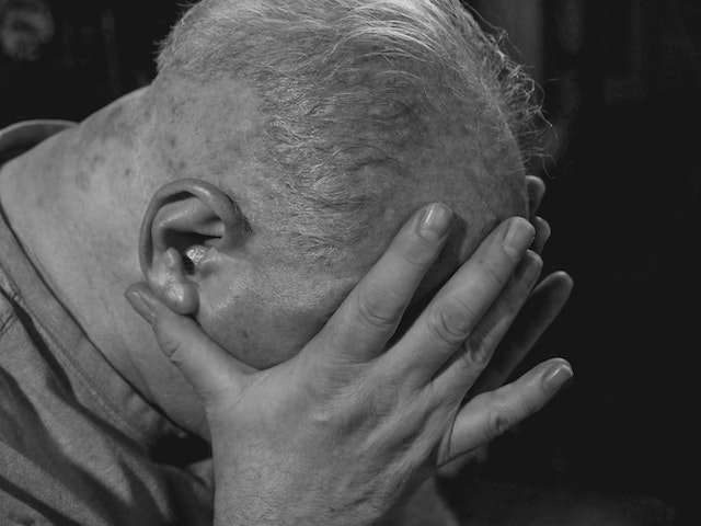
Alzheimer's

Recent research has discovered the possibility of using ultrasound to break up the plaques that form in the brains of Alzheimer’s patients, reducing the inflammation and damage that cause the disease. Research in this field is still in its early stages, but the possibility of a breakthrough treatment in the years to come is an exciting prospect.


Ultrasound Diagnostics

Of course, one of the primary uses of ultrasound in medicine is as a diagnostic tool. However, new studies are refining and expanding the possibilities of what ultrasound can detect.


For example, technicians now use ultrasound to locate and diagnose skin cancers and traditional mammography for breast cancer screening. Ultrasound also offers a non-invasive way to examine the heart and blood vessels in incredible detail, giving doctors a better understanding of the mechanisms of cardiovascular disease.


Ultrasound and Pain Management

In addition to being an incredible diagnostic tool, patients can use ultrasound to manage pain.


Ultrasound-guided nerve block injections can relieve patients suffering from chronic pain without the side effects of prolonged medication use. Studies are ongoing to refine this technique and expand its benefits beyond chronic pain.

pain

Other Applications of Ultrasound

The research and innovation happening in ultrasound are constantly expanding their possibilities.

For example, researchers are exploring ultrasound technology to treat kidney stones, sometimes allowing for less invasive procedures than traditional surgery.

They are also investigating ultrasound for use in regenerative medicine and even to monitor blood glucose levels in diabetic patients non-invasively.

The possibilities of ultrasound technology seem almost limitless, with new studies constantly uncovering ways to use this powerful tool for medical treatment and research.

From cancer treatment to pain management, diagnostics to disease prevention, ultrasound is a field that promises to revolutionize the face of medicine in the years to come. We can only look forward with enthusiasm and hope for what discoveries may come on this exciting frontier.


Teleradiology in Conjunction with Ultrasound


When you think about the role a teleradiology company like Vesta plays with healthcare providers, you might envision that the radiologists deal only with x-rays and MRI scans. We also work closely with hospitals for ultrasound readings, too. If you’re seeking an extra hand for preliminary and final ultrasound interpretations, please reach out to us today. We can handle any volume, large or small.

 

Reduced Radiation Exposure in Imaging

Medical imaging is a powerful tool for diagnosing and treating medical conditions; however, it can also expose patients to excess radiation levels. Fortunately, recent technological advancements have resulted in the development of new ways to reduce radiation exposure during imaging.

Here are a few ways these advancements are helping patients receive the care they need while reducing their radiation exposure.

New Guidelines

Clinicians have always used guidelines for the use of CT scans. Still, they recently developed new guidelines for the benefit of CT scans on the head, neck, chest, and abdomen/pelvis areas of the body–especially in pediatric practice.

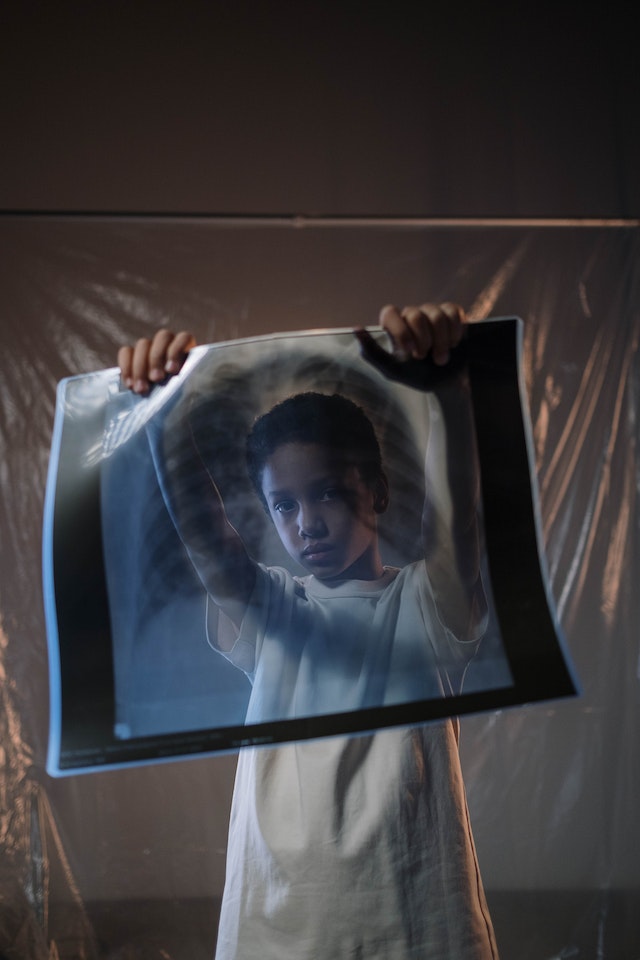
x-ray safety for kids
New guidelines for CT scans


Clinicians can reduce or eliminate the need for CT scans if the child is awake and alert, if the blood work is normal, and if the patient is not experiencing abdominal pain.

A study of 146 children not exposed to the excess CT scan radiation using the new guidelines showed no clinically significant missed injuries.

Smart Software

One of the most significant advancements in this area is using smart software to reduce radiation dose in computerized tomography (CT) scans. This software analyzes patient size, age, and other factors to determine the dosage necessary for an accurate scan that minimizes radiation exposure. In some cases, reducing radiation doses by up to 90 percent has been possible without any decrease in diagnostic accuracy.


Lower Radiation Levels in the New CT Machines

In addition to using innovative software to reduce doses, researchers are also looking at new technologies, such as low-dose CT machines, that can achieve similar results without any additional processing needed.


Technicians have designed these machines with unique components that limit X-ray emissions while still providing high-quality imaging. As a result, patients can receive the same level of care without worrying about unnecessary radiation exposure.


Reducing Radiation Exposure with Ultrasound

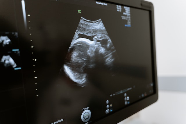
Finally, reducing radiation exposure through ultrasound technology is more progressive. For clinicians using sound waves instead of X-rays or other forms of energy, ultrasound technology can provide doctors with highly detailed imaging with no risk of exposing patients to dangerous radiation levels.

Ultrasound is an ideal option for pregnant women and children who may be more sensitive to the effects of radiation than adults.

vesta teleradiology for women's wellness centers
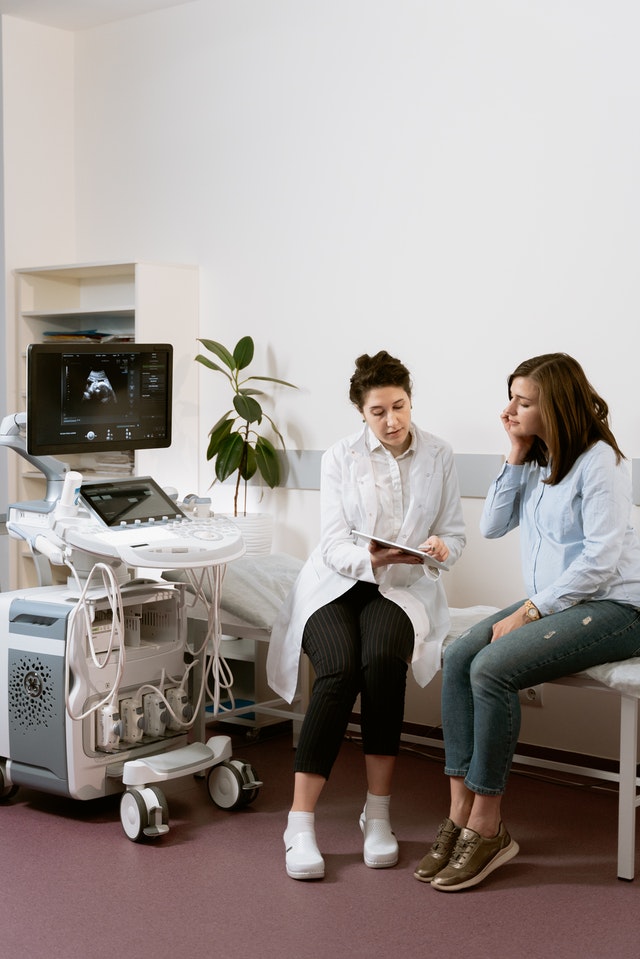
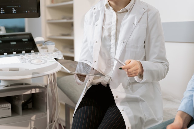
An ultrasound tech consults a patient

Recent advancements have made it easier for medical professionals to provide high-quality care while minimizing their patients’ exposure to harmful radiation.

With the medical profession redefining the guidelines, using low-dose scans, better x-ray machinery, and more ultrasounds, many options are now available for reducing radiation exposures during imaging procedures—all without sacrificing diagnostic accuracy or quality care.

That’s great news for anyone who needs an imaging scan but wants to ensure they’re taking steps to protect their health and safety!


Diagnostic Imaging Trends: Point of Care Ultrasound – POCUS

Point of care ultrasound (POCUS) is revolutionizing the healthcare industry and changing how doctors prescribe treatment for patients. POCUS is a diagnostic tool that utilizes ultrasound imaging to diagnose, monitor, and guide treatments for medical conditions.

This technology has been around for decades but is presently utilized more broadly throughout the healthcare system. Let’s take a closer look at POCUS and how it transforms patient care.


What Is Point of Care Ultrasound (POCUS)?

Point of Care Ultrasound (POCUS) is an ultrasound-based diagnostic tool used in clinical settings that uses sound waves to create images allowing doctors to see inside the body without having to do surgery or other invasive procedures.

POCUS can detect various medical conditions, such as heart defects, abdominal diseases, vascular diseases, musculoskeletal problems, and gynecological issues. POCUS can also be used in emergency settings to assess a patient’s condition quickly and determine if further intervention or testing is needed.


How Is Point of Care Ultrasound Transforming Healthcare?

One benefit of POCUS is its cost-effectiveness compared with other imaging tests, such as MRI or CT scans, and POCUS does not require expensive equipment as those tests do.


POCUS
Point of care ultrasound


Additionally, it can be done quickly and easily at the point of care, which reduces wait times for patients and increases accuracy in diagnosis, as well as reduces unnecessary treatments or hospital admissions. Furthermore, since it does not use radiation as other imaging tests do, there are no additional health risks associated with this technology, making it safer overall for patients.

Another advantage of POCUS is its ability to provide real-time data about a patient’s condition, which helps doctors make more informed decisions about treatment plans for their patients.

Additionally, because POCUS used in most circumstances does not require special training or expensive equipment, these systems are becoming increasingly available in low-resource areas where access to traditional diagnostic imaging may be limited. This benefit means more people have access to high-quality healthcare regardless of where they live or available resources.

The Gates Foundation recently provided financing to bring 1,000 handheld ultrasound devices to Africa. When low to mid-income nations can improve the accuracy of an efficient diagnosis–local doctors can save more lives.

Since 2012, however, emergency medicine program accreditation requires competency in POCUS. Competency assessment in this field includes demonstrations of technical skill and how it relates to the specific clinical practice.

Point of Care Ultrasound (POCUS) offers numerous benefits over traditional imaging tests. Its cost-effectiveness allows physicians to provide accurate diagnoses without breaking the bank. In contrast, its portability allows it to reach underserved populations who may not otherwise have access to quality healthcare services.

With further advances in technology coming soon, we could see even more widespread use of this powerful diagnostic tool across all areas of medicine in the near future.


Vesta Teleradiology: At the Forefront of Medical Technology

Vesta believes it is crucial to stay on top of technological trends that can benefit our hospital and healthcare facility partners. Vesta is always at the forefront of technological advances in order to help bring more efficiency and accuracy to imaging and radiological interpretations. For more information about Vesta’s teleradiology services, please contact us today.


How is Technology Helping with the Healthcare Labor Shortage?

The COVID-19 pandemic may seem never ending. While the exposure and infection numbers may be shrinking, the long-lasting effects of this illness are revealing themselves. One of the biggest and most concerning shortages is the labor shortage.

Of course, labor shortage can be vague. What industries are seeing these shortages, and how do those shortages affect customers? Many industries are seeing labor shortages, but one of the most concerning is the healthcare labor shortage.

staffing and labor shortage
Burnout has led many to quit their healthcare jobs

The Healthcare Labor Shortage

The COVID-19 pandemic proved to be tough on several fronts, especially for the first responders providing care and assistance to those suffering. Doctors and nurses were on the front lines treating patients, finding answers, and working long hours. Many healthcare workers were forced out for different physical and mental reasons like burnout. The pressures were so great that nearly 1.5 million healthcare workers left the profession in the first two months of the pandemic.

As the pandemic continues to wane, healthcare workers are returning to hospitals and doctors’ offices. The return is great, but the numbers are still down, and jobs are still left vacant. In fact, healthcare employment has still not returned to pre-pandemic numbers. Even in those who have returned, anecdotal evidence suggests many are thinking of leaving soon. This means sick people, some of the most vulnerable in society, will feel the consequences. 

Technology and the Healthcare Labor shortage

How do healthcare providers keep their invaluable workers and staff? How do they combat the pressures and stressors created and highlighted by the worldwide pandemic? The short answer is technology.

Healthcare providers can automate different tasks to allow healthcare providers – doctors and nurses – the freedom and space to care for patients. The best news is we live in the age of technology. There are dozens of different technological applications that can be used in these areas.

Inbound Calls

Hospitals and doctors’ offices are often overloaded by inbound calls, even when they are fully staffed. When these providers are understaffed, however, it can be time-consuming to field these ceaseless inbound calls. Patients can and should be encouraged to schedule their own appointments through web-based applications and portals. Not only will this open up more time and space for healthcare providers, but these tasks help empower patients to be more involved in their healthcare journey.


staffing in healthcare
Technology allows patients to book their appointments online

 Intake Process

The amount of paperwork in the healthcare industry is daunting. Technology, however, can limit the paperwork and streamline the intake process altogether. Mobile check-in and registration can make it easier for patients to check in, but it also limits the person-to-person contact that so easily spreads diseases.

 Access and Availability

Perhaps the best advantage of medicine is the access and availability afforded through telemedicine. Telemedicine isn’t necessarily “new,” but it has been brought to the forefront. Telemedicine is the ability to meet with medical professionals and healthcare workers to get information and establish treatment plans.

It’s especially beneficial when it comes to specialized medicine, like radiology. Teleradiology, the term widely used for this specific section, is a much more recent development. It helps patients get information and necessary access to radiology professionals.

Teleradiology allows a radiologist to get, review, and interpret CT or MRI images. Radiologists are able to communicate important information to patients who are desperate for that information. It means fewer radiologists can meet with more patients and get those patients the information and treatment they need.

Virtual Monitoring Systems

Telesitter programs help reduce the workload and potential burnout for nurses. With these systems, cameras are setup so that virtual monitoring can take place and track patient activity. Any time there are concerns or emergencies, staff would be notified.

telemonitoring
Telesitters offer virtual monitoring

The world is changing. It’s the one true constant. But technology offers us the chance to adapt and modify the ways we move about in the world. Technology can make things easier and fill in the gaps that form.

What to Expect During a Breast Cancer Screening at an Imaging Center

The best way to detect breast cancer in its early stages is by mammograms–merely an X-ray of the breast. There is no financial strain because health insurance plans are required to cover the costs of mammograms every one or two years.

When you reach your forties, you should begin conversing with your medical providers about when to start and how often you should get a mammogram.

breast cancer awareness
Take care to get your screenings


Specialists usually recommend the procedure for women between 50 to 74 years of age at average risk of breast cancer and performed every one or two years depending on the woman’s risk factors.

How to Prepare for a Mammogram

When your healthcare provider suggests scheduling a mammogram for the first time, there are some considerations.

If possible, you should consider making your appointments at a nearby facility that specializes in mammograms. In doing this, you can have technicians who are very familiar with the process and accurately compare your mammograms yearly.

mammogram technology
Mammogram screening

If you need to change facilities, it’s best to arrange to have all your records sent to the new clinic for X-ray comparison. It is also an excellent idea to bring all the dates and locations of any previous breast procedures done.

Specialists advise that women avoid making mammogram appointments the week before their periods. Breasts are more swollen and sensitive during this time which can cause the mammogram procedure to be uncomfortable for them and may interfere with taking clear X-rays.

Technicians will advise that women wear pants or a skirt with a top that they can easily remove along with their bra. They also recommend that women do not use any deodorant, lotions, or powders that may show up as spots on the X-ray.

When You Arrive at the Imaging Center

Most technicians are very considerate and sensitive to what women experience during mammograms. They will ask you to undress above the waist and will give you a wrap to wear.

Many clinics have private dressing areas where you undress and wait for your technician to escort you into the X-ray room. There will only be the two of you in this area which is helpful to reduce any anxieties you may have.

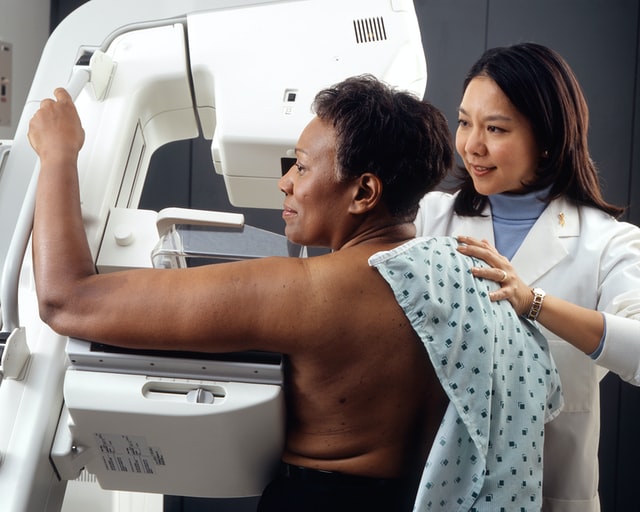
You will stand in front of the mammogram machine, and the technician will position your body and breast for the most accurate X-rays. Your breast will rest on the device, and a plastic upper plate will lower and compress your breast.

The process takes 10 to 15 seconds for each body position, and there are usually only two or three X-rays taken per breast. The whole procedure only takes about 20 minutes.

Your Mammography Results

Mammography produces black-and-white digital images of your breast tissue that will be sent to a physician specializing in radiology to interpret. An imaging center could also refer to a teleradiology company to have a radiologist do the interpretation. Most clinics offer 2D mammograms for breast screening, but many facilities now offer 3D mammograms.

The radiologist will then report their findings to your healthcare provider. This process will take approximately ten days but could take longer. The radiology report will determine if you will need further X-rays, possible MRIs, or treatments. Physicians call back about 10 to 13% of women for abnormal findings, but most of these findings are not breast cancer.

Regular mammograms are an easy, safe way to screen for breast cancer. The most challenging part about the process may be remembering to make your annual appointment, and many women make their birthdays a reminder to make their yearly appointment.

What can be more celebratory than scheduling what may be a life-saving procedure for your health on the most important day of the year for you, your friends, and your loved ones? 

Why is There a Radiologist Shortage and What Can Be Done?

Association of American Medical Colleges states that radiologist burnout and the demographics of the workforce have been critical for years. For example, 82% of the 20,950 currently active radiologists are over 45 and over, while 53%  are 55 or over. According to a recent Mayo Clinic study, radiologists ranked fifth out of 23 specialties that reported burnout rate, particularly among those screening for breast cancer. So not only are radiologists aging out, the newcomers aren’t lasting that long.

Why Are Radiologists Being Burned Out?

Radiology Business says radiologists mention things like “lack of respect” from administrators or colleagues as one of the leading causes of burnout 60%, along with lack of control or autonomy over one’s life, long work hours, too many non-essential tasks, and more. That tempered with COVID-related burnout and we are now facing a global shortage of radiologists and radiology trainees.

shortage of radiologists
Burnout has impacted healthcare staffing


According to a Diagnostic Imaging podcast, numerous factors result in radiologist burnout, from outdated and clunky software, sinking reimbursements, continuing education requirements, stressful imaging reports turnout expectations, to reduced interaction with colleagues related to the pandemic, all resulting in a slow, laborious system death.

Should You Consider Teleradiology/Outsourced Radiology Company?

If you have never heard of teleradiology or outsourcing radiology, you may be interested in learning that many clinics, offices, and hospitals are turning to medical outsourcing. Hospitals will likely start to engage more and more outsourced services that can include long-distance radiology and teleradiology. While there are pros and cons for hospitals and clinics using teleradiology, the fact is that this may quickly become a fact of life. Patient care can still be high-quality, immediate, and efficient, for example.

There are incredible benefits that come with radiology outsourcing companies and teleradiology. Many teleradiology practices employ board-certified and trained radiologists for neurological and Body Imaging needs. These radiologists are available for patient consultations to go over results, answer any questions, and much more. Plus, these services are often quick and done promptly. Many of these companies and services are coupled with a quality assurance program to maintain their ACR accreditation. Finally, these services tend to be more cost-effective than in-house services. Teleradiology and outsourced services are often used in the following practices:

  •     Private practices
  •     Urgent care clinics
  •     Imaging practices
  •     Mobile services
  •     Physician groups
  •     Independent diagnostic testing facilities
teleradiology
Teleradiology can assist with staffing shortages

What Does the Future of Radiology Look Like?

One popular solution that addresses the shortage of radiologists includes using a teleradiology/outsourced radiology company as mentioned above.

Another solution is using trained radiographers to double-read screening mammograms, extending their expertise and skill set to perform as radiologists when needed. Double reading has been used in Europe and has been incredibly efficient at detecting cancer. Training non-radiologists may end up becoming a necessity for hospitals and breast screening facilities without having to lose patient standards.

Radiology remains a solid and stable career, especially as medical professionals and facilities adapt to shortages, and potential pandemics, especially when considering teleradiology and outsourced radiology companies.

Vesta Teleradiology: For Full or Supportive Radiology Staffing Needs

Whether your healthcare facility need full-time support, or just coverage for nights and weekend radiology interpretations, Vesta is here for you! Vesta has dedicated 15 years to serving diagnostic imaging centers, physician’s offices, hospitals and other healthcare facilities with their radiology needs. Contact us for a free quote: 1-877-55-VESTA.


Ovarian Cancer: Encouraging Patients to Get Screened

It’s a topic not many people enjoy talking about, or even thinking about. Cancer, of any kind, is complicated, and ovarian cancer is, arguably, one of the most complicated and aggressive cancers there is. About 20% of women receive an early diagnosis, and of those detected early, 94% live longer than 5 years after their diagnosis, says the American Cancer Society. Encouraging patients to get screened for early detection, paying attention to the body’s signals, and regular exams are the biggest defenses we have against this deadly disease.


Ovarian cancer is difficult to detect, which is why it is paramount for patients to have regular pelvic exams. To help encourage patients to get the proper care for early detection, or with a new diagnosis, empower them with communication so they feel in charge, says Cancer Care. Suggesting the patient takes notes of the session will help, says the article, including dates, names, and discussion points. This will provide physical evidence of what went on during the appointment and a reference point for the possibly overwhelmed patient. Bringing a trusted friend or family member to the appointment can also ease the possible isolation or fear the patient may have. Another set of eyes and ears never hurts and the extra person may provide different questions and concerns the patient hadn’t thought of. Encouraging patients to write down questions or worries they may have before, during or after the appointment also gives the control back to the patient, says the article.


Persistent symptoms, even seemingly dismissible, should be examined. The fact is that ovarian cancer moves quickly, so before symptoms become worrisome, it’s important the patient knows her family history, says the American Cancer Society. If the patient has a strong family history of breast or ovarian cancer, has an inherited genetic syndrome, like Lynch syndrome, or a gene mutation such as BRCA, her high risk status must be presented and she must be heavily encouraged to get regular exams and to pay close attention to any changes within her body.


cancer awareness


The two most common screening tests for ovarian cancer are the transvaginal ultrasound (TVUS) and the CA-125 blood test, says American Cancer Society. The sound waves used during a TVUS detects abnormal shapes or measurements, says Healthline, and is about 75% effective, which is why you may order at CT scan, MRI, or a biopsy for further testing, says the article. The CA-125 blood test is not as reliable for ovarian cancer detection because high levels of the protein may not be an indicator of ovarian cancer, per se, but of inflammatory disease or endometriosis. Unfortunately, there aren’t many sure and simple tests to detect early ovarian cancer or recommend for your patient, says the CDC. Keeping your patients aware of these options may seem alarming, but they’ll know what to expect should the concern arise.


Telling your patients about genetic counseling is also a good option, says WebMd. For your high risk patients, genetic counseling will give them more concrete knowledge about their own bodies and family history. Should the patient’s test come back positive of a gene mutation, you will be aware of her risks and what to look out for during exams.


encourage your patients to get screened


Whether your patient is at high risk of ovarian cancer or not, urge them to consider regular exams and screening. There is research being done currently, says American Cancer Society, although the best and most proven way is through ultrasound and the CA-125 blood test. Reminding them you are always available if something in their body feels off, if they are in pain, or have been worried, is never a bad idea. Empower them to take charge of their health.

Teleradiology Interpretations for TVUS

Managing a healthcare practice means providing optimal care for your patients, and that includes providing the proper education and addressing patient concerns. We understand it is not always easy to balance running tests, interpretations and patient communication. That’s why Vesta has a team of US Board Certified radiologists who work with your team for preliminary and final interpretations – 24×7, nights, weekends and even holidays. Please contact us to learn more about our outsourced radiology services: 1-877-55-VESTA. 

Recent Advancements in Nuclear Medicine

The medical community is always looking for new and better ways to serve patients and save lives. Science, medicine, and technology often intersect to break barriers and create innovative new treatments – and nowhere is that truer than in the field of nuclear medicine.

What is Nuclear Medicine?

The National Institute of Biomedical Imaging and Bioengineering defines nuclear medicine as a specialty that uses radioactive tracers to diagnose and treat disease. Nuclear medicine is invaluable for patient care, as it can help detect disorders in the bones, gall bladder, heart, and much more.

advancements in nuclear medicine

This field has seen tremendous advancements in recent years, which offer the potential for incredible and life-saving benefits. Here are some of the latest developments in nuclear medicine.

Making AI More Effective

Artificial intelligence has been an integral part of medicine for decades, particularly in the realm of diagnostics. And now, new research suggests that nuclear medicine may make AI-based diagnostics even more effective.

radiology interpretations

For example, researchers in the Journal of Nuclear Medicine (JNM) suggest that nuclear imaging can help with machine learning and AI cancer diagnoses. This is because nuclear imaging creates a high contrast between tumors and normal tissue, making it much easier for the machine to identify abnormalities. Combining AI diagnostics with nuclear medicine can make the machines more accurate, which will ultimately result in better patient care over time.

Detecting Heart Disease

Radionuclide imaging has long been used to detect issues in patient heart function. However, researchers are beginning to explore new uses for this technology – including the examination of the heart’s very molecules.

Research from 2020 found that radionuclide imaging is successful at detecting cardiac amyloidosis, a rare condition in which a protein called amyloid is deposited in the cardiac muscle. Amyloid deposits can cause buildup over time and ultimately lead to heart failure, so it is very important to detect this condition as early as possible.

Discovering New Treatments

Nuclear medicine has many potential uses for imaging and diagnostics. However, it also offers many benefits for researchers.

For example, scientists at the Vanderbilt University Institute of Imaging Science recently used a radioligand (a radioactive substance used to study receptors in the body) to study whether an antioxidant called ERGO could penetrate the brain and protect against oxidative stress. The study successfully proved that ERGO can penetrate the brains of mice, which opens doors for further research on using this antioxidant to treat conditions like Alzheimer’s disease.

Nuclear medicine is always developing and advancing, and each advance makes it easier to give patients the care they deserve. 


Nuclear Radiology Readings


We are proud of our talented pool of teleradiologists who specialize in a variety of subspecialties, including nuclear radiology. If you’ would like to learn more about how we can integrate with your current workflow in order to provide preliminary and final interpretations, please contact us now at 1-877-55-VESTA

Bone Health with BMD and DEXA Scans

Bone density is the ratio of skeletal weight (mass) to the volume or area of the bones. The heavier the bones, the stronger they will be. It affects physical activity levels, menopause, nerve signals, and more. A bone mineral density (BMD) scan compares your bone mass to an established norm and produces a score unique to you. This is different than a bone scan that looks for infections or cancer, or the presence of a fracture. A BMD scan helps determine the presence of osteopenia, osteoporosis, and the probability of future falls and fractures. A BMD score, combined with personal and family medical history, can help doctors get a complete picture of bone health.


bone density x-ray
bone scan


The types of diagnostic imaging used to measure bone density have included ultrasonography, CT and MRI images, and central dual-energy x-ray absorptiometry (DXA or DEXA) tests. In 1988, the dual-energy x-ray absorptiometry (DEXA) scan was approved by the Food and Drug Administration (FDA) for clinical use. Since then, DEXA has become the gold standard for measuring bone mineral density. Its scan of the large bones at the lumbar spine and hips is most used. Shorter scan times and minimal radiation exposure makes it safe. DEXA transmits photons at two energy levels for soft tissue and cortical bone and aids in the diagnosing of osteopenia, osteoporosis, and fracture risk assessment. It is inexpensive and the most accurate imaging modality for assessing bone mass density and health.

Doctors and radiologists use the BMD score to comprise a T-score or Z-score, which is a comparison to a reference group on a standard deviation scale. T-scores are given to adults and are determined by comparison to a young gender-matched group with peak bone mass. Z-scores are given to children and are determined by comparison to an age-matched group. These scores are used in risk fracture assessment, low bone mass or osteoporosis diagnosis, patient criteria for clinical trials, and management guidelines for osteoporosis. It is crucial that BMD measurements are correct, as well as differences in T-score and Z-score population groups. Accurate documentation is necessary for dependable results. Any variation used in this process can affect the actual T-score and Z-score. Improvements in calculation methods are currently ongoing.

Maintaining strong bones is essential. Daily calcium, vitamin D supplements, and weight-bearing exercises can help slow bone loss. In addition, patients should have their BMD checked regularly. Patients should also be counseled on safety measures like fall prevention.

patient and doctor
Doctors should counsel their patients on bone health

Top Teleradiology Company: Vesta

At Vesta Teleradiology, our U.S. Board Certified Radiologists are able to read and interpret DEXA scans. If you need supporting staff to cover nights, weekends and holidays, please reach out to us today: 1-877-55-VESTA.

Benefits of Mobile Imaging for Outpatient Healthcare

Most of the healthcare provided by physicians involves some method of imaging. In the past, technicians have performed diagnostic services in the imaging departments of hospitals. Today, imaging services are brought to the patient with the help of mobile imaging.


Since there is such a high demand for x-rays, ultrasounds, MRI, CT scans, and EKGs, in-house hospital departments tend to be overwhelmed, and patients can have long wait times in crowded waiting rooms. Even getting an appointment for the imaging service can take a long time which delays treatment for the patient’s care.


With the emergence of mobile imaging, patients have more accessible, more efficient access to imaging services. With faster access to the patient, healthcare professionals can diagnose their patients more quickly and begin necessary treatments.

mobile radiology for assisted living
A man takes care of a resident in an assisted living facility

Senior Population and Imaging

The best example of the benefits of mobile imaging is with the senior population–many of whom reside in assisted living facilities and nursing homes. Persons over the age of 65 are a fragile population who need imaging services frequently.

The older age group has an increased fall rate, a higher rate of pneumonia and cancers, and compromised immune systems.  They need prompt diagnoses, but transportation to receive medical care is complicated.

The benefits are vast when mobile imaging can accommodate this population’s needs:

  • Overall, costs are reduced for the service because the facility doesn’t need to transport the patient to a hospital imaging department.
  • Understaffed facilities don’t need to assign an employee to accompany the patient for diagnostic imaging in a hospital.
  • There are fewer transfers between facility and hospital.
  • Mobile imaging services reduce anxiety in the elderly because transporting and waiting for imaging services accentuates fear in the undiagnosed.
  • There is a reduced need for hospitalizations and outpatient treatments of the patients because the assisted living facilities and nursing homes can provide prescribed care.
  • Mobile imaging can provide needed diagnostic information to the patient’s attending physician faster than an imaging department can, which expedites a treatment plan for the patient.
  • The patient can remain in familiar surroundings (and with people they know) while receiving diagnostic imaging services. This benefit is significant when the patient has difficulty understanding or processing information.

When people of any age are hurting, fearful, anxious, or lack understanding, their comfort is the most crucial factor in beneficial treatments. Mobile imaging comes to the patient and provides fast, efficient, accessible, and cost-effective diagnostics for the most fragile patients.

 Working with a Teleradiology Company

With Vesta Teleradiology, we work with mobile imaging centers and any health facility that provides this technology to their patients. We work with your workflow and integrate to your technology so sending and receiving scans is a breeze. Learn more about how we can help you now: call us at 1-877-55-VESTA.

teleradiology services
Vesta Teleradiology services include nighthawk, weekend and holiday interpretations