Summer 2025 Imaging Roundup: AI, New Modalities & Trends

The summer of 2025 has been packed with advancements in diagnostic imaging, from cutting-edge AI systems improving detection rates to emerging modalities pushing the boundaries of precision and speed. Here’s a look back at the most important developments from June through August that are shaping the future of radiology.

AI Is Reshaping Radiology Workflows

Generative AI Productivity Boost

In June, Northwestern Medicine unveiled a generative AI system capable of reducing radiologist reading time by up to 40% while identifying life-threatening conditions in milliseconds. This tool not only improves workflow efficiency but also offers a potential solution to the ongoing radiologist shortage (Northwestern Medicine).

ProFound AI for Mammography

A peer-reviewed study confirmed that iCAD’s ProFound AI significantly increases cancer detection rates, boosts diagnostic accuracy, and improves workflow for mammography screenings (ITN Online).

Aidoc’s $150M Expansion

July saw AI platform Aidoc raise $150 million in funding, led by NVIDIA and other major investors, aimed at expanding its reach into more hospitals and imaging centers globally (Aidoc).

Emerging Imaging Modalities and Research

Top Content Trends

Radiology publications in July spotlighted rising interest in abbreviated breast MRI, MRI-guided ultrasound for Parkinson’s disease, and dual-energy CT for understanding Long COVID-related lung changes (Diagnostic Imaging).

Photon-Counting CT and Whole-Body MRI

Photon-counting CT continues to gain attention for its ability to deliver higher resolution at lower doses, while whole-body MRI is increasingly used for cancer staging and early detection in high-risk populations (Radiology Business).

Multimodality Imaging at ACC.25

Cardiologists and radiologists at the ACC.25 conference explored how quantitative CT, functional cardiac MRI, and AI-enhanced echocardiography can bridge the gap between diagnostics and real-time therapy planning (American College of Cardiology).

August: A Month of Imaging Breakthroughs

AI-Native Imaging Viewers

Tech company New Lantern launched AI-native viewer modes for mammography and PET/CT, delivering sub-second load times and workflow automation (TMCNet).

Digital Radiography Gets Smarter

Advances in digital radiography are enhancing precision and speed, with newer systems providing better image quality at lower radiation doses (USA News).

ProCUSNet Ultrasound AI

Researchers at Stanford developed ProCUSNet, an AI tool that improved lesion detection by 44% and caught 82% of clinically significant prostate cancers on ultrasound—outperforming human interpretation (Becker’s Hospital Review).

DiffUS for Intraoperative Imaging

A new AI-based technique called DiffUS can create realistic ultrasound images from 3D MRI data, aiding in surgical planning and intraoperative navigation (arXiv).

Next-Gen PET Tracer

A novel PET tracer, Ga-68 Trivehexin, has shown promise in more accurately detecting breast cancer lesions and fibrotic lung tissue compared to traditional tracers (Journal of Nuclear Medicine).

Looking Ahead

The pace of innovation in diagnostic imaging this summer reinforces a clear trend: AI is no longer just an assistive tool—it’s becoming deeply embedded in clinical workflows. Coupled with emerging modalities like photon-counting CT and new PET tracers, radiology is entering an era of higher precision, speed, and accessibility.

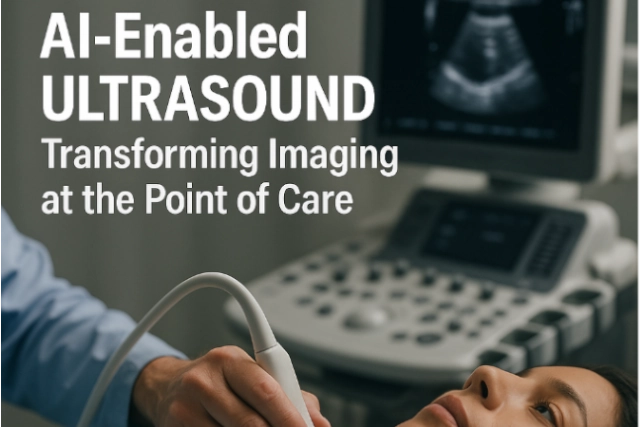
AI-Enabled Ultrasound: Transforming Imaging at the Point of Care


In today’s fast-paced healthcare environment, ultrasound is increasingly recognized not just for prenatal or cardiac assessment, but as a versatile diagnostic tool across specialties. Now, artificial intelligence (AI) is accelerating ultrasound’s impact — reducing operator dependency, improving diagnostic confidence, and enabling faster bedside care. For imaging leaders, especially in rural or underserved settings, AI-powered ultrasound technology paired with teleradiology support offers a compelling path for enhanced access and precision.

Innovations in AI-Ultrasound You Should Know

  1. FDA Clearance for AI Thyroid Ultrasound
    In 2024, See-Mode Technologies received FDA clearance for an AI-powered thyroid ultrasound system that can detect and classify nodules using the ACR TI-RADS scale. It has shown promising results in standardizing reporting and reducing unnecessary biopsies and follow-ups.
    Source: https://www.auntminnie.com
  2. Projected Market Growth
    The global AI ultrasound market is projected to grow at a compound annual growth rate (CAGR) of 22% through 2029. This rapid growth is fueled by the rising burden of chronic disease, limited radiologist availability, and the push for faster, more accessible diagnostics.

    Source: https://www.pharmiweb.com/

  3. Rural Potential with Point-of-Care AI
    A JAMA Cardiology viewpoint outlines how AI-assisted point-of-care ultrasound (POCUS) can enable more accurate cardiovascular assessments even when performed by generalists—especially valuable in remote areas without imaging specialists.
    Source: https://jamanetwork.com
  4. Clinician Enthusiasm and Challenges
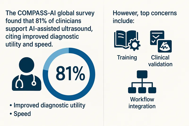
    The COMPASS-AI global survey found that 81% of clinicians support AI-assisted ultrasound, citing improved diagnostic utility and speed. However, top concerns include training, clinical validation, and workflow integration.

    Source: https://theultrasoundjournal.springeropen.com/

Infographic showing COMPASS-AI survey results on clinician support for AI-enabled ultrasound, benefits, and concernsWhy It Matters for Facilities and Radiology Teams

  • Reduces staffing burden: AI ultrasound reduces variability among operators, ideal for high-turnover or remote settings.
  • Speeds up decision-making: Frontline providers can quickly gather meaningful imaging data, while teleradiologists handle the interpretation.
  • Expands imaging reach: Portable, AI-powered ultrasound extends diagnostic capabilities to underserved regions.
  • Supports standardization: AI helps standardize image acquisition and reporting, improving overall workflow efficiency.

How Vesta Teleradiology Enhances AI-Ultrasound Value

While AI augments imaging workflows, expert interpretation is still essential. Vesta provides:

  • Subspecialty reads across thyroid, vascular, MSK, and more
  • 24/7 coverage with fast turnaround times
  • Seamless PACS/RIS integration for AI-acquired ultrasound data

Our radiologists help bridge the gap between frontline imaging and specialist analysis—ensuring that every AI-enabled ultrasound scan contributes to timely, confident patient care.

Bringing AI and Teleradiology Together

Whether you’re running a rural health center, a large outpatient clinic, or an emergency department, AI ultrasound paired with expert teleradiology interpretation helps:

  • Increase imaging access without compromising accuracy
  • Alleviate staffing constraints
  • Deliver faster diagnoses
  • Improve patient outcomes

AI in ultrasound is not replacing radiologists — it’s helping them focus on what matters most. With Vesta’s support, healthcare organizations can embrace innovation while maintaining high-quality, consistent imaging interpretation.


Photon-Counting CT: What Healthcare Facilities Need to Know Now

Photon-counting computed tomography (PCCT) is one of the most exciting breakthroughs in diagnostic imaging technology in recent years. Offering greater spatial resolution, reduced radiation dose, and improved tissue characterization, PCCT is quickly gaining attention from radiologists, imaging directors, and healthcare systems looking to stay ahead.

As the healthcare landscape evolves, staying informed about how new imaging technologies integrate with workflows and diagnostic goals is critical. Here’s what facilities need to know now about photon-counting CT—and how teleradiology can help maximize its impact.

What Is Photon-Counting CT?

Unlike conventional CT, which measures the total X-ray energy reaching the detector, photon-counting CT counts individual photons and measures their energy levels. This allows for:

  • Sharper images with better spatial resolution
  • Lower noise, especially in soft tissue
  • Multi-energy imaging from a single scan
  • Reduced radiation exposure

Siemens Healthineers introduced the first FDA-approved photon-counting CT system (NAEOTOM Alpha) in 2021, and adoption has slowly grown among academic and high-volume centers.

Clinical Benefits of PCCT

Photon-counting CT provides enhanced detail for a range of applications, including:

  • Cardiac imaging: Better visualization of stents and plaques
  • Pulmonary imaging: Improved nodule detection and perfusion data
  • Neuroimaging: Greater contrast at lower doses for brain scans
  • MSK imaging: Superior resolution for joint, bone, and soft tissue analysis

The ability to perform multi-energy imaging without dual-source CT equipment allows radiologists to generate virtual non-contrast images, improve lesion characterization, and reduce contrast agent use—benefiting both patients and providers.

Multi-energy CT image showing high-resolution internal anatomy used for virtual non-contrast imaging
Growing Market and Adoption

While still early in widespread adoption, the global photon-counting CT market is projected to grow rapidly. According to a recent report from Research and Markets, the global PCCT market is expected to reach over $800 million by 2030, driven by increasing demand for advanced diagnostic tools and a growing focus on radiation dose reduction.

As more vendors develop photon-counting detectors and more clinical use cases are validated, experts anticipate broader adoption beyond academic centers and into regional hospitals and imaging centers.

Source: Research and Markets, “Photon Counting CT Market – Forecast 2030”

How Teleradiology Supports Advanced CT Adoption

Deploying a photon-counting CT system requires more than just the hardware. Facilities must ensure they have access to radiologists who are:

  • Trained in multi-energy CT interpretation
  • Familiar with new artifact patterns and reconstructions
  • Able to optimize clinical workflows using new scan data types

That’s where teleradiology plays a critical role.

At Vesta Teleradiology, our radiologists stay at the forefront of imaging advances. With experience in multi-energy and advanced CT post-processing, we help facilities take full advantage of what photon-counting CT offers—delivering fast, accurate interpretations backed by subspecialty insight.

Integration and Workflow Considerations

Facilities considering photon-counting CT should think about:

  • PACS/RIS compatibility with new data formats
  • Training staff to understand and use spectral data
  • Building protocols for when and how to use PCCT scans
  • Collaborating with teleradiology teams for consistent interpretations

While the learning curve is real, the payoff is significant. Early adopters report better diagnostic confidence, fewer repeat scans, and more comprehensive patient evaluations.

Conclusion: Prepare for the Future of CT Imaging

Photon-counting CT represents the next leap in diagnostic precision. As this technology becomes more accessible, imaging leaders must evaluate how it fits into their long-term strategy. For facilities looking to stay competitive, offer premium diagnostics, and improve patient care, PCCT should be on the radar now—not later.

Partnering with a forward-thinking teleradiology provider like Vesta ensures you’re equipped with the expertise to unlock its full potential.


February AI News in Radiology

Brain Tumor Spotted on PET Imaging

An AI algorithm named “JuST_BrainPET” identified a glioblastoma in a patient that had been missed by physicians. This finding, reported in the Journal of Nuclear Medicine, underscores the potential of AI-based decision support in diagnostic and treatment planning. The algorithm automatically segments metabolic tumor volume from healthy tissue on brain PET imaging. In a case study, it detected a lesion in the frontoparietal region, not identified by an expert, which progressed to a small tumor. The AI tool’s early detection could have influenced diagnostic and treatment decisions.


Using Eye-Tracking

Researchers in Lisbon, Portugal, have pioneered a method to enhance AI interpretability in radiology by integrating eye-tracking data into deep learning algorithms. This innovative approach, outlined in the European Journal of Radiology, aims to align AI systems more closely with human understanding, marking a significant leap towards more human-centered AI technologies in radiology. By leveraging eye-gaze data, the researchers sought to bridge the gap between human expertise and AI computational power, anticipating that AI models could learn from the nuanced patterns of image analysis observed by radiologists.


This integration promises AI models that prioritize image characteristics relevant for diagnosis, potentially reducing the disparity between AI decision-making processes and human radiologists’ diagnostic approaches. The potential benefits of this research are vast, potentially leading to AI systems that are not only more effective in identifying pathologies but also more understandable to radiologists, thus fostering trust in AI-assisted diagnostics and accelerating their adoption in healthcare.


Review Paper on AI and Cancer Detection

Professor Pegah Khosravi and her team of researchers explore how artificial intelligence (AI) can enhance anomaly detection in MRI scans to advance precision medicine. Their comprehensive review, published in the Journal of Magnetic Resonance Imaging, focuses on AI techniques like machine learning and deep learning, particularly in identifying tumors in the brain, lungs, breast, and prostate.

The authors discuss several AI strategies for improving tumor detection, including a holistic approach that integrates data from various imaging techniques such as MRI, CT scans, and PET scans, along with genomic information and patient histories. This approach not only enhances anomaly detection accuracy but also facilitates personalized treatments based on comprehensive patient profiles.

Furthermore, the paper explores the use of ensemble methods in AI, which combine different AI models’ strengths to improve anomaly detection. By leveraging these methods, a more thorough analysis of MRI data is ensured. The authors advocate for AI systems that are accurate and transparent in their decision-making processes, fostering trust among healthcare professionals. They also stress the importance of collaboration among researchers, clinicians, and policymakers to effectively implement AI in medical imaging, guiding future advancements in the field.


Sources:

Auntminnie.com
bnnbreaking.com
gc.cuny.edu
openai.com