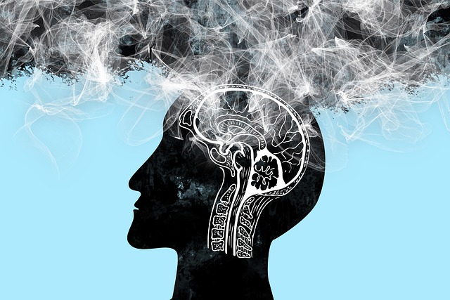
In recent years, awareness surrounding brain injuries has steadily risen, prompting significant strides in diagnostic technologies and treatment modalities. As we delve into the latest developments in this critical area of healthcare, it becomes increasingly apparent that advancements in medical imaging, particularly in the realm of neurological disorders, are poised to revolutionize the landscape of brain injury diagnosis and management.
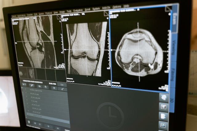
AI-based Quantitative Brain Imaging System
Philips and Synthetic MR have joined forces to advance the diagnosis of neurological disorders through cutting-edge quantitative brain imaging tools. Their collaboration introduces the Smart Quant Neuro 3D MRI software suite, combining Philips’ SmartSpeed image-reconstruction technology, the 3D SyntAc clinical application, and SyntheticMR’s SyMRI NEURO 3D software. This innovation employs AI to analyze brain tissues, enhancing the detection and analysis of conditions like multiple sclerosis, traumatic brain injuries, and dementia.
The rise of AI in diagnostic imaging, projected to reach $1.2bn by 2027, signifies a transformative shift in improving accuracy and patient outcomes. With the diagnostic imaging market expected to grow to $9.1bn by 2030, fueled by demand for early disease diagnosis and personalized medicine, this partnership underscores the crucial role of AI in enhancing medical imaging.
Read the press release here.
A New Way of Diagnosing Mild TBIs
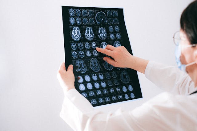
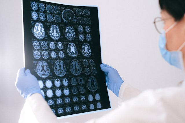
Researchers have developed a novel brain imaging method to diagnose mild traumatic brain injuries (mTBIs), which are often missed by standard techniques like MRI. This method involves loading gadolinium, a common MRI contrast agent, into micropatches attached to immune cells called macrophages. These cells migrate to areas of brain inflammation caused by mTBIs, enabling MRI detection. The technique, called M-GLAMs, was successfully tested in mice and pigs, showing promise for accurately diagnosing mTBIs. It also allows imaging at lower gadolinium doses, potentially benefiting patients with kidney issues. While unable to pinpoint injury locations, M-GLAMs could aid in identifying and treating brain inflammation. The researchers aim to bring this technology to clinical trials, with support from grants and intellectual property protection.
Read the study here.
New Imaging Tech that Captures Neuronal Activity Across the Brain During Recovery
Researchers at Tufts University School of Medicine have developed a novel imaging technology to monitor neuronal activity throughout the entire brain during the initial weeks of recovery from traumatic brain injury (TBI). Their study, published in Cerebral Cortex, reveals that TBI can induce changes in brain function beyond the injury site. Using a combination of fluorescent sensors and electrodes, they observed altered connectivity patterns in mice post-injury, even in regions distant from the impact. Despite the mice’s ability to perform physical tasks normally, their brain activity during both exercise and rest differed significantly from healthy brains. This impaired ability to switch between states suggests underlying brain state dysfunction post-injury. The findings highlight the brain’s plasticity in response to injury and have potential clinical implications for understanding TBI impacts and tailoring treatments. The researchers aim to further investigate long-term neural activity changes post-recovery and explore the technology’s potential in predicting specific dysfunctions or long-term outcomes of TBI.
Read the study here.
Sources:
Medicaldevice-network.com
Otd.harvard.edu
Scitechdaily.com
Openai.com