
In 2026, hospitals and imaging providers are looking beyond a vendor that can read studies after hours. They are looking for a teleradiology partner that can help protect turnaround times, expand subspecialty access, support strained radiology teams, and use AI responsibly to improve workflow without replacing radiologist judgment. That shift matters because radiology demand and workforce strain are still real, and healthcare organizations need solutions that are both scalable and clinically reliable. AAMC continues to project a broad U.S. physician shortage by 2036, while RSNA has highlighted ongoing radiologist workforce pressure and rising imaging volume.
So what should modern hospitals look for in a teleradiology company in the USA in 2026?
-
U.S.-Based, Board-Certified Radiologists
The foundation still matters most. A strong teleradiology company should offer U.S.-based, board-certified radiologists who understand clinical expectations, communication standards, and the realities of American hospital workflows. In a market where speed matters, quality cannot become an afterthought. Vesta partners with U.S. board-certified radiologists, nationwide coverage, and support for hospitals, imaging centers, and urgent care facilities.
-
Real Subspecialty Coverage, Not Just General Overflow
In 2026, hospitals should look beyond basic overnight reading coverage. They should ask whether a teleradiology company can support subspecialty interpretation when complexity rises. Neuro, body imaging, MSK, emergency imaging, and other focused reads can affect confidence, consistency, and downstream care decisions. Radiology workforce pressure is not evenly distributed, and subspecialty gaps can be especially difficult to fill.
That is why a modern teleradiology partner should be able to deliver both routine coverage and access to deeper expertise when needed.
-
24/7/365 Coverage That Holds Up Under Stress
Plenty of companies say they offer around-the-clock service. The better question is whether that coverage remains dependable on nights, weekends, holidays, and during sudden surges in volume. Hospitals should look for a partner with a proven operating model for continuous coverage, not just marketing language about availability. Vesta is proud to offer 24/7/365 support, preliminary and final interpretations, and scalable coverage across the U.S.
That kind of consistency matters because radiology delays can affect ED throughput, inpatient flow, and clinician satisfaction.
-
AI-Enhanced Workflow That Supports Radiologists
In 2026, AI is no longer a futuristic talking point. It is part of the decision set. But hospitals should be careful about how they evaluate it. The best teleradiology companies use AI to support workflow, triage, prioritization, consistency, and operational efficiency while keeping radiologists in control of interpretation. RSNA publications have noted that AI can improve productivity and support report generation and workflow efficiency, but they also stress that safe deployment, validation, and thoughtful integration are essential. FDA resources likewise show a growing U.S. landscape of AI-enabled medical devices and active regulatory guidance around lifecycle management and safety.
 Vesta has invested in AI-assisted imaging and workflow partnerships, including Qure.ai, Carpl.ai, and RadPair, as well as internal AI-based support tools that help staff retrieve protocols, schedules, credentialing information, and specialty details more efficiently. Vesta also states that it uses AI-driven prioritization and cloud-based workflow tools to help radiologists surface critical findings faster and return reports without delay.
For hospitals, the takeaway is simple: do not ask whether a teleradiology company uses AI. Ask how it uses AI, where it fits into workflow, and whether it strengthens speed and quality without weakening oversight.
-
Seamless Integration With Existing Systems
A teleradiology relationship should make operations easier, not harder. That means the company should be able to integrate with PACS, RIS, HL7, and related workflow infrastructure in a way that minimizes friction for staff. Fast onboarding, dependable communication, and technology compatibility should all be part of the evaluation process. Vesta offers HL7 integration, infrastructure support, managed implementation capabilities, and customizable IT solutions as part of its service mix.
The more seamless the operational fit, the faster a facility can realize value.
-
Support for Rural and Underserved Facilities
Hospitals in rural and underserved areas often feel imaging access problems first. AHRQ has noted that rural communities face provider shortages and may benefit significantly from telehealth-supported care models. Teleradiology can be especially valuable when geography and staffing limitations make local subspecialty access difficult.
Vesta uses AI-enabled radiology expansion as a way to support hospitals of every size, including rural and underserved communities.
-
Accreditation, Reliability, and Communication
Hospitals should also look for proof of organizational maturity. Accreditation, dependable service, and direct communication pathways all matter. Vesta is a Joint Commission-accredited provider and emphasizes timely, secure interpretations and direct service support.
In practical terms, a strong teleradiology company should be able to answer these questions clearly:
How fast can you onboard us?
Who reads our cases?
What subspecialties do you cover?
How do you handle critical findings?
How does your AI fit into workflow?
How do your radiologists communicate with our team?

The Bottom Line
In 2026, the top qualities to look for in a teleradiology company in the USA go well beyond basic night coverage. Hospitals should prioritize clinical quality, subspecialty depth, dependable 24/7/365 service, strong integration, and AI-enhanced workflow that improves efficiency while preserving radiologist oversight. For organizations trying to protect patient flow, reduce coverage risk, and modernize imaging operations, those qualities are no longer optional. They are the standard modern hospitals should expect from a serious teleradiology partner.
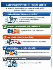
 2) Build priority tiers that match clinical urgency


 And for
