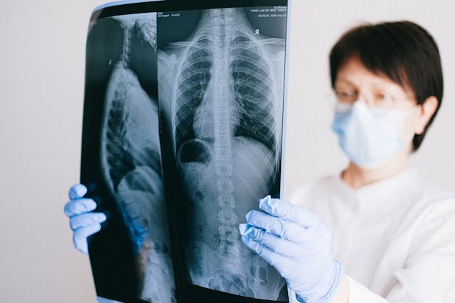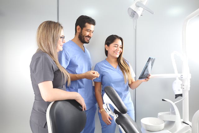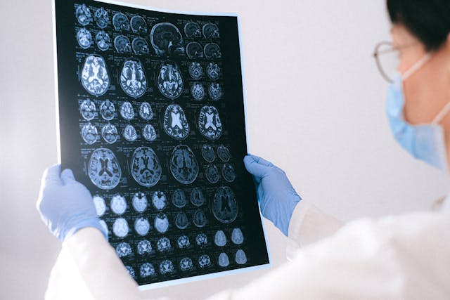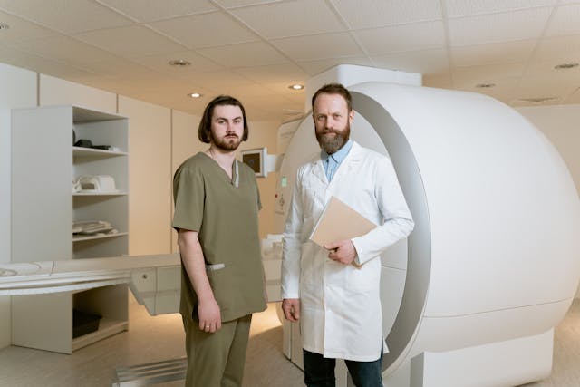
Outpatient radiology centers play a crucial role in the healthcare landscape by providing convenient, efficient, and cost-effective access to diagnostic imaging services for patients across a wide range of medical conditions. These services include X-rays, ultrasounds, MRIs, CT scans, mammography, and fluoroscopy, among others. Patients typically visit these centers for imaging tests prescribed by their healthcare providers to diagnose and monitor various medical conditions.
While these centers offer a convenient and efficient alternative to hospital-based imaging services, often providing faster appointments and reduced wait times, they do face challenges.
Issues with Outpatient Imaging Appointments
A recent study published in Academic Radiology reveals that nearly 24% of outpatient imaging appointments are missed, with the majority due to patient cancellations rather than no-shows. Factors such as younger age, being unwed, residing in disadvantaged neighborhoods, or lacking adequate insurance increase the likelihood of missing appointments. The study, conducted by researchers at the University of California, Irvine, analyzed data from their academic health center, finding that over 70% of cancellations were initiated by patients. Interventions are suggested to reduce missed appointments, such as self-scheduling, implementing checklists for necessary processes before imaging exams, and addressing health-related social risks like transportation access. Despite suggestions, limited research exists on reducing appointment cancellations in outpatient imaging.
Delays in MRI Orders
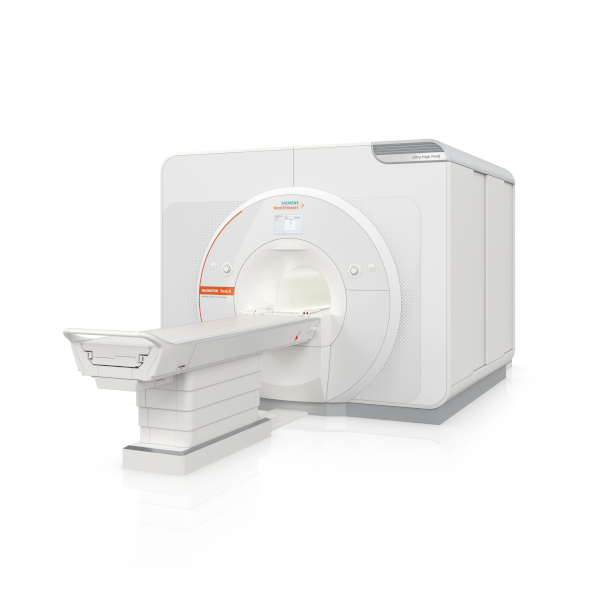
A recent study published in the Journal of the American College of Radiology reveals that nearly half of outpatient MRI orders experience significant delays, being performed more than 10 days from the date chosen by the referring provider. Led by Ronilda Lacson, MD, PhD, from Brigham and Women’s Hospital in Boston, the research emphasizes the critical importance of mitigating factors causing these delays, as they negatively impact patient care. Assessing over 97,000 outpatient MRI exams ordered between October 2021 and December 2022, the study identifies patient demographics, social determinants of health, and radiology practice- and community-level factors associated with delayed MR imaging. The study found that close to 50% of MRI orders had a prolonged order-to-performed interval, with factors such as higher Area Deprivation Index (ADI) scores contributing to delays. The authors stress the need for systemic approaches to address disparities in access to MRI examinations, including staff training, access to patient navigators, and programs tackling transportation barriers to outpatient imaging.
Other Challenges Outpatient Centers Face:
Technological Advancements: Keeping up with rapidly evolving imaging technologies requires significant investment and ongoing training for staff. Outpatient centers need to stay updated with the latest equipment and software to maintain competitiveness and provide accurate diagnostic services.

Regulatory Compliance: Compliance with healthcare regulations and standards, such as those related to patient privacy (HIPAA), radiation safety, and quality assurance, is essential but can be challenging to navigate. Failure to comply can result in fines, legal consequences, and damage to reputation.
Staffing and Workforce Management: Recruiting and retaining skilled radiologists, technicians, and support staff is crucial for maintaining quality and efficiency. Shortages in qualified personnel or high turnover rates can strain operations and affect patient care.
Integration with Healthcare Systems: Outpatient radiology centers need to effectively integrate with larger healthcare systems, including electronic health record (EHR) systems and referral networks. Seamless communication and coordination with referring physicians are essential for delivering comprehensive patient care.
Outpatient Centers Can Rely on Teleradiologists
In conclusion, outpatient radiology centers play a vital role in providing accessible, efficient, and high-quality diagnostic imaging services to patients. However, they face various challenges, including staffing shortages, which can impact their ability to deliver timely care. One solution to alleviate some of these challenges is the adoption of teleradiology services. Teleradiology services from reputable companies like Vesta, enables centers to access remote radiologists who can interpret images and provide diagnostic reports, helping to overcome staffing shortages and ensure continuous coverage. By embracing technology and innovative solutions like teleradiology, outpatient radiology centers can enhance their capabilities, improve patient care, and meet the evolving needs of healthcare delivery.
Sources:
Auntminnie.com
radiologybusiness.com
openai.com