February AI News in Radiology

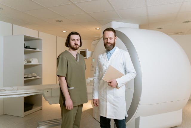
Brain Tumor Spotted on PET Imaging

An AI algorithm named “JuST_BrainPET” identified a glioblastoma in a patient that had been missed by physicians. This finding, reported in the Journal of Nuclear Medicine, underscores the potential of AI-based decision support in diagnostic and treatment planning. The algorithm automatically segments metabolic tumor volume from healthy tissue on brain PET imaging. In a case study, it detected a lesion in the frontoparietal region, not identified by an expert, which progressed to a small tumor. The AI tool’s early detection could have influenced diagnostic and treatment decisions.


Using Eye-Tracking

Researchers in Lisbon, Portugal, have pioneered a method to enhance AI interpretability in radiology by integrating eye-tracking data into deep learning algorithms. This innovative approach, outlined in the European Journal of Radiology, aims to align AI systems more closely with human understanding, marking a significant leap towards more human-centered AI technologies in radiology. By leveraging eye-gaze data, the researchers sought to bridge the gap between human expertise and AI computational power, anticipating that AI models could learn from the nuanced patterns of image analysis observed by radiologists.


This integration promises AI models that prioritize image characteristics relevant for diagnosis, potentially reducing the disparity between AI decision-making processes and human radiologists’ diagnostic approaches. The potential benefits of this research are vast, potentially leading to AI systems that are not only more effective in identifying pathologies but also more understandable to radiologists, thus fostering trust in AI-assisted diagnostics and accelerating their adoption in healthcare.


Review Paper on AI and Cancer Detection

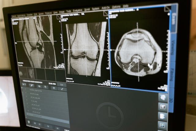
Professor Pegah Khosravi and her team of researchers explore how artificial intelligence (AI) can enhance anomaly detection in MRI scans to advance precision medicine. Their comprehensive review, published in the Journal of Magnetic Resonance Imaging, focuses on AI techniques like machine learning and deep learning, particularly in identifying tumors in the brain, lungs, breast, and prostate.

The authors discuss several AI strategies for improving tumor detection, including a holistic approach that integrates data from various imaging techniques such as MRI, CT scans, and PET scans, along with genomic information and patient histories. This approach not only enhances anomaly detection accuracy but also facilitates personalized treatments based on comprehensive patient profiles.

Furthermore, the paper explores the use of ensemble methods in AI, which combine different AI models’ strengths to improve anomaly detection. By leveraging these methods, a more thorough analysis of MRI data is ensured. The authors advocate for AI systems that are accurate and transparent in their decision-making processes, fostering trust among healthcare professionals. They also stress the importance of collaboration among researchers, clinicians, and policymakers to effectively implement AI in medical imaging, guiding future advancements in the field.


Sources:

Auntminnie.com
bnnbreaking.com
gc.cuny.edu
openai.com

How to Pick the Best Teleradiology Company

As the demand for healthcare services continues to surge and the shortage of healthcare workers persists, particularly in specialized fields, such as radiology, hospitals and healthcare centers find themselves facing the challenge of ensuring timely and accurate interpretations of medical imaging studies. The critical role of radiologists in diagnosing illnesses and guiding treatment decisions underscores the urgency of addressing this shortage. In response, many institutions are turning to teleradiology companies to bridge the gap and provide remote interpretation services. However, selecting the right teleradiology company is paramount to ensure high-quality patient care and seamless integration into existing workflows. In this discussion, we will explore the criteria for choosing a reputable teleradiology company, considering factors such as expertise, technology infrastructure, turnaround time, and adherence to regulatory standards. By making informed decisions in this regard, healthcare facilities can optimize their radiology services and meet the needs of patients efficiently.

remote radiology company
Rad tech and radiologist

Checklist for Choosing a Teleradiology Partner

Before selecting a teleradiology company, healthcare providers should consider several key factors to ensure they choose a partner that meets their needs and maintains high standards of service. Here are some important considerations:

  1. Quality and Expertise: Assess the qualifications and experience of the radiologists employed by the teleradiology company. Look for board-certified radiologists with expertise in relevant subspecialties. A recent survey of 2,749 radiologists from 108 countries reveals that while they read across almost five subspecialties daily, many lack confidence in certain areas. About 40% accept studies across all specialties, but less than half feel “very confident” in their current subspecialty, so it is vital to ensure the radiologists you work with have expertise in what you require.
  2. Technology and Infrastructure: Evaluate the teleradiology company’s technology infrastructure, including the software used for image transmission and reporting. Compatibility with existing systems and the ability to securely transmit images while maintaining patient privacy are crucial considerations.
  3. Turnaround Time: Timeliness is critical in radiology reporting. Consider the teleradiology company’s turnaround time for providing interpretations. Ideally, they should offer rapid reporting to facilitate prompt patient care and treatment decisions.
  4. 24/7 Availability: Healthcare facilities may require radiology services round-the-clock. Ensure that the teleradiology company offers 24/7 coverage (like at Vesta Teleradiology) to accommodate emergencies and provide continuous support.
  5. Communication and Collaboration: Effective communication between the teleradiology company and the healthcare facility is essential. Evaluate the company’s communication protocols, including how they handle urgent findings and facilitate collaboration between radiologists and onsite clinicians.
  6. Regulatory Compliance: Verify that the teleradiology company complies with all relevant regulatory standards, such as HIPAA (Health Insurance Portability and Accountability Act) regulations for patient data protection. They should also adhere to industry standards for image quality and reporting accuracy.Regulatory compliance
  7. Scalability and Flexibility: Consider the scalability of the teleradiology service to accommodate fluctuations in imaging volumes. Additionally, assess their flexibility in tailoring services to meet the specific needs of your healthcare facility.
  8. Cost and Value: While cost is a factor, prioritize value over price alone. Evaluate the overall value proposition of the teleradiology company, considering factors such as quality, reliability, and the ability to improve patient outcomes.

By thoroughly evaluating these factors and conducting due diligence, healthcare providers can make an informed decision when choosing a teleradiology company, ultimately enhancing the quality and efficiency of radiology services within their organization.

Partnering with a Top US Teleradiology Company—Vesta

Vesta serves as your dependable ally in radiology, extending support to various subspecialties—whether you’re a busy urban hospital or a private practice. We ensure swift processing for both urgent and routine studies. Recognizing the value of your staff’s time and well-being, our teleradiology services enable them to maintain a healthier work-life balance by covering shifts during nights, weekends, and holidays. We can also accommodate any volumes so please reach out to us to learn more.


Sources:

hcinnovationgroup.com
Radiologybusiness.com
openai.com


Healthcare Services for Native Americans (IHS)

In the pursuit of equitable healthcare in the United States, it is imperative to address the unique needs and challenges faced by all populations, including Native American communities. Despite advancements in healthcare delivery, disparities persist, particularly among Indigenous peoples.

Research additionally indicates that the life expectancy of Native Americans falls short by 5.5 years compared to the national average. Like the general populace, prevalent causes of mortality within Native American communities encompass heart disease, cancer, and accidents. Nevertheless, Native Americans face more than a threefold increase in mortality from diabetes-related complications, are over six times more prone to succumb to alcoholism, and exhibit a mortality rate from liver diseases exceeding four times that of the general population.


Reasons for Improved Healthcare for Natives

Higher Rates of Chronic Diseases: Native Americans experience higher rates of chronic diseases such as diabetes, cardiovascular disease, and obesity compared to the general population. These health issues are often linked to socioeconomic factors, limited access to nutritious foods, and inadequate healthcare services.

Barriers to Healthcare Access: Many Native American communities are located in rural or remote areas with limited access to healthcare facilities. Additionally, cultural and language barriers may deter individuals from seeking medical care.

Cultural Sensitivity: Traditional Western healthcare systems may not always be culturally sensitive to the needs of Native American communities. Culturally tailored healthcare services can improve patient outcomes and foster trust between healthcare providers and patients.


Indian Health Services: Staff Physician Vacancies

The American Medical Association (AMA) acknowledges the severe physician shortage within the Indian Health Service (IHS), with a vacancy rate of 25% in 2018. They advocate for strategies to address this shortage and ensure that American Indians, Alaska Natives, and Native Hawaiians receive adequate healthcare. The AMA’s recommendations include raising physician compensation, modernizing IHS facilities, promoting educational opportunities at IHS facilities, and establishing partnerships with academic medical centers. They stress the importance of addressing regulatory and licensure barriers for physicians interested in serving these communities. Overall, the AMA is committed to long-term solutions to alleviate the physician shortage and improve healthcare access for Indigenous populations.

Teleradiology for Indian Health Services

As a premier teleradiology company, Vesta understands the critical importance of reliable and efficient diagnostic imaging services, especially in regions facing shortages. With our state-of-the-art technology and a team of highly skilled U.S. Board Certified radiologists, we are committed to bridging the gap by offering accurate and timely readings. Whether it’s X-rays, MRIs, mammograms, CT scans, or other subspecialty, our streamlined process ensures swift delivery of results without compromising on quality. By partnering with us, Indian Health Services can confidently meet the demands of patient care, ensuring every individual receives the prompt and precise diagnoses they deserve.


Sources:

Ncbi.nlm.nih.gov
ama-assn.org
Openai.com

New FDA Clearances: Imaging Technology

AI Enabled Software Program for Lung CT Scans:

The Food and Drug Administration (FDA) has granted 510(k) clearance for LungQ 3.0.0., an updated version of an artificial intelligence (AI)-enabled software platform designed for lung computed tomography (CT) scans. Developed by Thirona and currently utilized in over 600 hospitals, the enhanced software offers improved visualization and assessment of lung structures, including lobes and subsegments, aiding in the diagnosis and treatment of conditions such as emphysema and COVID-19. Thirona highlights that LungQ 3.0.0. enhances precision and efficiency in interventional procedures, ranging from lung segmentectomy and ablation to lung cancer biopsies and lung volume reduction. The software’s validation in over 200 global publications underscores its significance in advancing personalized treatment for lung patients. Eva van Rikxoort, the founder and CEO of Thirona, emphasizes the role of solutions like LungQ in ushering in a new era of personalized and less invasive procedures for lung diseases.


Strongest-Ever Gradient System: Siemens Magnetom Cima.X  3 Tesla

Siemens Healthineers has received FDA clearance for the Magnetom Cima.X 3 Tesla (3T) magnetic resonance imaging whole-body scanner. This scanner features the industry’s strongest gradient system for a clinically released whole-body MR scanner, providing improved visibility of smaller structures in the body and faster image capture. The Magnetom Cima.X also includes new features aimed at enhancing scientific research and addressing imaging challenges related to cancer and other diseases. The high gradient level of the Gemini Gradients, with an amplitude of 200 mT/m and a slew rate of 200 T/m/s, allows for better study of neurodegenerative diseases and can be leveraged across the body. The scanner introduces Physiologging for precise, time-stamped physiological data during functional brain imaging and the Open Recon platform for immediate image reconstruction using custom algorithms directly on the scanner. Additionally, key features such as BioMatrix technology, Deep Resolve reconstruction technology, and myExam Companion workflow solution contribute to improved clinical performance, increased productivity, and a better patient experience. Katie Grant, Vice President of Magnetic Resonance at Siemens Healthineers North America, highlights the scanner’s ability to deliver new insights into oncologic, cardiac, and neurodegenerative diseases.


First FDA-Cleared Portable Magnetic Resonance Brain Imaging System

Hyperfine, Inc. has launched the eighth generation of its Swoop® system software, featuring an AI-powered diffusion-weighted imaging (DWI) denoising feature that received FDA clearance in October 2023. The software enhances the image quality of the Swoop® system, the world’s first FDA-cleared portable magnetic resonance brain imaging system. The update introduces ease-of-use features, including real-time assistance for precise patient loading and positioning, as well as a streamlined image upload process. With CE and UKCA certifications, Hyperfine is positioned for international expansion, aiming to reach a broader global market. The software focuses on improving image quality in the DWI sequence, crucial for stroke imaging. The limited market release phase received positive feedback, with users praising DWI image quality and workflow efficiencies. The update allows clinicians to view each series in real-time, communicate with technologists, and share critical findings with clinicians without waiting for the entire study to be completed.

Sources:

Hyperfine.io
itnonline.com
diagnosticimaging.com
openai.com

How Does Teleradiology Actually Work?

In today’s rapidly evolving healthcare landscape, the prominence and necessity of teleradiology have surged to the forefront. With advancements in technology and the growing demand for efficient, timely, and accurate diagnostic services, especially in light of staffing shortages, teleradiology has emerged as a pivotal solution. Teleradiology not only addresses the increasing demand for imaging interpretations but also bridges geographical gaps, enabling swift access to specialized radiologists regardless of location. Its rising popularity stems from its ability to enhance healthcare delivery by offering remote interpretations, thereby improving patient outcomes and streamlining diagnostic processes in a progressively interconnected world.

Teleradiology operates by leveraging digital communication technologies to transmit medical images, such as X-rays, MRIs, CT scans, and ultrasounds, from one location to another for interpretation and diagnosis.

Here’s a breakdown of how teleradiology works in real time:

Image Capture: Medical images are taken at a healthcare facility using specialized imaging equipment, creating digital files.

Image Transmission: These digital images are securely transmitted over networks, often utilizing Picture Archiving and Communication Systems (PACS) or secure internet connections.

Remote Interpretation: Radiologists, often located at a different site or working remotely, receive these images. They access the images through specialized software, review them in real time, and provide interpretations, diagnoses, and reports.

Consultation and Reporting: Upon reviewing the images, radiologists generate detailed reports containing their findings, interpretations, and recommendations. They may also engage in consultations with healthcare providers or specialists as needed.

Communication: The reports and findings are transmitted back to the referring healthcare facility, where they become part of the patient’s medical record. This allows for prompt decision-making and treatment planning by the attending physicians.

how teleradiology works
Physicians go over a patient’s scan

Quality Control: Teleradiology services often have quality assurance measures in place to ensure accurate and timely interpretations, adhering to industry standards and protocols.


Throughout this process, encryption and secure transmission protocols are employed to safeguard patient data and comply with healthcare privacy regulations (such as HIPAA in the United States). The real-time nature of teleradiology facilitates quicker diagnoses, especially in emergency situations, and enables access to specialized expertise irrespective of geographical barriers.


Top Teleradiology Company: Vesta is Here for You 24/7/365

Vesta is here to be your supporting partner in radiology, even for subspecialties. We offer fast turnaround for both STAT and Routine studies. Your staff is important–they don’t always work around the clock and shouldn’t have to. With our teleradiology services, you can allow your staff a better work-life balance while we fill in those gaps whether it’s during night hours, weekends, holidays.


Sources:

Acr.org
openai.com


What You Missed at RSNA 2023

The RSNA annual meeting draws tens of thousands of healthcare professionals in medical imaging, offering a comprehensive platform for unveiling groundbreaking innovations and fostering discussions among industry leaders. This year’s conference just ended, so if you didn’t get to join, we’ll be highlighting some interesting takeaways from this amazing event.

New Technology

Royal Philips introduces the BlueSeal MR Mobile, a groundbreaking mobile MRI system featuring helium-free operations, marking a significant advancement in diagnostic imaging technology. This pioneering device, equipped with the industry’s first fully sealed 1.5T magnet, provides patient-centric MRI services, offering agility and flexibility in placement, especially near hospital entrances for patient convenience. Developed initially for Akumin, the first unit to be showcased at the event, this innovation extends Philips’ BlueSeal magnet technology, having saved over 1.5 million liters of helium since 2018. The helium-free mobile unit expands access to MRI exams sustainably, catering to more patients in diverse locations, addressing resource constraints, and enhancing healthcare delivery, as highlighted by Ruud Zwerink, General Manager Magnetic Resonance at Philips. Notably, the BlueSeal MR Mobile’s reduced helium requirements improve operational efficiency and connect to Philips’ Radiology Operations Command Center (ROCC), enabling real-time remote support for imaging experts, ensuring quality care delivery.

Radpair, a pioneering platform in radiology innovation, unveiled its cutting-edge generative AI-driven technology at the conference. This groundbreaking system, described by Avez Rizvi, Radpair’s CEO, as a revolutionary advancement, promises to reshape radiology reporting and elevate patient care standards. Positioned as the first of its kind, Radpair’s web-based and user-friendly platform utilizes generative AI in clinical settings to automate radiology report generation, streamlining radiologists’ workflow and enhancing efficiency while prioritizing patient care. Vesta Teleradiology is proud to collaborate with Radpair, with Vesta CEO, Vijay Vonguru stating, “This partnership propels us to the forefront of innovation in radiology. The synergy between Radpair’s advanced generative AI technology and Vesta’s robust teleradiology platform and onsite Radiology will redefine the standards of care we provide, ensuring high-quality, swift, and more nuanced radiological interpretations.”

Radpair and Vesta Telereadiology

Addressing the People

Dr. Pedram Keshavarz from UCLA presented findings indicating widespread burnout symptoms among radiologists and trainees. Emotional exhaustion and depersonalization were prevalent, particularly among residents and trainees who exhibited the highest rates of low personal accomplishment. These symptoms are considered warning signs for potential professional dropout or retirement. The study reviewed multiple contributing factors to burnout, including sleep deprivation, heavy workloads, low salaries, and various responsibilities. Analyzing nine studies with over 15,000 participants, the research highlighted different rates of burnout across radiology subspecialties, linking factors like having a partner, child, and lower debt levels to reduced emotional exhaustion and higher personal accomplishment. The presentation emphasized the need for future research to focus on interventions to alleviate burnout symptoms, potentially exploring the impact of remote work and other aspects on radiologists’ well-being. Large cross-sectional studies were suggested to further understand and address burnout progression among radiologists.


Sources:

Auntminnie.com
itnonline.com
Phillips.com
Openai.com


Winter Influx of Patients: What Hospitals Can Expect This Year

Every year, hospitals experience an influx of patients during winter due to respiratory illnesses, falls, and accidents. However, with the ongoing pandemic, the winter season brings extra challenges for the medical staff.

Preparing ahead is crucial to ensure that hospitals can cope with the surge in the winter season.

What to Expect

The winter season often increases respiratory illnesses such as pneumonia and bronchitis. Hospital admissions for these conditions can increase by up to 25% during winter. Medical staff should expect to see more patients with respiratory illnesses in 2023/2024.

Imaging procedures such as X-rays and CT scans can help to diagnose and monitor these conditions. Therefore, hospitals should ensure enough imaging technicians, equipment, and supplies.

Hospitals can also expect more patients with falls and accidents. Icy roads and pavements can cause slips, trips, and falls, leading to broken bones and head injuries.

slip and fall winter accidents

Imaging procedures such as X-rays and MRI scans can help to diagnose fractures and internal injuries. Therefore, hospitals should ensure that they have enough imaging equipment to diagnose and monitor these injuries.

They should also provide an imaging safety committee to promote safety in the imaging department.

With the ongoing pandemic, hospitals should expect a surge in COVID-19 patients during the winter season. The increase in COVID-19 cases will put additional pressure on hospital resources.

Imaging procedures such as chest X-rays and CT scans can help to diagnose and monitor COVID-19 patients. Therefore, hospitals should ensure enough imaging equipment to diagnose and monitor COVID-19 patients.

They should also have policies and procedures to protect staff and patients from infection.

How to Prepare

The winter season not only brings a surge in patients but also a surge in demand for medical staff. Hospitals should expect medical staff shortages to increase during the winter months due to staff illness, vacation time, and extra medical staff needed to manage the surge.

Therefore, hospitals should consider hiring temporary medical staff during winter to cope with the increased demand. They should also provide ongoing training for new and temporary staff, including imaging technicians.

Vesta Teleradiology to fill in Gaps and Radiologist Shortages

In conclusion, the winter influx of patients can be challenging for hospitals, especially with the ongoing pandemic. Hospitals should prepare to cope with the patient surge and medical staff shortages.

Imaging procedures can help to diagnose and monitor respiratory illnesses, falls and accidents, and COVID-19 patients. Therefore, hospitals should ensure that they have enough imaging equipment, technicians and of course radiologists for interpretations. Vesta Teleradiology provides US Board Certified radiologists who will work with you day and night, 24/7.

They should also have policies and procedures to protect staff and patients from infection. By preparing ahead, hospitals can ensure that they provide the best possible care for their patients during winter.


ChatGPT in Radiology: Is it a Pro or Con?

The emergence of ChatGPT in the medical field, particularly in radiology, has generated a mix of excitement and concern about its role. But is it accurate enough to put into use? Can we trust artificial intelligence (AI) with the health of our patients?

How Could ChatGPT be Used?

An article in Diagnostic and Interventional Imaging discusses various ways in which radiologists can leverage ChatGPT. It highlights applications for clinical radiologists, such as implementing ChatGPT as a chatbot for patient inquiries, supporting clinical decision-making with information and analysis assistance, and enhancing patient communication and follow-up care by simplifying radiology reports and crafting tailored recommendations. Academic radiologists can benefit from ChatGPT by receiving suggestions for impactful research article titles, assistance with structuring and formatting academic papers, and help in formatting citations for bibliographies. The article emphasizes that the best use of ChatGPT in radiology depends on individual needs and goals, potentially paving the way for a more intelligent future in the field.  It notes that while ChatGPT offers valuable support, it’s crucial to fact-check its answers and review its output to ensure accuracy and relevance.

What Radiologists Have to Say

In RSNA’s article, The Good, the Bad and the Ugly of Using ChatGPT, various radiologists give their opinions on the use of this AI. Dr. Som Biswas, who published an article in Radiology entirely written by ChatGPT, believes that its potential benefits in reducing the workload and improving efficiency in radiology outweigh its limitations, which could be especially valuable in addressing the growing demand for medical imaging and reports in the face of a radiologist shortage.

Yiqiu Shen, MS, a researcher at New York University’s Center for Data Science, remarked, “In general, it’s ok to use ChatGPT as a language aid or to provide a template, but it’s dangerous to rely on ChatGPT to make a clinical decision.”


Urologic Imaging and AI: A Study

A study published in Current Problems in Diagnostic Radiology compared the performance of OpenAI’s ChatGPT and Google Bard in suggesting appropriate urologic imaging methods based on American College of Radiology (ACR) criteria. Both chatbots demonstrated an appropriate imaging modality rate of over 60%, with no significant difference between them in the proportion of correct imaging modality selected. However, the researchers noted that both chatbots lacked consistent accuracy and further development is needed for clinical implementation. The study found that while the chatbots were not entirely consistent in their responses, they hold promise in assisting healthcare providers in determining the best imaging modality, potentially improving clinical workflows in the future. ChatGPT provided shorter responses and had a slightly longer response time compared to Bard, which was faster but struggled with determining appropriate imaging modalities in a few scenarios.


Vesta: A Tech-Forward Company

Vesta Teleradiology looks forward to a future integrating AI with medicine. Click here to read more about Vesta Teleradiology Partners with MIT for AI Research


Sources:

radiologybusiness.com
rsna.org
Auntminnie.com
openai.com

Healthcare Strikes Can Burden Hospitals this Fall

Around 75,000 healthcare workers, including radiology professionals, were on strike at Kaiser Permanente across five states and Washington, D.C. This strike is considered one of the largest in U.S. healthcare history. Unions had been negotiating since April and overwhelmingly voted to authorize the strike if no resolution was reached by September 30. The strike affected regions in California, Oregon, Washington, Colorado, Virginia, and Washington, D.C. On October 7, the strike ended without a resolution after three days per federal rules.

Why Do Healthcare Strikes Like This Happen?

The Kaiser Permanente workers were on strike due to pay as well as for ensuring increases in staffing levels and protections against job outsourcing. Just a week ago, 600 registered nurses and medical support staff from St. Francis Medical Center issued a 10-day strike notice warning of walking off the job October 9 through October 13 if the hospital fails to deliver a contract for safe staffing levels. As of today, healthcare workers from St. Francis Medical Center and three other Southern California medical facilities initiated a five-day strike to protest what they perceive as unfair labor conditions and unsafe patient care practices. The strike involves nurses and other medical staff at St. Francis, Centinela Hospital Medical Center, Garden Grove Hospital Medical Center, and Encino Hospital Medical Center, all of which are under Prime Healthcare’s management. The unions representing approximately 1,800 workers, UNAC/UHCP and SEIU-UHWH, argue that chronic understaffing has led to hazardous patient care situations, exacerbated by layoffs resulting from Prime Healthcare’s acquisition of St. Francis during the pandemic.

labor strike

Other reasons healthcare strikes occur:

Workplace Safety: Workers may strike when they feel that their safety is compromised due to inadequate safety protocols, insufficient personal protective equipment (PPE), or exposure to hazardous conditions, such as infectious diseases.

Contract Disputes: Labor unions representing healthcare workers negotiate employment contracts with healthcare facilities. If these negotiations fail to address the concerns of workers, strikes may occur.

Patient Care: Healthcare workers are often deeply committed to patient well-being. Strikes may result from concerns that cost-cutting measures or management decisions compromise patient care quality.

Workload and Burnout: Heavy workloads, excessive overtime, and insufficient breaks contribute to burnout among healthcare workers. Strikes can be a way to address these issues and improve work-life balance.

staffing and labor shortage


Retirement and Pension Plans: Disagreements over retirement benefits and pension plans can lead to labor disputes among healthcare workers, particularly as they plan for their future financial security.

Lack of Resources: Inadequate resources, including medical supplies, equipment, and technology, can hinder healthcare workers’ ability to provide quality care. Strikes may aim to secure better resources.

Job Security: Concerns about job security may arise due to outsourcing, facility closures, or layoffs. Healthcare workers may strike to protect their employment.

Union Organizing Rights: Workers may go on strike to assert their rights to form or join labor unions, address unfair labor practices, or challenge anti-union policies and actions by employers.


It’s important to note that healthcare worker strikes can have significant implications for patient care and public health. Patient care cannot be compromised so if your hospital or healthcare center is in immediate need of radiologists to fill any shortages or gaps, please reach out to Vesta Teleradiology today.


Sources:

Radiologybusiness.com
npr.org
Medscape.com
healthcaredive.com
abc7.com
Openai.com


Breast Cancer Awareness Month Kicks Off Now: The Latest in Breast Cancer Studies

October is Breast Cancer Awareness Month, a time when individuals and organizations around the world unite to raise awareness about one of the most prevalent and potentially life-threatening diseases affecting women and, in some cases, men. Throughout this month, campaigns, events, and educational initiatives aim to promote early detection, support those impacted by breast cancer, and advance research efforts. In this article, we will delve into the latest news and developments in the field of breast cancer awareness and research, highlighting the ongoing efforts to combat this disease and improve the lives of those affected by it.


Migraine and Breast Cancer: Is There a Link?

Migraine, a debilitating neurological disorder affecting 14-15% of the global population, has been associated with various health risks, including stroke, high blood pressure, epilepsy, tinnitus, and irritable bowel syndrome (IBS). Recent research has explored a potential link between migraines and breast cancer, both influenced by estrogen levels. While some studies suggest a higher breast cancer risk in individuals with migraines, others indicate the opposite or mixed results.

woman with a migraine

A study by researchers from the Cancer Center at West China Hospital of Sichuan University in China delved into this connection, utilizing genetic data from genome-wide association studies (GWAS). Their Mendelian randomization analysis revealed that women with any type of migraine face an increased risk of overall breast cancer and estrogen receptor (ER)-positive breast cancer. Notably, women experiencing migraine headaches without aura showed a heightened risk of ER-negative breast cancer, with suggestive associations for overall breast cancer risk.

However, medical experts caution that this study is retrospective and associative, requiring replication in diverse populations to establish a causal relationship. The degree of increased risk is relatively small compared to other genetic factors influencing breast cancer risk. Nevertheless, this research opens the door to future investigations into the complex interplay between migraines, genetics, and breast cancer, shedding light on potential contributors to this disease.


Are Older Women At Risk for Overdiagnosis?

A study involving 54,635 women aged 70 and older found that continuing breast cancer screening after age 70 carries a significant risk of overdiagnosis, which is the detection and treatment of cancers that would not have caused harm in a person’s lifetime. Over 80% of women aged 70-84 and over 60% of women aged 85 and older continued screening. The study showed that overdiagnosis estimates ranged from 31% of breast cancer cases in the 70-74 age group to 54% in the 85 and older group. However, there was no statistically significant reduction in breast cancer-specific death associated with screening in any age group. Overdiagnosis was primarily driven by detecting in situ and localized invasive breast cancer, not advanced cases. The study emphasizes the importance of considering patient preferences, risk tolerance, comfort with uncertainty, and willingness to undergo treatment when making screening decisions for older women. The study’s limitations include the potential misclassification of diagnostic mammograms as screening and the inability to adjust for certain breast cancer risk factors.

Vesta Teleradiology: Mammogram Interpretations, Day and Night

In conclusion, as healthcare practices navigate the intricacies of mammogram interpretations, our company is here to provide unwavering support. We understand the importance of accurate diagnoses in breast health, which is why our dedicated team is available day and night, even during holidays, to assist healthcare professionals. Your commitment to patient care is our priority, and we’re here to ensure that you have the expertise and assistance you need for precise mammogram interpretations.