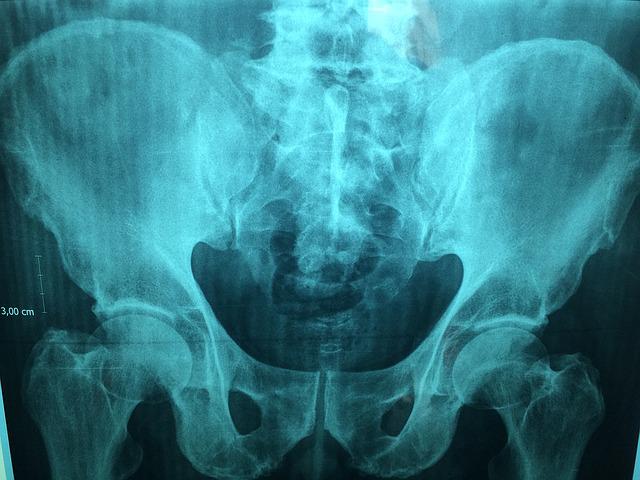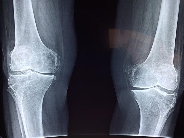
Association of American Medical Colleges states that radiologist burnout and the demographics of the workforce have been critical for years. For example, 82% of the 20,950 currently active radiologists are over 45 and over, while 53% are 55 or over. According to a recent Mayo Clinic study, radiologists ranked fifth out of 23 specialties that reported burnout rate, particularly among those screening for breast cancer. So not only are radiologists aging out, the newcomers aren’t lasting that long.
Why Are Radiologists Being Burned Out?
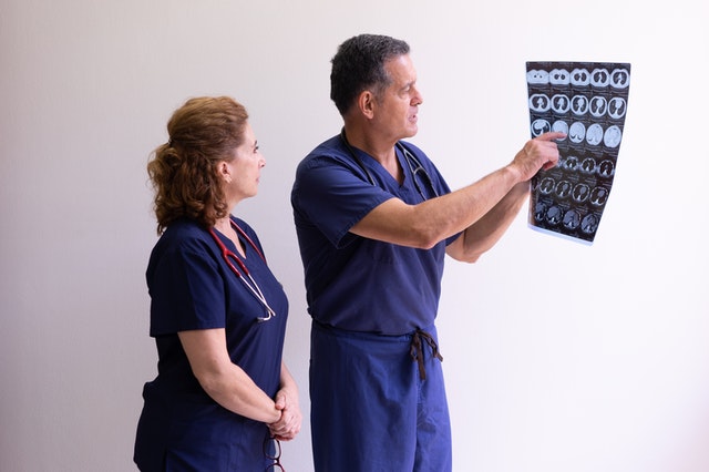
Radiology Business says radiologists mention things like “lack of respect” from administrators or colleagues as one of the leading causes of burnout 60%, along with lack of control or autonomy over one’s life, long work hours, too many non-essential tasks, and more. That tempered with COVID-related burnout and we are now facing a global shortage of radiologists and radiology trainees.

According to a Diagnostic Imaging podcast, numerous factors result in radiologist burnout, from outdated and clunky software, sinking reimbursements, continuing education requirements, stressful imaging reports turnout expectations, to reduced interaction with colleagues related to the pandemic, all resulting in a slow, laborious system death.
Should You Consider Teleradiology/Outsourced Radiology Company?
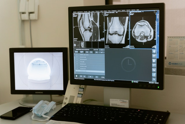
If you have never heard of teleradiology or outsourcing radiology, you may be interested in learning that many clinics, offices, and hospitals are turning to medical outsourcing. Hospitals will likely start to engage more and more outsourced services that can include long-distance radiology and teleradiology. While there are pros and cons for hospitals and clinics using teleradiology, the fact is that this may quickly become a fact of life. Patient care can still be high-quality, immediate, and efficient, for example.
There are incredible benefits that come with radiology outsourcing companies and teleradiology. Many teleradiology practices employ board-certified and trained radiologists for neurological and Body Imaging needs. These radiologists are available for patient consultations to go over results, answer any questions, and much more. Plus, these services are often quick and done promptly. Many of these companies and services are coupled with a quality assurance program to maintain their ACR accreditation. Finally, these services tend to be more cost-effective than in-house services. Teleradiology and outsourced services are often used in the following practices:
- Private practices
- Urgent care clinics
- Imaging practices
- Mobile services
- Physician groups
- Independent diagnostic testing facilities

What Does the Future of Radiology Look Like?
One popular solution that addresses the shortage of radiologists includes using a teleradiology/outsourced radiology company as mentioned above.
Another solution is using trained radiographers to double-read screening mammograms, extending their expertise and skill set to perform as radiologists when needed. Double reading has been used in Europe and has been incredibly efficient at detecting cancer. Training non-radiologists may end up becoming a necessity for hospitals and breast screening facilities without having to lose patient standards.
Radiology remains a solid and stable career, especially as medical professionals and facilities adapt to shortages, and potential pandemics, especially when considering teleradiology and outsourced radiology companies.
Vesta Teleradiology: For Full or Supportive Radiology Staffing Needs
Whether your healthcare facility need full-time support, or just coverage for nights and weekend radiology interpretations, Vesta is here for you! Vesta has dedicated 15 years to serving diagnostic imaging centers, physician’s offices, hospitals and other healthcare facilities with their radiology needs. Contact us for a free quote: 1-877-55-VESTA.