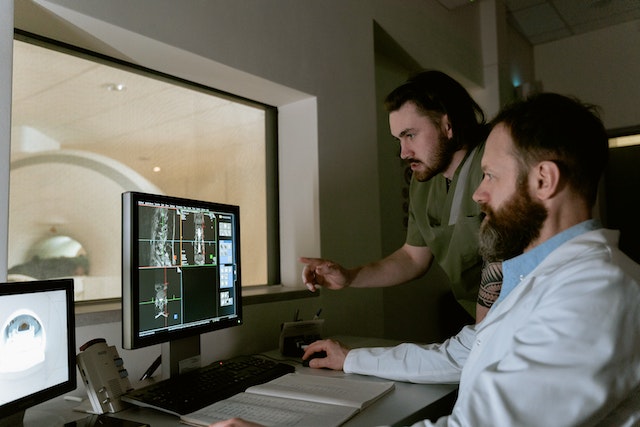
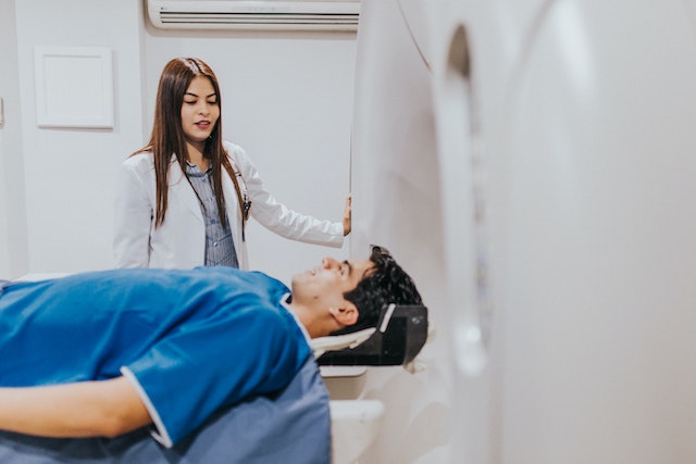
As we bid adieu to the final moments of 2023, it’s a great time to reflect on advancements and studies that have redefined the world of imaging this year. In this article, we’ll delve into the hottest news and breakthroughs in imaging, highlighting the remarkable strides that have made the headlines.
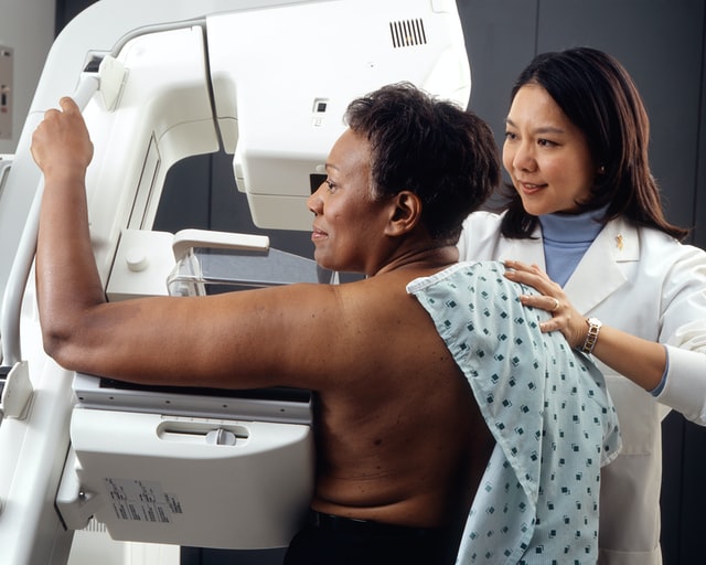
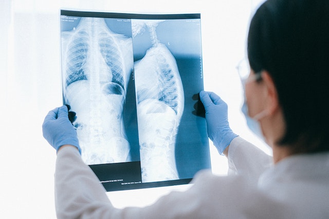
Study Suggest that Cancer Death Risk From Low-Dose Radiation Is Underestimated
A recent study featured in the British Medical Journal unveils concerning associations between extended exposure to low-dose radiation, commonly experienced by nuclear industry workers, and amplified cancer-related mortality. Drawing insights from the International Nuclear Workers Study (INWORKS) encompassing data from over 300,000 workers, researchers discovered a stark reality: for each cumulative unit of radiation exposure, the risk of death from solid cancer surged by 52%. Even at the lowest cumulative doses, this risk doubled, challenging the assumption that low-dose exposures present less carcinogenic hazard. While the absolute risk remains small, these findings prompt reconsideration of safety limits for workers and call for further studies to confirm the accelerated risk of cancer with ionizing radiation exposure. The hope is that regulatory bodies will integrate these insights into revising protection standards for individuals exposed to low-dose radiation.
Long COVID
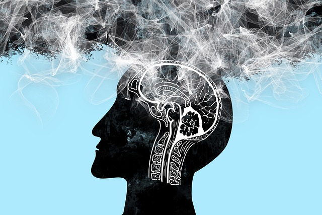
In a study published in Medical Hypotheses, a French group presented a theory regarding the brain fog experienced in long COVID, based on brain patterns identified in patient PET scans. They propose that inflammation triggered by COVID-19 disrupts astrocyte cells’ regulation of glutamate, impacting energy metabolism and leading to cognitive fatigue. The authors suggest targeting this malfunction with therapies focused on astrocytic glutamate regulation as a potential way to alleviate long-COVID neurological symptoms. They highlight the lack of mental clarity, difficulty concentrating, and cognitive strain characterizing long COVID, affecting up to 15% of patients after three months of the initial infection. This study builds on previous findings of hypometabolism patterns in long COVID patients’ brain images and explores cellular mechanisms, including links between glutamate dysregulation and cognitive fatigue from other studies. Drawing parallels with “chemo-fog” in cancer patients and cognitive impairment in Parkinson’s disease, the authors suggest therapeutic strategies targeting the identified brain patterns, citing examples from epilepsy treatments and a recent study using medication to improve cognitive function in long-COVID patients. However, the authors stress the need for further research, proposing PET imaging studies using specific markers to comprehend astrocyte function and glutamate regulation for a comprehensive understanding of long COVID’s underlying mechanisms.
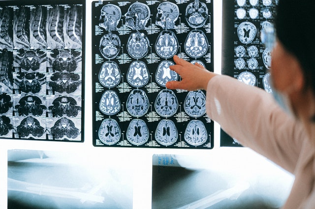
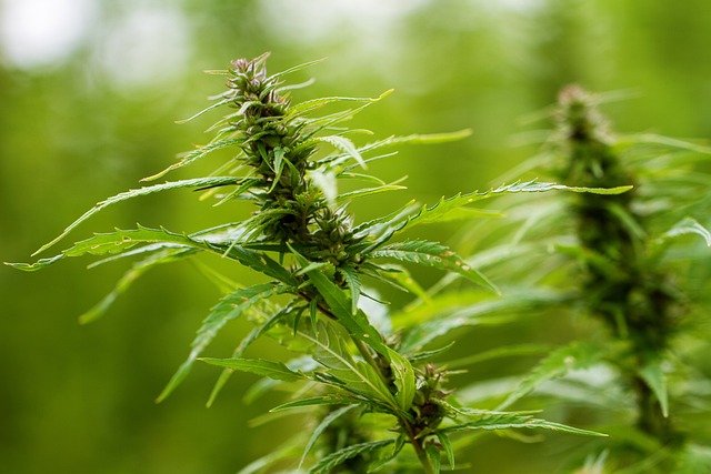
MRIs and Past Cannabis Users
At the International Society for Magnetic Resonance in Medicine (ISMRM) annual meeting, New Zealand researchers presented findings on heavy cannabis use in adolescence to early adulthood and its correlation with brain structure differences in hippocampus and amygdala subregions. The study, led by medical physicist Rebecca Lee and colleagues from the University of Otago in Christchurch, indicated volumetric disparities in these brain regions among heavy cannabis users compared to non-using controls. Notably, past cannabis users showed smaller volumes in specific hippocampal and amygdala subregions. However, the research did not find detectable differences in cerebral blood flow or white-matter tract integrity related to cannabis use, suggesting potential transient brain changes or no long-term effect on these properties. The study, conducted using MRI techniques, emphasized the need for longitudinal studies to clarify the causation and long-term functional impacts of these structural brain changes associated with heavy cannabis use. Despite revealing structural brain changes linked to cannabis use, the study does not definitively establish a causal relationship between these changes and cannabis consumption. Further prospective longitudinal MRI studies are essential to elucidate causality in this context.
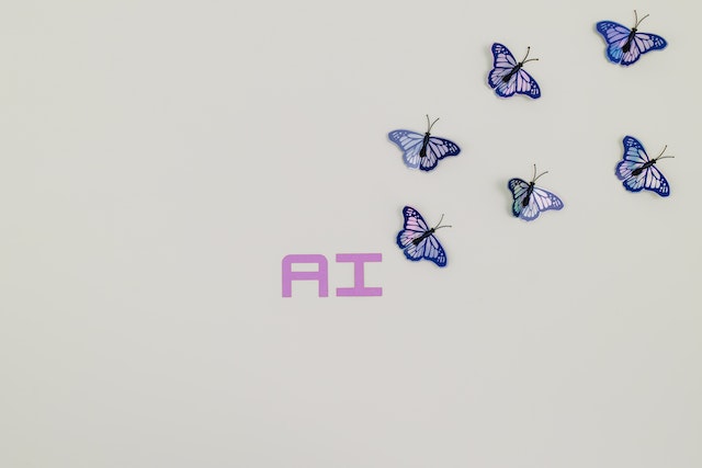
All About AI
We’d be remiss to not mention how artificial intelligence has shaped the industry this year. Check out our previous articles highlighting the impact that ChatGPT and Bard have made in 2023.
Sources:
technologynetworks.com
auntminnie.com
Openai.com


 Sources: