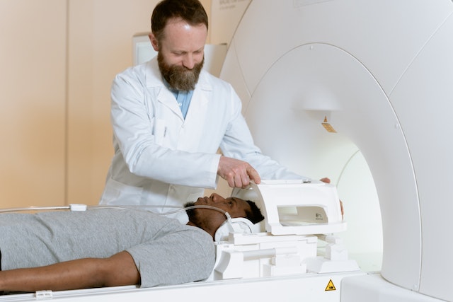
A healthcare facility – hospital, private practice, imaging center – may choose to utilize the services of a teleradiology company to enhance their radiology capabilities and streamline their diagnostic imaging processes. Teleradiology services offers numerous advantages, such as increased access to specialized radiologists, expedited turnaround times, reduced waiting time for patients, and improved patient care. By partnering with a reputable teleradiology company, healthcare facilities can leverage advanced technology, secure data transmission, and a network of skilled radiologists to obtain accurate and timely radiology preliminary and final interpretations. This collaboration allows healthcare providers to optimize their resources, extend their coverage, and ultimately deliver high-quality diagnostic imaging services to their patients, regardless of time or location.
Teleradiology Reports Delivery
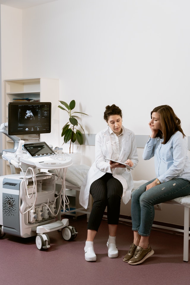
Before engaging with an outsourced radiology company, you may wonder how reports are delivered. Teleradiology specialists deliver their reports through secure electronic means, utilizing advanced technology and secure communication platforms.

When you partner with Vesta, a Gateway application is installed remotely to allow your systems direct access to our PACS. Once linked, your technicians send exams straight from your machine to our PACS. Vesta account specialists assign it to the proper physician who read and dictate the report, it’s uploaded to your branded template where it’s easily accessible for your facility to access and download!
These customized reports can also be distributed electronically, and any critical findings are immediately shared with the facility via phone, email, or text. We also facilitate callbacks between the physician and radiologist and provide an unlimited amount of customized report templates with your facility’s information and logo. It’s a seamless experience which allows you to focus on what matters most, the patient experience.
Safety and Security: HIPAA
Teleradiology companies like Vesta prioritize the security and confidentiality of patient data throughout the entire process. They employ strict data encryption, comply with privacy regulations (such as HIPAA in the United States), and implement robust security measures to protect patient information during transmission and storage.

Looking to outsource your radiology interpretations? Please reach out to Vesta to learn more. Vesta Teleradiology can accommodate any type of volume, large, medium and small.
Sources
https://www.ncbi.nlm.nih.gov
https://openai.com