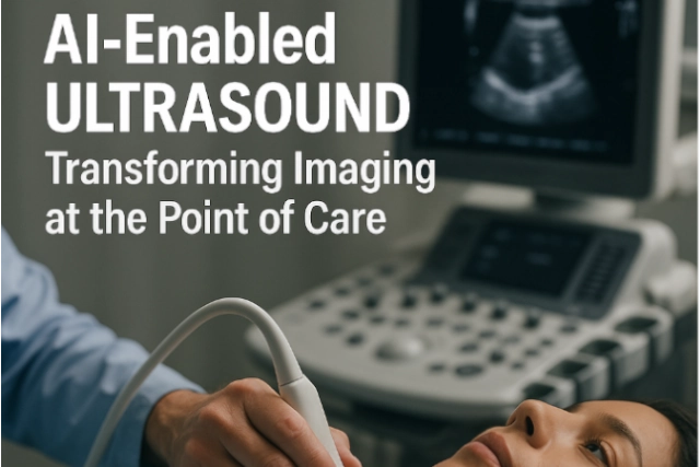
AI-Enabled Ultrasound: Transforming Imaging at the Point of Care


In today’s fast-paced healthcare environment, ultrasound is increasingly recognized not just for prenatal or cardiac assessment, but as a versatile diagnostic tool across specialties. Now, artificial intelligence (AI) is accelerating ultrasound’s impact — reducing operator dependency, improving diagnostic confidence, and enabling faster bedside care. For imaging leaders, especially in rural or underserved settings, AI-powered ultrasound technology paired with teleradiology support offers a compelling path for enhanced access and precision.

Innovations in AI-Ultrasound You Should Know

  1. FDA Clearance for AI Thyroid Ultrasound
    In 2024, See-Mode Technologies received FDA clearance for an AI-powered thyroid ultrasound system that can detect and classify nodules using the ACR TI-RADS scale. It has shown promising results in standardizing reporting and reducing unnecessary biopsies and follow-ups.
    Source: https://www.auntminnie.com
  2. Projected Market Growth
    The global AI ultrasound market is projected to grow at a compound annual growth rate (CAGR) of 22% through 2029. This rapid growth is fueled by the rising burden of chronic disease, limited radiologist availability, and the push for faster, more accessible diagnostics.

    Source: https://www.pharmiweb.com/

  3. Rural Potential with Point-of-Care AI
    A JAMA Cardiology viewpoint outlines how AI-assisted point-of-care ultrasound (POCUS) can enable more accurate cardiovascular assessments even when performed by generalists—especially valuable in remote areas without imaging specialists.
    Source: https://jamanetwork.com
  4. Clinician Enthusiasm and Challenges
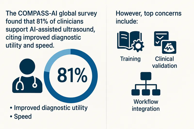
    The COMPASS-AI global survey found that 81% of clinicians support AI-assisted ultrasound, citing improved diagnostic utility and speed. However, top concerns include training, clinical validation, and workflow integration.

    Source: https://theultrasoundjournal.springeropen.com/

Infographic showing COMPASS-AI survey results on clinician support for AI-enabled ultrasound, benefits, and concernsWhy It Matters for Facilities and Radiology Teams

  • Reduces staffing burden: AI ultrasound reduces variability among operators, ideal for high-turnover or remote settings.
  • Speeds up decision-making: Frontline providers can quickly gather meaningful imaging data, while teleradiologists handle the interpretation.
  • Expands imaging reach: Portable, AI-powered ultrasound extends diagnostic capabilities to underserved regions.
  • Supports standardization: AI helps standardize image acquisition and reporting, improving overall workflow efficiency.

How Vesta Teleradiology Enhances AI-Ultrasound Value

While AI augments imaging workflows, expert interpretation is still essential. Vesta provides:

  • Subspecialty reads across thyroid, vascular, MSK, and more
  • 24/7 coverage with fast turnaround times
  • Seamless PACS/RIS integration for AI-acquired ultrasound data

Our radiologists help bridge the gap between frontline imaging and specialist analysis—ensuring that every AI-enabled ultrasound scan contributes to timely, confident patient care.

Bringing AI and Teleradiology Together

Whether you’re running a rural health center, a large outpatient clinic, or an emergency department, AI ultrasound paired with expert teleradiology interpretation helps:

  • Increase imaging access without compromising accuracy
  • Alleviate staffing constraints
  • Deliver faster diagnoses
  • Improve patient outcomes

AI in ultrasound is not replacing radiologists — it’s helping them focus on what matters most. With Vesta’s support, healthcare organizations can embrace innovation while maintaining high-quality, consistent imaging interpretation.


Photon-Counting CT: What Healthcare Facilities Need to Know Now

Photon-counting computed tomography (PCCT) is one of the most exciting breakthroughs in diagnostic imaging technology in recent years. Offering greater spatial resolution, reduced radiation dose, and improved tissue characterization, PCCT is quickly gaining attention from radiologists, imaging directors, and healthcare systems looking to stay ahead.

As the healthcare landscape evolves, staying informed about how new imaging technologies integrate with workflows and diagnostic goals is critical. Here’s what facilities need to know now about photon-counting CT—and how teleradiology can help maximize its impact.

What Is Photon-Counting CT?

Unlike conventional CT, which measures the total X-ray energy reaching the detector, photon-counting CT counts individual photons and measures their energy levels. This allows for:

  • Sharper images with better spatial resolution
  • Lower noise, especially in soft tissue
  • Multi-energy imaging from a single scan
  • Reduced radiation exposure

Siemens Healthineers introduced the first FDA-approved photon-counting CT system (NAEOTOM Alpha) in 2021, and adoption has slowly grown among academic and high-volume centers.

Clinical Benefits of PCCT

Photon-counting CT provides enhanced detail for a range of applications, including:

  • Cardiac imaging: Better visualization of stents and plaques
  • Pulmonary imaging: Improved nodule detection and perfusion data
  • Neuroimaging: Greater contrast at lower doses for brain scans
  • MSK imaging: Superior resolution for joint, bone, and soft tissue analysis

The ability to perform multi-energy imaging without dual-source CT equipment allows radiologists to generate virtual non-contrast images, improve lesion characterization, and reduce contrast agent use—benefiting both patients and providers.

Multi-energy CT image showing high-resolution internal anatomy used for virtual non-contrast imaging
Growing Market and Adoption

While still early in widespread adoption, the global photon-counting CT market is projected to grow rapidly. According to a recent report from Research and Markets, the global PCCT market is expected to reach over $800 million by 2030, driven by increasing demand for advanced diagnostic tools and a growing focus on radiation dose reduction.

As more vendors develop photon-counting detectors and more clinical use cases are validated, experts anticipate broader adoption beyond academic centers and into regional hospitals and imaging centers.

Source: Research and Markets, “Photon Counting CT Market – Forecast 2030”

How Teleradiology Supports Advanced CT Adoption

Deploying a photon-counting CT system requires more than just the hardware. Facilities must ensure they have access to radiologists who are:

  • Trained in multi-energy CT interpretation
  • Familiar with new artifact patterns and reconstructions
  • Able to optimize clinical workflows using new scan data types

That’s where teleradiology plays a critical role.

At Vesta Teleradiology, our radiologists stay at the forefront of imaging advances. With experience in multi-energy and advanced CT post-processing, we help facilities take full advantage of what photon-counting CT offers—delivering fast, accurate interpretations backed by subspecialty insight.

Integration and Workflow Considerations

Facilities considering photon-counting CT should think about:

  • PACS/RIS compatibility with new data formats
  • Training staff to understand and use spectral data
  • Building protocols for when and how to use PCCT scans
  • Collaborating with teleradiology teams for consistent interpretations

While the learning curve is real, the payoff is significant. Early adopters report better diagnostic confidence, fewer repeat scans, and more comprehensive patient evaluations.

Conclusion: Prepare for the Future of CT Imaging

Photon-counting CT represents the next leap in diagnostic precision. As this technology becomes more accessible, imaging leaders must evaluate how it fits into their long-term strategy. For facilities looking to stay competitive, offer premium diagnostics, and improve patient care, PCCT should be on the radar now—not later.

Partnering with a forward-thinking teleradiology provider like Vesta ensures you’re equipped with the expertise to unlock its full potential.


Celebrating National Health Center Week: The Frontline of Community Care

Every August, National Health Center Week (August 3-9 2025) recognizes the critical role community health centers play in delivering affordable, high-quality healthcare across the United States. These centers serve more than 30 million patients annually, many of whom live in medically underserved or rural regions. But as demand for comprehensive care grows, so does the need for accessible diagnostic imaging—an area where teleradiology is helping bridge the gap.

The Imaging Gap in Rural and Underserved Areas

Access to diagnostic imaging remains a persistent challenge for many community health centers. Facilities in rural or low-resource areas often face:

  • Limited access to on-site radiologists
  • Delays in turnaround times for imaging reads
  • Difficulty recruiting or retaining subspecialty radiologists
  • Rising imaging volumes due to expanded preventive care

These barriers can compromise patient outcomes, especially in time-sensitive cases involving stroke, cancer screening, or trauma. Imaging is a critical step in diagnosis—and delays in radiology reports can delay treatment.

Teleradiology: A Scalable Solution for Imaging Access

Teleradiology enables healthcare facilities to send medical images (like X-rays, CT scans, MRIs, and mammograms) electronically to off-site, board-certified radiologists for interpretation. For community health centers, this technology is transformative.

Here’s how teleradiology supports health centers during National Health Center Week and year-round:

  1. 24/7 Coverage, Including Nights and Holidays
    Teleradiology ensures that community health centers can offer imaging services around the clock—even if there’s no radiologist physically on-site. This is especially important for urgent care and emergency settings in rural hospitals.

  2. Access to Subspecialty Reads
    Facilities may not always have access to neuroradiologists, MSK radiologists, or breast imaging specialists. Vesta Teleradiology offers access to subspecialty reads, ensuring every case is interpreted by the right expert.

  3. Faster Turnaround Times
    With cloud-based image transfer and structured reporting, teleradiology reduces delays and improves turnaround times. That means faster results, quicker clinical decisions, and better patient care.

  4. Support for Preventive Imaging Initiatives
    Community health centers are expanding their use of imaging for preventive care—particularly for breast cancer screening, lung health, and cardiovascular risk. Teleradiology provides scalable support during screening campaigns or high-volume periods.

    Female patient undergoing a mammogram with a radiologic technologist in a medical exam room

  5. Cost-Effective Radiology Staffing
    Teleradiology helps optimize budgets by supplementing in-house radiologists or replacing expensive on-call coverage. Flexible pricing models ensure services align with facility needs and patient volume.

Why Imaging Access Matters More Than Ever

The need for diagnostic imaging continues to rise in 2025. According to recent projections from the Harvey L. Neiman Health Policy Institute, demand for imaging will grow at a faster rate than the radiologist workforce through 2055. In rural and medically underserved areas, the shortage is even more pronounced.

Community health centers are on the front lines of closing this gap. But without reliable imaging access, they face limitations in diagnosis, monitoring, and treatment planning.

How Vesta Teleradiology Helps Health Centers Thrive

At Vesta, we understand the pressures community health centers face. That’s why we offer:

  • Fully customizable radiology services tailored to your patient population
  • Rapid onboarding and seamless PACS integration
  • Weekend, holiday, and night coverage
  • A team of U.S.-based, board-certified radiologists
  • Subspecialty interpretations across all major imaging fields

Whether you’re a rural clinic needing full radiology coverage or a mid-sized health center looking for overflow support, our teleradiology solutions are built to help you scale—without compromising care quality.

Join the Movement: National Health Center Week

National Health Center Week is more than a celebration. It’s a reminder that access, equity, and quality care start with supporting the providers who serve our most vulnerable populations. Teleradiology is a powerful tool to help meet that mission.

If your health center is planning to expand imaging services or looking for reliable radiology coverage, Vesta is here to help.

Let’s build healthier communities—one accurate read at a time.


Supporting Women’s Health with Subspecialty Teleradiology: National Women’s Health Week 2025

Each May, National Women’s Health Week serves as a reminder of the importance of preventive care, early detection, and access to high-quality medical services for women across the country. Among these essential services, breast imaging stands out as a cornerstone of women’s health — and timely, accurate interpretation of mammograms plays a vital role in early detection of breast cancer.

But what happens when a facility doesn’t have immediate access to a subspecialty-trained breast radiologist?

That’s where teleradiology steps in.

At Vesta Teleradiology, we support women’s health initiatives year-round by providing reliable, fast, and compliant mammography interpretations, especially for facilities that may not have in-house specialists available.

The Need for Expert Mammography Interpretation

According to the CDC, breast cancer is the second most common cancer among women in the U.S., and regular mammograms are the best way to detect breast cancer early, when it’s easier to treat and before symptoms appear1. The American College of Radiology (ACR) also notes that interpretation by radiologists trained in breast imaging can improve detection rates and reduce false positives2.

However, many imaging centers and rural hospitals don’t have a dedicated breast radiologist on-site — and delays in interpretation can lead to gaps in care or unnecessary anxiety for patients.

Female patient undergoing a mammogram with a radiologic technologist in a medical exam roomThis challenge is magnified by a growing shortage of radiologists, particularly those specializing in breast imaging. A 2023 workforce survey from the Association of American Medical Colleges (AAMC) highlighted that more than 50% of practicing radiologists are over the age of 55, and retirements are outpacing new entrants. Breast imaging — already a subspecialty with fewer practitioners — is feeling the strain. Many facilities are facing longer turnaround times or are unable to offer advanced imaging interpretation consistently.

In addition, burnout remains a real concern. Breast radiologists face high volumes and frequent callbacks, which can affect accuracy and job satisfaction. Teleradiology can help balance the workload by offering overflow and relief coverage, supporting both the health system and the radiologists themselves.

How Teleradiology Closes the Gap

Vesta Teleradiology provides healthcare facilities with remote access to subspecialty-trained radiologists, including experts in breast imaging. This allows imaging centers, OB/GYN clinics, and hospitals to meet women’s health needs without overextending in-house teams.

Here’s how we help:

  • Full MQSA-compliant interpretations
  • Support for both screening and diagnostic mammograms
  • 24/7/365 availability, including STAT and overflow reads
  • Subspecialty reads in breast MRI, ultrasound, and 3D mammography (tomosynthesis)
  • Seamless PACS integration and secure data exchange

Whether your site needs full-time coverage or help during vacation season, we ensure that your patients receive timely, high-quality reads.

National Women’s Health Week Is the Perfect Time to Prioritize Imaging Readiness

National Women’s Health Week 2025 runs from May 12–18 and encourages women to schedule important preventive screenings — including mammograms.

Facilities should be prepared for increased volume during this time and throughout Breast Cancer Awareness campaigns later in the year (October). Having a trusted teleradiology partner means you can handle increased demand without sacrificing quality or turnaround times.

Vesta Teleradiology: Your Partner in Women’s Imaging

At Vesta, we believe in supporting facilities that support women. Our flexible coverage options and experienced radiologists help ensure that women’s health screenings — including mammograms — are interpreted accurately, securely, and quickly.

Whether you’re preparing for Women’s Health Week or looking for year-round coverage, we’re here to help you deliver the care your patients deserve.

Let’s improve access, together. Contact us to learn more about how Vesta can support your women’s imaging services.


New CMS-Approved MRI Standards: Enhancing Safety in Remote Scanning and Portable Imaging

The Centers for Medicare & Medicaid Services (CMS) has approved new MRI standards introduced by the Intersocietal Accreditation Commission (IAC), focusing on remote scanning and portable imaging technologies. These updates, effective immediately, aim to enhance patient safety and adapt to advancements in MRI practices.

Key Updates in MRI Standards

Remote Scanning Protocols: The revised standards mandate that a registered technologist must always be present with the patient during remote MRI scans. This ensures immediate care availability if needed. Facilities are also required to implement policies addressing potential challenges such as equipment or communication failures, internet instability, and power outages.

Portable MRI Scanning: The IAC has introduced standards for portable MRI technology, distinguishing it from mobile MRI units. This inclusion acknowledges the growing use of portable MRI devices in various healthcare settings and emphasizes the need for specific guidelines to ensure their safe and effective operation.

Contrast Administration and Supervision: Recognizing the challenges in meeting physician supervision requirements for contrast injections, the IAC has revised its policies to ensure a safe environment for patients. The new standards emphasize the presence of appropriately trained nonphysician personnel during contrast administration.


Implications for Healthcare Providers

These updates reflect the IAC’s commitment to quality improvement and patient safety in MRI services. Facilities must comply with the new standards to maintain accreditation, which may involve updating protocols, training staff, and investing in new technologies. The emphasis on remote scanning and portable MRI acknowledges the evolving landscape of medical imaging and the need for standards that keep pace with technological advancements.

Industry Response

The introduction of these standards has been met with support from industry stakeholders. For instance, Hyperfine, a manufacturer of portable MRI devices, noted that the new guidelines pave the way for their Swoop® Portable MR Imaging® system to be available in neurology offices and clinics. This development enables physicians to obtain diagnostic-quality MR brain images within their clinics, providing patients with timely and convenient MRI access at the point of care.


Conclusion

The CMS-approved MRI standards introduced by the IAC represent a significant step forward in ensuring patient safety and adapting to technological advancements in medical imaging. Healthcare providers are encouraged to familiarize themselves with these updates and implement the necessary changes to comply with the new accreditation requirements. As the medical imaging landscape continues to evolve, such proactive measures are essential to maintain high standards of care and patient safety.


Sources:
radiologybusiness.com
auntminnie.com
openai.com


MRI Explosions and Safety Measures Your Facility Needs to Make

An explosion occurred in the MRI suite at Pietersburg Provincial Hospital in Limpopo, South Africa, injuring three individuals: two hospital employees and a technician from a private service provider. The explosion happened while the technician was decommissioning the MRI machine, leading to moderate injuries and significant damage to the radiology department, which has temporarily suspended services. MRI safety expert Tobias Gilk explained that such incidents, though rare, can occur during the servicing or de-installation of MRI scanners due to pressure build-up from liquid helium turning into gas. The exact cause of the explosion is still under investigation, and patients requiring radiology services are being redirected to alternative facilities.

How Common are MRI Machine Explosions?

MRI machine explosions are extremely rare but not unheard of. These incidents typically occur during the servicing, decommissioning, or de-installation of the machines. The main risk comes from the liquid helium used to cool the MRI magnets. If the helium warms up even slightly, it can expand rapidly as it turns from liquid to gas, increasing pressure inside the machine. If this expanding gas is trapped, it can lead to an explosion, often referred to as a “quench-plosion.”

While MRI machines are designed with safety mechanisms to prevent such events, errors during maintenance or de-installation can sometimes lead to these accidents. However, because of strict safety protocols and the inherent design of MRI systems, these explosions are considered very uncommon.

In November of 2023, another significant incident occurred at Kaiser Permanente’s Redwood City Medical Center in California. While not an explosion, this accident involved a nurse being crushed by a bed pulled into an MRI machine due to its strong magnetic force. This highlights the potential dangers associated with MRI machines, even though such accidents remain uncommon​

Hospitals and facilities can take several precautions to prevent MRI-related accidents, including explosions or other incidents involving the powerful magnetic field. Check out the ACR Manual on MR Safety for more details.


Comprehensive Safety Training: All MRI staff should undergo detailed training on MRI safety, focusing on understanding the dangers associated with the magnetic field, proper patient and equipment screening, and emergency procedures. Regular training refreshers and certifications help maintain a high level of safety awareness.


Strict Screening Protocols: Implementing rigorous screening for ferromagnetic materials is crucial. This includes ensuring that no metal objects, such as gurneys, tools, or even some medical implants, enter the MRI suite. Facilities can use ferromagnetic detectors to enhance this process.


Proper Maintenance and Decommissioning: When servicing or decommissioning MRI machines, it is vital to follow manufacturer guidelines carefully. This includes safely managing liquid helium, which cools the MRI magnets, to prevent pressure build-up that could lead to an explosion.

Emergency Quench Procedures: Facilities should have clear protocols for initiating a quench, which rapidly shuts down the magnetic field in an emergency. Staff should be trained on when and how to use this feature.

Monitoring Systems: Installing monitoring systems and alarms that detect abnormal conditions, such as excessive pressure within the machine, can provide early warnings and prevent dangerous situations.

Restricted Access: Limiting access to the MRI suite to trained personnel only, with clear signage warning of the magnetic field’s dangers, is another key precaution.

By implementing these precautions, hospitals can significantly reduce the risk of MRI-related accidents, ensuring the safety of both patients and staff.


Sources:
independent.co.uk
acr.org
healthimaging.com
openai.com

Growth in Demand for Imaging Procedures Will Increase Need for Teleradiology

The healthcare market is experiencing a shift towards outpatient care, driven by reimbursement changes, pandemic effects, and patient preferences, particularly prominent in the U.S. Providers are diversifying into sub-specializations like neurology and oncology, raising the demand for advanced imaging like MRI and CT. This trend has led to increased utilization of outpatient imaging and teleradiology services.

By the NuMbers

Diagnostic imaging is becoming increasingly crucial in healthcare, with the market projected to reach $31.9bn in 2023 and grow at a 4.8% CAGR to $45.8bn in 2030. The rise is driven by chronic diseases, an aging population, and post-Covid-19 demand recovery. To meet this demand, companies are focusing on advanced and accessible technologies, such as handheld ultrasounds. About 1,949 imaging devices are in development, with 112 expected to gain approval in 2023.

imaging device

According to Fortune Business Insights, in 2022, the computed tomography (CT) segment held the largest market share due to a rise in CT scan procedures and higher average pricing. For instance, OECD data for 2021 showed 84.5 million CT scan procedures in the U.S., up by 15.8% from the previous year. The growing geriatric population has also contributed to increased demand for CT scans.

Key Players

Key players include GE Healthcare, Philips, Siemens Healthineers, and more. Challenges include high equipment costs and a shortage of skilled personnel, impacting accessibility and patient care quality. Opportunities arise from the growing demand for imaging services, especially for chronic diseases, and the development of new modalities like 3D mammography and MRI-guided focused ultrasound. Emerging economies like India, China, and Brazil are pivotal, driven by rising chronic diseases. Major players like Siemens Healthineers are expanding in these markets. Additionally, teaching hospitals are increasing demand for advanced imaging methods to enhance patient care.

Your Dedicated Radiology Partner: Vesta

Partnering with Vesta as your radiology partner ensures access to accurate and timely imaging interpretations and readings for subspecialties. Whether you are an outpatient imaging center or traditional hospital, our collaboration offers a seamless and efficient experience. Trust us to be your reliable radiology partner, empowering you with the insights and tools needed for improved healthcare outcomes.


radiology peer reviewSources:

Itnonline.com
Medicaldevices-network.com
fortunebusinessinsights.com
openai.com


How Does Working With a Teleradiology Company Enhance Diagnostic Accuracy?

A teleradiology company is a healthcare organization that provides remote radiology services using telecommunication technology. It connects radiologists with healthcare facilities, allowing them to interpret and analyze medical images remotely, regardless of geographical locations. These companies facilitate the transmission of medical images and reports, enabling timely diagnoses, expert consultations, and improved access to radiology services.

Working with a teleradiology company can enhance diagnostic accuracy in several ways

Access to Specialized Radiologists: Teleradiology companies often have a network of highly specialized radiologists who can provide expertise in specific areas, such as musculoskeletal radiology, neuroradiology, or pediatric radiology. This specialization allows for more accurate and precise interpretations of complex cases.

Peer Review and Collaboration

Teleradiology platforms facilitate peer review and collaboration among radiologists. This enables multiple experts to review and discuss challenging cases, leading to a more accurate diagnosis. Radiologists can consult with their peers, share knowledge, and seek second opinions, which can significantly enhance diagnostic accuracy.

Reduced Turnaround Time

Teleradiology companies typically offer quick turnaround times for reporting. Faster access to radiology reports means timely diagnoses, enabling prompt patient management decisions. Rapid reporting reduces the chances of missed diagnoses or delayed treatment, improving overall diagnostic accuracy.

24/7 Coverage

Teleradiology services often provide round-the-clock coverage, allowing healthcare facilities to access radiology expertise at any time, including nights, weekends, and holidays. This availability ensures that critical cases receive immediate attention, leading to accurate and timely diagnoses.

day and night coverage

Subspecialty Interpretations

Teleradiology companies can match specific cases with radiologists who specialize in the relevant subspecialties. This ensures that complex or rare cases are interpreted by radiologists with extensive experience and knowledge in those areas. Subspecialty interpretations enhance diagnostic accuracy by reducing the likelihood of misdiagnosis or overlooking important findings.

Quality Assurance Programs

Teleradiology companies usually have robust quality assurance programs in place. These programs include regular audits, peer reviews, and adherence to strict reporting standards. By maintaining high-quality standards, teleradiology companies ensure accurate and reliable interpretations.

Advanced Technology and Tools

Teleradiology companies often employ advanced technologies and tools to enhance diagnostic accuracy. These include computer-aided detection (CAD) systems, advanced visualization software, and access to a comprehensive database of previous imaging studies for comparison. These tools assist radiologists in identifying subtle abnormalities, improving diagnostic accuracy. Another benefit is that teleradiology offers is the ability for the radiologist to manipulate the images in ways that cannot be achieved solely with film. As a result, it allows for the extraction of additional clinically significant information from the images and leads to more precise diagnoses and clinical decisions.


Vesta Teleradiology: Over 16 Years of Excellence

By leveraging the expertise of specialized board certified radiologists, promoting collaboration, offering fast turnaround times, providing subspecialty interpretations, implementing quality assurance programs, and utilizing advanced technology, teleradiology companies like Vesta can significantly enhance diagnostic accuracy and contribute to improved patient care.

Sources

Ncbi.nlm.nih.gov
Sciencedirect.com
Openai.com

How is Teleradiology and AI Impacting the Medical Industry Today?

Artificial Intelligence (AI) is revolutionizing the medical industry, transforming the way healthcare is delivered, diagnosed, and managed. With its ability to analyze vast amounts of data quickly and accurately, AI is reshaping various aspects of healthcare. From aiding in disease diagnosis to personalized treatment recommendations, AI is enhancing the precision and efficiency of medical practices. Moreover, AI-powered technologies are streamlining administrative tasks, optimizing resource allocation, and improving patient outcomes. As AI continues to advance, it holds immense potential to revolutionize healthcare delivery, foster medical innovations, and ultimately improve the quality of patient care on a global scale.

Teleradiology has had a profound impact on healthcare by enabling remote access to radiology expertise, bridging geographical barriers, and ensuring timely diagnoses. It has improved patient care by providing faster turnaround times, facilitating collaboration among radiologists, and increasing access to specialized interpretations, ultimately enhancing diagnostic accuracy and treatment outcomes. Going even further, a latest white paper from One Call describes how teleradiology and AI are helping reduce the strain of the radiology shortage.

artificial intelligence

Teleradiology and AI in Action

Medical imaging vendor, Nanox, is looking to address heath disparities and lack of access care with a new x-ray system which would be offered to countries in Africa, Asian and South American using a pay-per-scan model. The potential of combining cold cathode X-ray technology with teleradiology and artificial intelligence (AI) to enhance diagnostic capabilities and improve healthcare economics. Cold cathode X-ray systems offer advantages such as reduced energy consumption and improved image quality. When integrated with teleradiology, these systems can enable remote interpretation of X-rays, leading to faster diagnoses and improved patient care. Additionally, the use of AI algorithms in conjunction with cold cathode X-ray technology has the potential to enhance image analysis, automate certain tasks, and optimize resource allocation, offering cost-saving opportunities in healthcare settings.

diagnostic imaging
A teleradiologist examines a chest x-ray

There are plans to roll out AI-powered teleradiology by the “Screen for Life” program at the Primary Health Care Corporation in Qatar, aimed at early detection and prevention of cancer in the United Arab Emirates. The program plans to utilize AI algorithms to analyze radiology images, enhancing the accuracy and efficiency of cancer screening. The integration of AI in teleradiology will help automate image interpretation, expedite diagnoses, and reduce the workload on radiologists. The implementation of AI teleradiology in the “Screen for Life” program is expected to improve cancer detection rates, streamline healthcare processes, and ultimately save lives by identifying cancers at earlier stages.

Vesta Teleradiology

Looking to outsource your radiology interpretations using an expert Teleradiology company that is at the forefront of technology including AI?  Please reach out to Vesta to learn more. Vesta Teleradiology can accommodate any type of volume, large, medium and small.

Sources:

Radiologybusiness.com
menafn.com
openai.com
cdc.gov

Practice Management: How to Soothe Patient Concerns over Diagnostic Imaging

Being told that you need diagnostic imaging can be scary. Healthcare professionals deal with diagnostic imaging every day, so they may become immune to the concerns of patients.

Still, amidst patients’ fear of diagnostic imaging, healthcare professionals can set their minds at ease.

The main thing to remember is to simply treat your patients like humans. Slow down, treat them with kindness, and really listen to their concerns. Doing these simple things can go a long way in setting your patients’ minds at ease.

What Concerns Do Patients Have About Diagnostic Imaging?

One of the most common concerns amongst patients is the claustrophobia caused by MRI machines. Dealing with the tight space of an MRI machine can be very stressful for patients dealing with claustrophobia and anxiety.

Regarding X-rays, patients are often concerned about their radiation exposure. They fear that the imaging may put them at increased risk for cancer.

Treat Your Patient Like a Person

One of the most common complaints from patients is that they don’t feel like the person working with them is treating them with care and understanding.


talking to your patients
Address the patients concerns

Smile and say hello when you first enter the room. Something as small as a smile can instantly put a person’s mind at ease, especially in a clinical setting that is scary for many patients.

Always refer to the patient by their name. Never refer to them as “the patient” while they are in earshot. Doing so sounds cold, while using their name makes the interaction feel more personal.

Maintain eye contact with your patient. Don’t look at their chart so much that you forget to make them feel like they’re being heard. Eye contact can make them less anxious, especially if they’ve been waiting a long time or have specific fears.

Make sure you sit — standing over a seated patient can be intimidating.

Listen & Understand

Health Management emphasizes how important it is to listen to your patient and ask questions. Let them explain what is going on before you begin to speak. Don’t interrupt them. If they have something to add while you’re talking, allow them to interject and listen to what they say.

Giving your patient space to speak can clarify any confusion on both parts.

After they have finished, reiterate what they’ve said to ensure you understand their concerns. Doing so shows the patient that you are interested in helping them and have respect for their situation. It also ensures that both the patient and healthcare provider are on the same page with treatment going forward.

Also, remember to slow down. Many patients feel that healthcare providers are in a rush, that they’re wasting their time, and that the provider would rather be elsewhere. Patients deserve to feel that their time is valued. Slowing down also helps patients to feel like they are heard and understood.

Watch Your Tone

Patients often come to healthcare providers with sensitive information that can make them feel vulnerable. Health Management encourages professionals to speak to patients with a warm, calm tone of voice. It can do a lot to set their mind at ease. Speaking more slowly can do the same.

Educate Your Patients

Lawrence T. Dauer et al. say professionals should educate their patients. They should know exactly what’s happening during the imaging and what effects the imaging may have on their bodies.

For example, many patients fear how much radiation they’re getting. Explain that they are receiving very little radiation exposure.

Don’t lie to them. Professionals know that repeated exposure, a person’s age, and other factors may increase risk. Patients deserve to know that, but you can set their minds at ease by explaining their risks.

Patients should always have informed consent. They should know precisely what they’re going into and be okay with it. Consent is not just about getting the patient to sign a form. It ensures they are adequately educated about their procedure and entirely on board.

Be Clear

As a healthcare professional, you know a lot of medical and technical jargon that the patient likely does not. Speak straightforwardly, using familiar words. It is crucial that the patient understands what is going on with their treatment.

At the same time, Health Management warns you to be careful not to come across as patronizing. Although patients may not know medical jargon, it doesn’t mean they’re stupid.


Reducing MRI Claustrophobia Concerns

The University of Virginia says the main reason patients are fearful of MRI machines is because they lack understanding of them. Many professionals can ease patients’ minds by informing them that MRI machines are well-lit and open at both ends. They are not closed off and dark, as many patients fear.


patient anxiety
Understand some people may have claustrophobia


Professionals can teach their patients easy breathing and meditation techniques to help keep them calm during the procedure. Another option is to tell them to count to keep their minds busy or to go to their “happy place.”

Provide them with a towel or washcloth they can drape over their eyes so they can’t see what’s going on.

Provide patients with headphones and allow them to listen to their music of choice.

Talk to your patients — about anything — get their minds off the procedure.

Conclusion

Diagnostic imaging can be scary for patients, especially those dealing with anxiety. Medical professionals are critical in setting a patient’s mind at ease, and it’s not hard to do.

Slow down, treat your patients with kindness and respect, and ensure they are properly informed. These simple things can help calm your patients and help you build a lasting professional relationship.