The Latest in Brain Imaging News

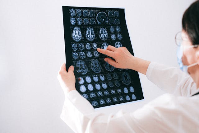
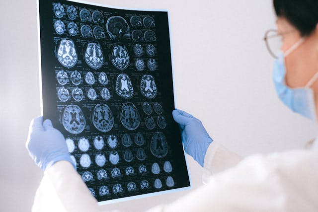
In recent years, awareness surrounding brain injuries has steadily risen, prompting significant strides in diagnostic technologies and treatment modalities. As we delve into the latest developments in this critical area of healthcare, it becomes increasingly apparent that advancements in medical imaging, particularly in the realm of neurological disorders, are poised to revolutionize the landscape of brain injury diagnosis and management.


AI-based Quantitative Brain Imaging System

Philips and Synthetic MR have joined forces to advance the diagnosis of neurological disorders through cutting-edge quantitative brain imaging tools. Their collaboration introduces the Smart Quant Neuro 3D MRI software suite, combining Philips’ SmartSpeed image-reconstruction technology, the 3D SyntAc clinical application, and SyntheticMR’s SyMRI NEURO 3D software. This innovation employs AI to analyze brain tissues, enhancing the detection and analysis of conditions like multiple sclerosis, traumatic brain injuries, and dementia.

The rise of AI in diagnostic imaging, projected to reach $1.2bn by 2027, signifies a transformative shift in improving accuracy and patient outcomes. With the diagnostic imaging market expected to grow to $9.1bn by 2030, fueled by demand for early disease diagnosis and personalized medicine, this partnership underscores the crucial role of AI in enhancing medical imaging.

Read the press release here.


A New Way of Diagnosing Mild TBIs

Researchers have developed a novel brain imaging method to diagnose mild traumatic brain injuries (mTBIs), which are often missed by standard techniques like MRI. This method involves loading gadolinium, a common MRI contrast agent, into micropatches attached to immune cells called macrophages. These cells migrate to areas of brain inflammation caused by mTBIs, enabling MRI detection. The technique, called M-GLAMs, was successfully tested in mice and pigs, showing promise for accurately diagnosing mTBIs. It also allows imaging at lower gadolinium doses, potentially benefiting patients with kidney issues. While unable to pinpoint injury locations, M-GLAMs could aid in identifying and treating brain inflammation. The researchers aim to bring this technology to clinical trials, with support from grants and intellectual property protection.

Read the study here.

tbi

New Imaging Tech that Captures Neuronal Activity Across the Brain During Recovery

Researchers at Tufts University School of Medicine have developed a novel imaging technology to monitor neuronal activity throughout the entire brain during the initial weeks of recovery from traumatic brain injury (TBI). Their study, published in Cerebral Cortex, reveals that TBI can induce changes in brain function beyond the injury site. Using a combination of fluorescent sensors and electrodes, they observed altered connectivity patterns in mice post-injury, even in regions distant from the impact. Despite the mice’s ability to perform physical tasks normally, their brain activity during both exercise and rest differed significantly from healthy brains. This impaired ability to switch between states suggests underlying brain state dysfunction post-injury. The findings highlight the brain’s plasticity in response to injury and have potential clinical implications for understanding TBI impacts and tailoring treatments. The researchers aim to further investigate long-term neural activity changes post-recovery and explore the technology’s potential in predicting specific dysfunctions or long-term outcomes of TBI. 

Read the study here.


Sources:

Medicaldevice-network.com
Otd.harvard.edu
Scitechdaily.com
Openai.com


How to Pick the Best Teleradiology Company

As the demand for healthcare services continues to surge and the shortage of healthcare workers persists, particularly in specialized fields, such as radiology, hospitals and healthcare centers find themselves facing the challenge of ensuring timely and accurate interpretations of medical imaging studies. The critical role of radiologists in diagnosing illnesses and guiding treatment decisions underscores the urgency of addressing this shortage. In response, many institutions are turning to teleradiology companies to bridge the gap and provide remote interpretation services. However, selecting the right teleradiology company is paramount to ensure high-quality patient care and seamless integration into existing workflows. In this discussion, we will explore the criteria for choosing a reputable teleradiology company, considering factors such as expertise, technology infrastructure, turnaround time, and adherence to regulatory standards. By making informed decisions in this regard, healthcare facilities can optimize their radiology services and meet the needs of patients efficiently.

remote radiology company
Rad tech and radiologist

Checklist for Choosing a Teleradiology Partner

Before selecting a teleradiology company, healthcare providers should consider several key factors to ensure they choose a partner that meets their needs and maintains high standards of service. Here are some important considerations:

  1. Quality and Expertise: Assess the qualifications and experience of the radiologists employed by the teleradiology company. Look for board-certified radiologists with expertise in relevant subspecialties. A recent survey of 2,749 radiologists from 108 countries reveals that while they read across almost five subspecialties daily, many lack confidence in certain areas. About 40% accept studies across all specialties, but less than half feel “very confident” in their current subspecialty, so it is vital to ensure the radiologists you work with have expertise in what you require.
  2. Technology and Infrastructure: Evaluate the teleradiology company’s technology infrastructure, including the software used for image transmission and reporting. Compatibility with existing systems and the ability to securely transmit images while maintaining patient privacy are crucial considerations.
  3. Turnaround Time: Timeliness is critical in radiology reporting. Consider the teleradiology company’s turnaround time for providing interpretations. Ideally, they should offer rapid reporting to facilitate prompt patient care and treatment decisions.
  4. 24/7 Availability: Healthcare facilities may require radiology services round-the-clock. Ensure that the teleradiology company offers 24/7 coverage (like at Vesta Teleradiology) to accommodate emergencies and provide continuous support.
  5. Communication and Collaboration: Effective communication between the teleradiology company and the healthcare facility is essential. Evaluate the company’s communication protocols, including how they handle urgent findings and facilitate collaboration between radiologists and onsite clinicians.
  6. Regulatory Compliance: Verify that the teleradiology company complies with all relevant regulatory standards, such as HIPAA (Health Insurance Portability and Accountability Act) regulations for patient data protection. They should also adhere to industry standards for image quality and reporting accuracy.Regulatory compliance
  7. Scalability and Flexibility: Consider the scalability of the teleradiology service to accommodate fluctuations in imaging volumes. Additionally, assess their flexibility in tailoring services to meet the specific needs of your healthcare facility.
  8. Cost and Value: While cost is a factor, prioritize value over price alone. Evaluate the overall value proposition of the teleradiology company, considering factors such as quality, reliability, and the ability to improve patient outcomes.

By thoroughly evaluating these factors and conducting due diligence, healthcare providers can make an informed decision when choosing a teleradiology company, ultimately enhancing the quality and efficiency of radiology services within their organization.

Partnering with a Top US Teleradiology Company—Vesta

Vesta serves as your dependable ally in radiology, extending support to various subspecialties—whether you’re a busy urban hospital or a private practice. We ensure swift processing for both urgent and routine studies. Recognizing the value of your staff’s time and well-being, our teleradiology services enable them to maintain a healthier work-life balance by covering shifts during nights, weekends, and holidays. We can also accommodate any volumes so please reach out to us to learn more.


Sources:

hcinnovationgroup.com
Radiologybusiness.com
openai.com


New FDA Clearances: Imaging Technology

AI Enabled Software Program for Lung CT Scans:

The Food and Drug Administration (FDA) has granted 510(k) clearance for LungQ 3.0.0., an updated version of an artificial intelligence (AI)-enabled software platform designed for lung computed tomography (CT) scans. Developed by Thirona and currently utilized in over 600 hospitals, the enhanced software offers improved visualization and assessment of lung structures, including lobes and subsegments, aiding in the diagnosis and treatment of conditions such as emphysema and COVID-19. Thirona highlights that LungQ 3.0.0. enhances precision and efficiency in interventional procedures, ranging from lung segmentectomy and ablation to lung cancer biopsies and lung volume reduction. The software’s validation in over 200 global publications underscores its significance in advancing personalized treatment for lung patients. Eva van Rikxoort, the founder and CEO of Thirona, emphasizes the role of solutions like LungQ in ushering in a new era of personalized and less invasive procedures for lung diseases.


Strongest-Ever Gradient System: Siemens Magnetom Cima.X  3 Tesla

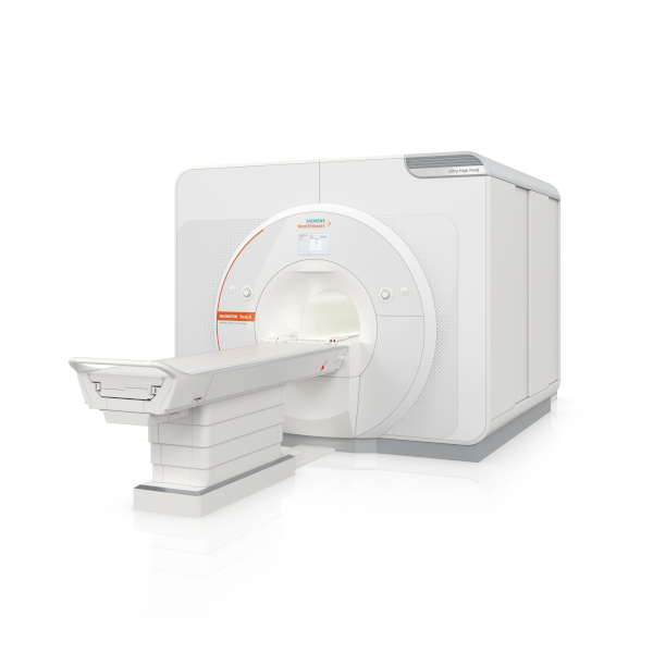
Siemens Healthineers has received FDA clearance for the Magnetom Cima.X 3 Tesla (3T) magnetic resonance imaging whole-body scanner. This scanner features the industry’s strongest gradient system for a clinically released whole-body MR scanner, providing improved visibility of smaller structures in the body and faster image capture. The Magnetom Cima.X also includes new features aimed at enhancing scientific research and addressing imaging challenges related to cancer and other diseases. The high gradient level of the Gemini Gradients, with an amplitude of 200 mT/m and a slew rate of 200 T/m/s, allows for better study of neurodegenerative diseases and can be leveraged across the body. The scanner introduces Physiologging for precise, time-stamped physiological data during functional brain imaging and the Open Recon platform for immediate image reconstruction using custom algorithms directly on the scanner. Additionally, key features such as BioMatrix technology, Deep Resolve reconstruction technology, and myExam Companion workflow solution contribute to improved clinical performance, increased productivity, and a better patient experience. Katie Grant, Vice President of Magnetic Resonance at Siemens Healthineers North America, highlights the scanner’s ability to deliver new insights into oncologic, cardiac, and neurodegenerative diseases.


First FDA-Cleared Portable Magnetic Resonance Brain Imaging System

Hyperfine, Inc. has launched the eighth generation of its Swoop® system software, featuring an AI-powered diffusion-weighted imaging (DWI) denoising feature that received FDA clearance in October 2023. The software enhances the image quality of the Swoop® system, the world’s first FDA-cleared portable magnetic resonance brain imaging system. The update introduces ease-of-use features, including real-time assistance for precise patient loading and positioning, as well as a streamlined image upload process. With CE and UKCA certifications, Hyperfine is positioned for international expansion, aiming to reach a broader global market. The software focuses on improving image quality in the DWI sequence, crucial for stroke imaging. The limited market release phase received positive feedback, with users praising DWI image quality and workflow efficiencies. The update allows clinicians to view each series in real-time, communicate with technologists, and share critical findings with clinicians without waiting for the entire study to be completed.

Sources:

Hyperfine.io
itnonline.com
diagnosticimaging.com
openai.com

Top Imaging News of 2023

As we bid adieu to the final moments of 2023, it’s a great time to reflect on advancements and studies that have redefined the world of imaging this year. In this article, we’ll delve into the hottest news and breakthroughs in imaging, highlighting the remarkable strides that have made the headlines.

Study Suggest that Cancer Death Risk From Low-Dose Radiation Is Underestimated

A recent study featured in the British Medical Journal unveils concerning associations between extended exposure to low-dose radiation, commonly experienced by nuclear industry workers, and amplified cancer-related mortality. Drawing insights from the International Nuclear Workers Study (INWORKS) encompassing data from over 300,000 workers, researchers discovered a stark reality: for each cumulative unit of radiation exposure, the risk of death from solid cancer surged by 52%. Even at the lowest cumulative doses, this risk doubled, challenging the assumption that low-dose exposures present less carcinogenic hazard. While the absolute risk remains small, these findings prompt reconsideration of safety limits for workers and call for further studies to confirm the accelerated risk of cancer with ionizing radiation exposure. The hope is that regulatory bodies will integrate these insights into revising protection standards for individuals exposed to low-dose radiation.

Long COVID

In a study published in Medical Hypotheses, a French group presented a theory regarding the brain fog experienced in long COVID, based on brain patterns identified in patient PET scans. They propose that inflammation triggered by COVID-19 disrupts astrocyte cells’ regulation of glutamate, impacting energy metabolism and leading to cognitive fatigue. The authors suggest targeting this malfunction with therapies focused on astrocytic glutamate regulation as a potential way to alleviate long-COVID neurological symptoms. They highlight the lack of mental clarity, difficulty concentrating, and cognitive strain characterizing long COVID, affecting up to 15% of patients after three months of the initial infection. This study builds on previous findings of hypometabolism patterns in long COVID patients’ brain images and explores cellular mechanisms, including links between glutamate dysregulation and cognitive fatigue from other studies. Drawing parallels with “chemo-fog” in cancer patients and cognitive impairment in Parkinson’s disease, the authors suggest therapeutic strategies targeting the identified brain patterns, citing examples from epilepsy treatments and a recent study using medication to improve cognitive function in long-COVID patients. However, the authors stress the need for further research, proposing PET imaging studies using specific markers to comprehend astrocyte function and glutamate regulation for a comprehensive understanding of long COVID’s underlying mechanisms.

chemo fog
Study on brain fog experienced in long COVID

MRIs and Past Cannabis Users

At the International Society for Magnetic Resonance in Medicine (ISMRM) annual meeting, New Zealand researchers presented findings on heavy cannabis use in adolescence to early adulthood and its correlation with brain structure differences in hippocampus and amygdala subregions. The study, led by medical physicist Rebecca Lee and colleagues from the University of Otago in Christchurch, indicated volumetric disparities in these brain regions among heavy cannabis users compared to non-using controls. Notably, past cannabis users showed smaller volumes in specific hippocampal and amygdala subregions. However, the research did not find detectable differences in cerebral blood flow or white-matter tract integrity related to cannabis use, suggesting potential transient brain changes or no long-term effect on these properties. The study, conducted using MRI techniques, emphasized the need for longitudinal studies to clarify the causation and long-term functional impacts of these structural brain changes associated with heavy cannabis use. Despite revealing structural brain changes linked to cannabis use, the study does not definitively establish a causal relationship between these changes and cannabis consumption. Further prospective longitudinal MRI studies are essential to elucidate causality in this context.

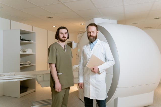
MRI study

All About AI

We’d be remiss to not mention how artificial intelligence has shaped the industry this year. Check out our previous articles highlighting the impact that ChatGPT and Bard have made in 2023.


Sources:

technologynetworks.com
auntminnie.com
Openai.com

How Does Teleradiology Actually Work?

In today’s rapidly evolving healthcare landscape, the prominence and necessity of teleradiology have surged to the forefront. With advancements in technology and the growing demand for efficient, timely, and accurate diagnostic services, especially in light of staffing shortages, teleradiology has emerged as a pivotal solution. Teleradiology not only addresses the increasing demand for imaging interpretations but also bridges geographical gaps, enabling swift access to specialized radiologists regardless of location. Its rising popularity stems from its ability to enhance healthcare delivery by offering remote interpretations, thereby improving patient outcomes and streamlining diagnostic processes in a progressively interconnected world.

Teleradiology operates by leveraging digital communication technologies to transmit medical images, such as X-rays, MRIs, CT scans, and ultrasounds, from one location to another for interpretation and diagnosis.

Here’s a breakdown of how teleradiology works in real time:

Image Capture: Medical images are taken at a healthcare facility using specialized imaging equipment, creating digital files.

Image Transmission: These digital images are securely transmitted over networks, often utilizing Picture Archiving and Communication Systems (PACS) or secure internet connections.

Remote Interpretation: Radiologists, often located at a different site or working remotely, receive these images. They access the images through specialized software, review them in real time, and provide interpretations, diagnoses, and reports.

Consultation and Reporting: Upon reviewing the images, radiologists generate detailed reports containing their findings, interpretations, and recommendations. They may also engage in consultations with healthcare providers or specialists as needed.

Communication: The reports and findings are transmitted back to the referring healthcare facility, where they become part of the patient’s medical record. This allows for prompt decision-making and treatment planning by the attending physicians.

how teleradiology works
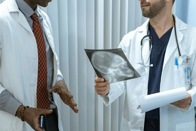
Physicians go over a patient’s scan

Quality Control: Teleradiology services often have quality assurance measures in place to ensure accurate and timely interpretations, adhering to industry standards and protocols.


Throughout this process, encryption and secure transmission protocols are employed to safeguard patient data and comply with healthcare privacy regulations (such as HIPAA in the United States). The real-time nature of teleradiology facilitates quicker diagnoses, especially in emergency situations, and enables access to specialized expertise irrespective of geographical barriers.


Top Teleradiology Company: Vesta is Here for You 24/7/365

Vesta is here to be your supporting partner in radiology, even for subspecialties. We offer fast turnaround for both STAT and Routine studies. Your staff is important–they don’t always work around the clock and shouldn’t have to. With our teleradiology services, you can allow your staff a better work-life balance while we fill in those gaps whether it’s during night hours, weekends, holidays.


Sources:

Acr.org
openai.com


RSNA 2023 Annual Meeting Highlights and Special Exhibits

The RSNA Annual Meeting stands as a pivotal event in the realm of radiology, serving as a dynamic nexus where cutting-edge advancements, emerging trends, and transformative innovations converge. This annual gathering not only showcases the latest breakthroughs in medical imaging but also fosters a collaborative environment where radiology professionals from various subspecialties can engage with state-of-the-art technologies, exchange insights, and navigate the intricate landscape of healthcare’s future.

Trending topics for RSNA 2023

This year’s meeting will be held on November 26-30. It will feature diverse trends across radiology subspecialties, with a notable emphasis on AI and photon-counting CT applications. Theranostics and the use of large language models are also gaining popularity. The event will offer non-interpretive sessions focusing on skills development and addressing diversity, equity, and inclusion in healthcare. The RSNA Annual Meeting Learning Center will provide a comprehensive experience, including the latest research, CME opportunities, and insights for improving DEI and health equity. Attendees can access a quick-reference guide to navigate trending topics and recommended sessions by subspecialty.

Some notable plenary sessions include:

Leading Through Technology: Valuing Artificial and Human Intelligence led by Elizabeth S. Burnside, MD, MPH; The Future of Healthcare Delivery: Considerations for Patients and Providers led by Vin Gupta, MD and Understanding and Revitalizing the Radiology Workforce led by Jocelyn D. Chertoff, MD, MS.

Technical Exhibits

The RSNA 2023 Technical Exhibits promise an immersive experience at the forefront of medical imaging innovation, boasting nearly 700 leading manufacturers, suppliers, and technology developers. The event showcases the latest advancements in AI through the AI Showcase, featuring over 90 companies presenting AI solutions, software demonstrations, and product displays. Attendees can delve into the RSNA AI Theater to explore hot topics in AI, machine learning, and deep learning, with special recognition for winners of AI challenges. The 3D Printing & Mixed Reality Showcase in the North Hall offers networking opportunities in 3D medical printing, augmented reality, and virtual reality.


Learn more at RSNA.org

Sources:
itnonline.com
rsna.org
openai.com


Growth in Demand for Imaging Procedures Will Increase Need for Teleradiology

The healthcare market is experiencing a shift towards outpatient care, driven by reimbursement changes, pandemic effects, and patient preferences, particularly prominent in the U.S. Providers are diversifying into sub-specializations like neurology and oncology, raising the demand for advanced imaging like MRI and CT. This trend has led to increased utilization of outpatient imaging and teleradiology services.

By the NuMbers

Diagnostic imaging is becoming increasingly crucial in healthcare, with the market projected to reach $31.9bn in 2023 and grow at a 4.8% CAGR to $45.8bn in 2030. The rise is driven by chronic diseases, an aging population, and post-Covid-19 demand recovery. To meet this demand, companies are focusing on advanced and accessible technologies, such as handheld ultrasounds. About 1,949 imaging devices are in development, with 112 expected to gain approval in 2023.

imaging device

According to Fortune Business Insights, in 2022, the computed tomography (CT) segment held the largest market share due to a rise in CT scan procedures and higher average pricing. For instance, OECD data for 2021 showed 84.5 million CT scan procedures in the U.S., up by 15.8% from the previous year. The growing geriatric population has also contributed to increased demand for CT scans.

Key Players

Key players include GE Healthcare, Philips, Siemens Healthineers, and more. Challenges include high equipment costs and a shortage of skilled personnel, impacting accessibility and patient care quality. Opportunities arise from the growing demand for imaging services, especially for chronic diseases, and the development of new modalities like 3D mammography and MRI-guided focused ultrasound. Emerging economies like India, China, and Brazil are pivotal, driven by rising chronic diseases. Major players like Siemens Healthineers are expanding in these markets. Additionally, teaching hospitals are increasing demand for advanced imaging methods to enhance patient care.

Your Dedicated Radiology Partner: Vesta

Partnering with Vesta as your radiology partner ensures access to accurate and timely imaging interpretations and readings for subspecialties. Whether you are an outpatient imaging center or traditional hospital, our collaboration offers a seamless and efficient experience. Trust us to be your reliable radiology partner, empowering you with the insights and tools needed for improved healthcare outcomes.


radiology peer reviewSources:

Itnonline.com
Medicaldevices-network.com
fortunebusinessinsights.com
openai.com


Mammography: Is AI Better than Humans?

In recent years, artificial intelligence (AI) has made remarkable strides in revolutionizing the landscape of the medical field, offering unprecedented opportunities for enhanced patient care, diagnosis, and treatment. From accelerating the analysis of medical imagery to predicting disease outcomes with unparalleled accuracy, AI-powered technologies have swiftly established themselves as indispensable tools for healthcare professionals. Beyond diagnostics, AI has played a pivotal role in drug discovery, streamlining clinical trials, and personalizing patient interventions. As AI continues to evolve, its potential to transform healthcare systems globally is becoming increasingly evident, promising not only improved medical outcomes but also cost-effective solutions and optimized resource allocation. The fusion of AI’s computational prowess with medical expertise heralds a new era of medical advancements that hold the potential to alleviate the burden on healthcare systems, save lives, and redefine the standards of patient well-being.

In the United States alone, it is estimated that around 40 million mammograms were performed each year. Mammograms are crucial as they are the primary method for early detection of breast cancer, enabling timely intervention and improving survival rates. By detecting small abnormalities and tumors that may not be palpable, mammograms help identify potential breast cancer cases in their earliest stages, allowing for more effective and less invasive treatment options.

Abnormal mammogram

Radiologists often find themselves overwhelmed due to the increasing volume of medical images requiring analysis, coupled with a shortage of radiology specialists. The demand for accurate and timely diagnoses, especially in fields like mammography, can lead to extended work hours and heightened stress levels among radiologists. Introducing AI technologies can alleviate this burden by assisting in image analysis, enabling radiologists to focus on complex cases and ensuring more efficient patient care.

How AI Helps in Mammography

A recent study published in The Lancet Oncology suggests that artificial intelligence (AI) may outperform trained doctors in detecting breast cancer from mammogram images. Mammograms face challenges due to factors like breast density, leading to missed cancer cases. The study analyzed 80,000 mammograms from Swedish women, finding that AI-assisted readings detected 20% more cancers compared to human radiologists. While not a standalone solution, AI could alleviate doctors’ workloads, enhancing accuracy without increasing false negatives. Despite FDA-approved AI technologies, integration with conventional methods is likely, aiding radiologists in managing a growing workload. The balance between AI and human expertise remains essential, ensuring optimal patient care and early cancer detection.

Healthcare experts, including the NHS and the Royal College of Radiologists, acknowledge AI’s promise in enhancing efficiency, decision-making, and prioritizing critical cases.

mammograms

Vesta Teleradiology

AI applied to diagnostic imaging holds the potential to significantly enhance the level of patient care. We eagerly anticipate further progress in this field. However, we maintain the viewpoint that presently, no machine can effectively substitute for the expertise of a skilled human observer for interpretations. At Vesta, we offer the services of radiologists who are US Board Certified, dedicated to delivering precise preliminary and final analyses. Discover how we can bolster your radiology department by reaching out to us today.


Sources:

Criver.com
health.com
theguardian.com
openai.com


How is Teleradiology and AI Impacting the Medical Industry Today?

Artificial Intelligence (AI) is revolutionizing the medical industry, transforming the way healthcare is delivered, diagnosed, and managed. With its ability to analyze vast amounts of data quickly and accurately, AI is reshaping various aspects of healthcare. From aiding in disease diagnosis to personalized treatment recommendations, AI is enhancing the precision and efficiency of medical practices. Moreover, AI-powered technologies are streamlining administrative tasks, optimizing resource allocation, and improving patient outcomes. As AI continues to advance, it holds immense potential to revolutionize healthcare delivery, foster medical innovations, and ultimately improve the quality of patient care on a global scale.

Teleradiology has had a profound impact on healthcare by enabling remote access to radiology expertise, bridging geographical barriers, and ensuring timely diagnoses. It has improved patient care by providing faster turnaround times, facilitating collaboration among radiologists, and increasing access to specialized interpretations, ultimately enhancing diagnostic accuracy and treatment outcomes. Going even further, a latest white paper from One Call describes how teleradiology and AI are helping reduce the strain of the radiology shortage.

artificial intelligence

Teleradiology and AI in Action

Medical imaging vendor, Nanox, is looking to address heath disparities and lack of access care with a new x-ray system which would be offered to countries in Africa, Asian and South American using a pay-per-scan model. The potential of combining cold cathode X-ray technology with teleradiology and artificial intelligence (AI) to enhance diagnostic capabilities and improve healthcare economics. Cold cathode X-ray systems offer advantages such as reduced energy consumption and improved image quality. When integrated with teleradiology, these systems can enable remote interpretation of X-rays, leading to faster diagnoses and improved patient care. Additionally, the use of AI algorithms in conjunction with cold cathode X-ray technology has the potential to enhance image analysis, automate certain tasks, and optimize resource allocation, offering cost-saving opportunities in healthcare settings.

diagnostic imaging
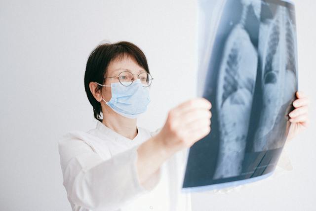
A teleradiologist examines a chest x-ray

There are plans to roll out AI-powered teleradiology by the “Screen for Life” program at the Primary Health Care Corporation in Qatar, aimed at early detection and prevention of cancer in the United Arab Emirates. The program plans to utilize AI algorithms to analyze radiology images, enhancing the accuracy and efficiency of cancer screening. The integration of AI in teleradiology will help automate image interpretation, expedite diagnoses, and reduce the workload on radiologists. The implementation of AI teleradiology in the “Screen for Life” program is expected to improve cancer detection rates, streamline healthcare processes, and ultimately save lives by identifying cancers at earlier stages.

Vesta Teleradiology

Looking to outsource your radiology interpretations using an expert Teleradiology company that is at the forefront of technology including AI?  Please reach out to Vesta to learn more. Vesta Teleradiology can accommodate any type of volume, large, medium and small.

Sources:

Radiologybusiness.com
menafn.com
openai.com
cdc.gov

Teleradiology FAQ: How Are Teleradiology Reports Delivered?

A healthcare facility – hospital, private practice, imaging center – may choose to utilize the services of a teleradiology company to enhance their radiology capabilities and streamline their diagnostic imaging processes. Teleradiology services offers numerous advantages, such as increased access to specialized radiologists, expedited turnaround times, reduced waiting time for patients, and improved patient care. By partnering with a reputable teleradiology company, healthcare facilities can leverage advanced technology, secure data transmission, and a network of skilled radiologists to obtain accurate and timely radiology preliminary and final interpretations. This collaboration allows healthcare providers to optimize their resources, extend their coverage, and ultimately deliver high-quality diagnostic imaging services to their patients, regardless of time or location.

Teleradiology Reports Delivery

Before engaging with an outsourced radiology company, you may wonder how reports are delivered. Teleradiology specialists deliver their reports through secure electronic means, utilizing advanced technology and secure communication platforms.

telerad

When you partner with Vesta, a Gateway application is installed remotely to allow your systems direct access to our PACS. Once linked, your technicians send exams straight from your machine to our PACS. Vesta account specialists assign it to the proper physician who read and dictate the report, it’s uploaded to your branded template where it’s easily accessible for your facility to access and download!

These customized reports can also be distributed electronically, and any critical findings are immediately shared with the facility via phone, email, or text. We also facilitate callbacks between the physician and radiologist and provide an unlimited amount of customized report templates with your facility’s information and logo. It’s a seamless experience which allows you to focus on what matters most, the patient experience.

Safety and Security: HIPAA

Teleradiology companies like Vesta prioritize the security and confidentiality of patient data throughout the entire process. They employ strict data encryption, comply with privacy regulations (such as HIPAA in the United States), and implement robust security measures to protect patient information during transmission and storage.

hipaa compliance
Look to Vesta for secure and compliant reports

Looking to outsource your radiology interpretations? Please reach out to Vesta to learn more. Vesta Teleradiology can accommodate any type of volume, large, medium and small.

Sources

https://www.ncbi.nlm.nih.gov
https://openai.com