Breast Imaging 2025–26: Risk Models, CEM/MRI Momentum — RSNA Preview

RSNA 2025 is putting real energy behind risk-adjusted screening and the evolving roles of contrast-enhanced mammography (CEM) and breast MRI. For breast programs, the takeaway is practical: risk tools are moving from the research poster to the reading room, and CEM/MRI decisions are becoming operational levers you can plan around—especially for dense-breast pathways and overflow routing to subspecialists.

What’s new at RSNA: risk from the image itself

RSNA’s breast-imaging preview highlights sessions on image-only, 5-year breast cancer risk models, external validation work, and how MRI adds value in multi-modal AI. It also calls out global screening updates and a deeper look at background parenchymal enhancement (BPE) on MRI. RSNA

In parallel, the FDA granted De Novo authorization to the first image-only AI risk platform that predicts 5-year risk directly from a screening mammogram—an inflection point that makes risk-adjusted pathways far more scalable. Coverage from Radiology Business and BCRF explains the authorization and clinical intent. Radiology Business

Why it matters: average-risk guidance in the U.S. now begins screening at age 40 (USPSTF, 2024). Programs can layer image-based risk on top of that baseline to triage who needs annual vs. short-interval follow-up and who merits supplemental imaging. USPSTF

CEM is earning a seat next to MRI

Expect exhibits and sessions positioning CEM as a cost-effective, accessible adjunct—particularly for dense-breast populations and diagnostic workups. RSNA News recently framed CEM as a practical alternative to MRI in some screening/diagnostic scenarios, and new peer-review literature is refining technique (e.g., lower volume/higher-iodine contrast while preserving diagnostic performance). RSNA

On outcomes, the RACER trial in The Lancet Regional Health – Europe reported that using CEM as primary imaging for recalled women improved the accuracy and efficiency of the work-up compared with conventional imaging—evidence that will influence protocols beyond the show floor. The Lancet

MRI still leads for sensitivity—BPE is your underused signal

Breast MRI remains the sensitivity champion for high-risk patients and for problem solving. This year’s RSNA content spotlights BPE—how the level of background enhancement relates to tumor biology and outcomes. Recent reviews (2024–2025) synthesize BPE’s predictive/prognostic value, including associations with pathologic complete response after neoadjuvant therapy and survival in certain subtypes. SpringerLink

Practical move: standardize how you document BPE and incorporate it into structured reports and risk conferences; it’s becoming more than a descriptive footnote.

What to ask vendors at RSNA

  1. Risk engine proof: “Show external validation and calibration plots by density and race; how does your image-only model integrate into our mammography worklist and letters?”
  2. CEM logistics: “Demonstrate CEM acquisition workflows, contrast protocols, and how your viewer handles subtraction/kinetics alongside priors.”
  3. MRI + BPE analytics: “Can we standardize BPE capture in structured reports and trend it across treatment?”

As risk-first screening, CEM, and MRI gain real traction, the winners will be the programs that operationalize them quickly and consistently. If you’re planning your 2026 breast-imaging playbook, stop by Vesta at RSNA to see how our subspecialists, standardized templates, and overflow routing make risk-adjusted pathways usable on day one.

Imaging the Individual — In the Trenches: AI, Personalization & Equity at RSNA 2025

RSNA’s 2025 theme, Imaging the Individual, isn’t just about futuristic science—it’s about doing the basics better for each patient, every day. The official Trending Topics preview highlights three threads cutting across subspecialties: AI you can deploy, personalized care you can operationalize, and equity you can measure. This guide translates those themes into practical checkpoints hospitals and imaging centers can use right now. RSNA

1) AI that graduates from pilot to practice

This year’s agenda emphasizes real outcomes over proofs of concept: reader-in-the-loop tools, bias monitoring, and governance. In breast imaging alone, RSNA previews spotlight external validation for image-only risk models and integration of MRI signals into multimodal AI—clear signals that “personalization” is landing in routine workflows. Bring vendor questions that force specifics: external validation cohorts, drift detection, and how metrics (TAT, recalls, rework) appear in your dashboard. RSNA

What to set up before RSNA: define 3–5 outcome metrics and insist every demo shows pre/post performance tied to those measures. Use QIBA concepts to push for standardized inputs/outputs so results are reproducible across scanners and sites. QIBA Wiki

2) Personalization that reaches the reading room

Personalization isn’t only radiogenomics. RSNA’s preview points to risk-stratified pathways you can actually run: e.g., image-only 5-year breast cancer risk at the point of screening to route patients into annual vs. short-interval follow-up or supplemental imaging (CEM/MRI). That pairs well with updated U.S. recommendations: screening beginning at age 40 for average-risk women, then adjusting based on risk and local policy. Build routing rules, templates, and letters now, so RSNA demos can plug into your plan.

Operational checklist:

  • Map risk thresholds → next steps (annual vs. short-interval, CEM/MRI).
  • Standardize templates so risk outputs appear consistently in reports and patient letters.
  • Decide who reviews outlier risk flags and how quickly (SLA).

3) Equity you can instrument—not just endorse

RSNA is foregrounding health equity, with sessions on encoding equity in AI and addressing access gaps for underserved communities. Equity becomes real when you can see it in your data: turnaround times by language, missed-appointment patterns by zip code, recall rates by screening site, and AI performance by subgroup. Build those slices into your analytics now; then ask vendors to show subgroup performance in their dashboards.

Practical moves:

  • Add demographic and language filters to your TAT and recall reports.
  • Require AI vendors to show calibration and error analysis by subgroup.
  • Stand up multilingual patient letter templates to support new screening starts at 40. USPSTF

4) CEM/MRI momentum: choose the lever that fits your service line

RSNA coverage calls out CEM as an increasingly practical adjunct—especially useful for dense-breast populations and diagnostic workups where capacity or cost limits MRI. The RACER trial reported higher accuracy and efficiency for CEM as the primary exam for recalled women vs. conventional imaging—evidence that can justify protocol changes and equipment planning. Meanwhile, MRI retains the sensitivity crown, with renewed attention on background parenchymal enhancement (BPE) as a signal worth documenting consistently.


Action items:

  • Decide where CEM fits: diagnostic recall pathway, dense-breast supplemental strategy, or both.
  • Add BPE level to structured MRI reports and trend it during therapy response clinics.

5) Governance, not guesswork

If personalization is the “what,” governance is the “how.” Use QIBA ideas—claim definitions, acquisition standards, and profile adherence—to control variability across devices and shifts. Tie RSNA learnings to a written governance plan with three parts: 1) protocol book (who owns it, update cadence), 2) quality book (metrics, subgroup views), and 3) AI book (approval process, monitoring, rollback).

6) Where teleradiology extends your capacity

Personalization increases complexity at peaks (recalls, dense-breast seasons, MR backlogs). A teleradiology partner helps you keep individualized pathways moving: standardized templates, subspecialty over-reads, and after-hours coverage that adheres to your risk rules and equity metrics—so “Imaging the Individual” doesn’t stop at 5 p.m.

Headed to RSNA?


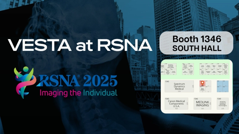
Visit Vesta at Booth 1346 (South Hall) to see how we make “Imaging the Individual” work in real clinics—then enter to win a 1-year Medality CME subscription. Don’t wait: email “RSNA CME Entry” to info@vestarad.com now for a reserved entry, and show your confirmation at the booth for a bonus entry.

Powering Quality and Efficiency Through AI

Elevating Radiology. Expanding Access. Enhancing Care.

Vesta Teleradiology is redefining radiology delivery by integrating artificial intelligence (AI) into our diagnostic and operational workflows – helping hospitals of every size achieve higher quality, faster turnaround, and greater consistency in patient care.

Through our newly launched partnerships with Qure.ai and Carpl.ai, Vesta is bringing the benefits of AI assisted imaging to both large health systems and rural or underserved communities across the nation. This innovation enhances the speed, accuracy, and accessibility of radiology services – ensuring clinical excellence reaches every patient, everywhere.

AI Partnerships Driving Clinical Quality and Efficiency

Vesta now integrates Qure.ai’s FDA cleared AI solutions directly into our reading workflow to support both CT and X-ray imaging. For CT Brain (Non-Contrast), the AI automatically detects intracranial hemorrhages, fractures, and mass effect to improve triage and accelerate emergency response times. For Chest X-rays, it identifies nodules, effusions, and acute pulmonary findings to strengthen diagnostic consistency and enable earlier intervention. These tools work as a co-pilot for radiologists – helping prioritize critical studies, standardize interpretations, and deliver higher-quality reports with precision and speed.

Vesta also leverages Carpl.ai’s enterprise grade AI platform for musculoskeletal (MSK) fracture detection, enabling faster identification of subtle skeletal injuries that are often missed under high volume workloads. This integration enhances both radiologist efficiency and patient safety by improving consistency, turnaround times, and workflow throughput.

Expanding AI Across Vesta’s Clinical and Operational Ecosystem

In addition to our partnerships with Qure.ai and Carpl.ai, Vesta continues to implement AI across the organization to enhance both clinical quality and operational efficiency. Through RadPair, Vesta improves dictation accuracy, peer review workflows, and reporting analytics for radiologists – driving consistency and precision across the reading process.

On the operations side, Vesta has developed and launched an AI based support platform that allows staff to instantly retrieve internal protocols, radiologist schedules, credentialing data, and study specialty details from a centralized location. These tools streamline communication, improve turnaround time, and strengthen coordination across departments – supporting faster, more efficient service for clients and radiologists alike.

AI with a Purpose: Clinical Quality Care for All

Vesta’s mission has always been clear – to combine technology, compassion, and clinical excellence to improve access to quality radiology care. By implementing these AI partnerships and innovations, we’re ensuring faster turnaround for emergent and high acuity studies, improved diagnostic accuracy through validated AI support, greater access for rural and underserved hospitals, and consistent quality across every facility, 24/7/365.

These advancements reaffirm Vesta’s leadership as a trusted partner in AI driven radiology innovation, bringing cutting edge technology to the frontlines of patient care while optimizing the systems that support it.

About Vesta Teleradiology

Vesta Teleradiology is a Joint Commission-Accredited, 24/7/365 radiology provider serving hospitals, imaging centers, and healthcare systems nationwide. Our team of board-certified radiologists delivers timely, accurate, and secure interpretations – now further enhanced by AI technology to support faster decisions, higher quality, and better outcomes.

Interested in learning how Vesta’s AI powered radiology can support your hospital or health system?
Contact us at info@vestarad.com or visit www.vestarad.com/contact to schedule a demo or consultation.

Attribution:
Vesta Teleradiology integrates third party AI technologies through collaborations with Qure.ai, Carpl.ai, and RadPair. Descriptions of imaging and workflow capabilities in this publication are based on publicly available clinical use cases and are provided for informational purposes only. All content and messaging on this page are original to Vesta Teleradiology.

Vesta Teleradiology Heads to RSNA 2025: AI + Expertise = Faster, Smarter Imaging Coverage


Every year, the Radiological Society of North America (RSNA) brings together innovators shaping the future of medical imaging. This November 30–December 3, 2025, the Vesta Teleradiology team is proud to join that community at RSNA 2025 in Chicago — showcasing how AI and human expertise combine to deliver faster, smarter imaging coverage for hospitals and imaging centers nationwide.

Meet Vesta at Booth 1346 — South Hall

At Booth 1346, attendees can discover how Vesta helps healthcare facilities overcome some of today’s biggest radiology challenges — from staffing shortages to increasing imaging volumes — without compromising patient care.

Vesta’s solutions are designed to help your organization:

  • Gain 24/7 radiology coverage without the burnout
  • Access fellowship-trained subspecialists across all modalities
  • Deliver faster turnaround times with AI-assisted workflow tools
  • Scale imaging services without adding staff
  • Rely on dependable IT services and seamless PACS integration

How Vesta Combines AI + Human Expertise

Teleradiology isn’t just about remote reads — it’s about precision, speed, and collaboration. Vesta’s radiologists use advanced AI-assisted workflow technology to prioritize cases, enhance diagnostic consistency, and streamline communication with hospitals and imaging centers.

AI tools don’t replace radiologists; they empower them. By automating repetitive tasks and highlighting critical findings faster, AI allows Vesta’s board-certified radiologists to focus where their expertise matters most — delivering accurate interpretations and improving patient outcomes around the clock.

Dependable Excellence, Every Time

Since its founding, Vesta has remained committed to providing dependable, high-quality radiology coverage that healthcare organizations can trust. Whether you need overnight support, overflow assistance, or full departmental coverage, Vesta’s network of U.S.-based, fellowship-trained subspecialists ensures that every scan gets the attention it deserves — anytime, anywhere.

Join Us in Chicago

If you’re attending RSNA 2025, we’d love to meet you in person. Stop by Booth 1346 in the South Hall to see how Vesta’s combination of human insight and artificial intelligence is helping healthcare facilities achieve diagnostic excellence — without adding to their workload.

RSNA 2025 — Chicago, IL
November 30 – December 3, 2025
VESTARAD.COM

Vizamyl’s New PET Label: Quantify & Monitor Amyloid—What It Means for Imaging Teams


What changed—and why it matters

The FDA has expanded the label for flutemetamol F 18 (Vizamyl), enabling quantification of amyloid plaque burden and long-term therapy monitoring in Alzheimer’s disease. This shift moves amyloid PET beyond a qualitative “positive/negative” decision toward objective, longitudinal assessment that can inform treatment choice, dose intervals, and discontinuation decisions. Business Wire

Professional groups report the update aligns amyloid PET with the clinical era of disease-modifying anti-amyloid therapies (e.g., lecanemab, donanemab), clarifying roles for baseline confirmation, on-treatment monitoring, and response tracking in routine care. Notably, SNMMI stated the FDA granted supplemental indications—including quantitative measurement and use for therapy monitoring—to three amyloid PET agents (flutemetamol F-18/Vizamyl, florbetapir F-18, and florbetaben F-18). SNMMI

Operational updates for radiology leaders

  • Protocols & quant pipelines: Build or validate a quant workflow (SUVr or comparable metrics) that’s scanner-calibrated and reproducible across sites. If you operate multi-vendor fleets, document harmonization steps in your SOPs.
  • Structured reports: Add fields for quantified burden at baseline, change from baseline, and interpretive guidance tied to therapeutic decisions (initiation, continuation, or discontinuation).
  • Scheduling & throughput: Expect rising referral volume from neurology and geriatrics as therapy monitoring enters routine practice; protect access with extended hours or overflow capacity.
  • Quality & governance: Define thresholds for biologically meaningful change, reader training for quant review, and reconciliation rules when quant and visual impressions diverge.

For additional context, trade coverage underscores that the updated label formally removes previous limitations around therapy monitoring and permits quant analysis in routine reporting. Empr

How Vesta Teleradiology helps

Vesta’s subspecialty neuro and nuclear medicine radiologists provide:

  • Amyloid PET expertise: Visual+quant reads with structured templates aligned to your therapy pathway.
  • Coverage when you need it: After-hours, weekends, or daytime overflow—without sacrificing turnaround time.
  • Interoperability: Seamless delivery to your PACS/RIS and EMR; clear flags for therapy decisions and recall intervals.
  • QA you can see: Peer review, consistency checks across readers, and optional double-reads during program ramp-up.

If you’re standing up or scaling amyloid PET services, we can supply immediate subspecialty coverage and templates tuned to your neurologists’ needs.


FDA’s 2025 AI Draft Guidance: A Buyer’s Checklist for Imaging Leaders

In January 2025, the U.S. Food and Drug Administration released a draft guidance for AI-enabled medical devices that lays out expectations across the total product life cycle—design, validation, bias mitigation, transparency, documentation, and post-market performance monitoring. For imaging leaders, it’s a clear signal to tighten procurement criteria and operational guardrails before piloting AI in CT, MRI, mammo, ultrasound, or PET.

As teams lock in Q4 budgets and head into RSNA season, the FDA’s AI lifecycle draft (Jan 2025) and the now-final PCCP (Dec 2024) have reset what buyers should expect from AI in imaging—devices, software, and workflows. Vendors are updating claims and governance; this issue distills a practical buyer’s checklist—multisite validation with subgroup results, drift monitoring and version control, clear in-viewer transparency—and how pairing those tools with Vesta’s subspecialty coverage and QA turns promise into measurable gains across CT/MRI/US/mammography.

A practical buyer’s checklist

Use this when evaluating AI for your service lines:

  1. Intended use fit: Verify indications, inputs/outputs, and claims match your pathway and patient mix.
  2. Validation depth: Prefer multisite, diverse datasets; stratified results; pre-specified endpoints; documented data lineage and splits.
  3. Bias mitigation: Demand subgroup performance (sex, age, race/ethnicity when available), scanner/vendor variability analyses, and site-transfer testing.
  4. TPLC plan: Require drift monitoring, retraining triggers, versioning, and how updates are communicated.
  5. Human factors & transparency: Ensure limitations, failure modes, and interpretable outputs are presented in-viewer without slowing reads.
  6. Security & support: Patch cadence, vulnerability disclosure, SOC2/ISO posture, uptime SLAs, and rollback paths for version issues.
  7. Governance: Define metrics owners, review cadence, and thresholds to pause or roll back a model.

Implementation playbook: pilot → scale without disruption

Start with a 60–90 day pilot in one high-impact line (e.g., ED stroke CT or mammography triage) and lock in baselines: median TAT, positive/negative agreement, recall rate, PPV/NPV, and discrepancy rate. Set guardrails—when to auto-triage vs. force human review—and document escalation paths for model failures. Require case-level confidence and structured outputs your radiologists can verify quickly. Stand up a model governance huddle (modality lead, QA, IT security, and your teleradiology partner) that meets biweekly to review drift signals, subgroup performance, and near-misses. Bake in a rollback plan (version pinning) and a quiet-hours change window so updates don’t collide with peak volumes. As results stabilize, scale by cohort (e.g., expand to non-contrast head CT, then CTA) and keep training “micro-bursts” for techs/readers—short videos or checklists in-workflow. Tie vendor SLAs to uptime, support response, and clinical KPIs so the AI program stays accountable to operational value.

Where teleradiology fits

AI only delivers when it’s welded to coverage, quality, and speed. A teleradiology partner should provide:

  • 24/7 subspecialty + surge capacity: Vesta absorbs volume peaks so AI never becomes a bottleneck.
  • QA you can see: We benchmark pre/post-AI performance, add targeted second looks for edge cases, and feed variance data back to your team.
  • Standardized outputs: Structured reports that integrate model outputs with radiologist findings—no black-box surprises.
  • Smooth rollout: Pilot by service line (stroke CT, mammo triage, PE workups), then scale with tracked KPIs (TAT, PPV, recalls).
  • Interoperability & security: Seamless PACS/RIS/EMR integration with strict access controls, audit trails, and support for change-controlled updates.

Bottom line: Pairing AI with Vesta Teleradiology gives you round-the-clock subspecialty reads, measurable QA, and operational breathing room while you pilot and scale responsibly. If you’re mapping your AI roadmap under the FDA’s 2025 draft guidance, we’ll be your coverage and quality backbone—so your clinicians see faster answers and your patients see safer care. Visit vestarad.com to get started.


Summer 2025 Imaging Roundup: AI, New Modalities & Trends

The summer of 2025 has been packed with advancements in diagnostic imaging, from cutting-edge AI systems improving detection rates to emerging modalities pushing the boundaries of precision and speed. Here’s a look back at the most important developments from June through August that are shaping the future of radiology.

AI Is Reshaping Radiology Workflows

Generative AI Productivity Boost

In June, Northwestern Medicine unveiled a generative AI system capable of reducing radiologist reading time by up to 40% while identifying life-threatening conditions in milliseconds. This tool not only improves workflow efficiency but also offers a potential solution to the ongoing radiologist shortage (Northwestern Medicine).

ProFound AI for Mammography

A peer-reviewed study confirmed that iCAD’s ProFound AI significantly increases cancer detection rates, boosts diagnostic accuracy, and improves workflow for mammography screenings (ITN Online).

Aidoc’s $150M Expansion

July saw AI platform Aidoc raise $150 million in funding, led by NVIDIA and other major investors, aimed at expanding its reach into more hospitals and imaging centers globally (Aidoc).

Emerging Imaging Modalities and Research

Top Content Trends

Radiology publications in July spotlighted rising interest in abbreviated breast MRI, MRI-guided ultrasound for Parkinson’s disease, and dual-energy CT for understanding Long COVID-related lung changes (Diagnostic Imaging).

Photon-Counting CT and Whole-Body MRI

Photon-counting CT continues to gain attention for its ability to deliver higher resolution at lower doses, while whole-body MRI is increasingly used for cancer staging and early detection in high-risk populations (Radiology Business).

Multimodality Imaging at ACC.25

Cardiologists and radiologists at the ACC.25 conference explored how quantitative CT, functional cardiac MRI, and AI-enhanced echocardiography can bridge the gap between diagnostics and real-time therapy planning (American College of Cardiology).

August: A Month of Imaging Breakthroughs

AI-Native Imaging Viewers

Tech company New Lantern launched AI-native viewer modes for mammography and PET/CT, delivering sub-second load times and workflow automation (TMCNet).

Digital Radiography Gets Smarter

Advances in digital radiography are enhancing precision and speed, with newer systems providing better image quality at lower radiation doses (USA News).

ProCUSNet Ultrasound AI

Researchers at Stanford developed ProCUSNet, an AI tool that improved lesion detection by 44% and caught 82% of clinically significant prostate cancers on ultrasound—outperforming human interpretation (Becker’s Hospital Review).

DiffUS for Intraoperative Imaging

A new AI-based technique called DiffUS can create realistic ultrasound images from 3D MRI data, aiding in surgical planning and intraoperative navigation (arXiv).

Next-Gen PET Tracer

A novel PET tracer, Ga-68 Trivehexin, has shown promise in more accurately detecting breast cancer lesions and fibrotic lung tissue compared to traditional tracers (Journal of Nuclear Medicine).

Looking Ahead

The pace of innovation in diagnostic imaging this summer reinforces a clear trend: AI is no longer just an assistive tool—it’s becoming deeply embedded in clinical workflows. Coupled with emerging modalities like photon-counting CT and new PET tracers, radiology is entering an era of higher precision, speed, and accessibility.

AI-Enabled Ultrasound: Transforming Imaging at the Point of Care


In today’s fast-paced healthcare environment, ultrasound is increasingly recognized not just for prenatal or cardiac assessment, but as a versatile diagnostic tool across specialties. Now, artificial intelligence (AI) is accelerating ultrasound’s impact — reducing operator dependency, improving diagnostic confidence, and enabling faster bedside care. For imaging leaders, especially in rural or underserved settings, AI-powered ultrasound technology paired with teleradiology support offers a compelling path for enhanced access and precision.

Innovations in AI-Ultrasound You Should Know

  1. FDA Clearance for AI Thyroid Ultrasound
    In 2024, See-Mode Technologies received FDA clearance for an AI-powered thyroid ultrasound system that can detect and classify nodules using the ACR TI-RADS scale. It has shown promising results in standardizing reporting and reducing unnecessary biopsies and follow-ups.
    Source: https://www.auntminnie.com
  2. Projected Market Growth
    The global AI ultrasound market is projected to grow at a compound annual growth rate (CAGR) of 22% through 2029. This rapid growth is fueled by the rising burden of chronic disease, limited radiologist availability, and the push for faster, more accessible diagnostics.

    Source: https://www.pharmiweb.com/

  3. Rural Potential with Point-of-Care AI
    A JAMA Cardiology viewpoint outlines how AI-assisted point-of-care ultrasound (POCUS) can enable more accurate cardiovascular assessments even when performed by generalists—especially valuable in remote areas without imaging specialists.
    Source: https://jamanetwork.com
  4. Clinician Enthusiasm and Challenges
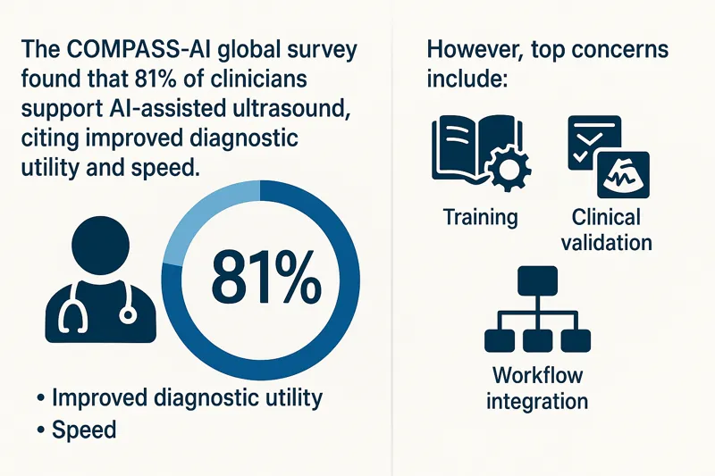
    The COMPASS-AI global survey found that 81% of clinicians support AI-assisted ultrasound, citing improved diagnostic utility and speed. However, top concerns include training, clinical validation, and workflow integration.

    Source: https://theultrasoundjournal.springeropen.com/

Infographic showing COMPASS-AI survey results on clinician support for AI-enabled ultrasound, benefits, and concernsWhy It Matters for Facilities and Radiology Teams

  • Reduces staffing burden: AI ultrasound reduces variability among operators, ideal for high-turnover or remote settings.
  • Speeds up decision-making: Frontline providers can quickly gather meaningful imaging data, while teleradiologists handle the interpretation.
  • Expands imaging reach: Portable, AI-powered ultrasound extends diagnostic capabilities to underserved regions.
  • Supports standardization: AI helps standardize image acquisition and reporting, improving overall workflow efficiency.

How Vesta Teleradiology Enhances AI-Ultrasound Value

While AI augments imaging workflows, expert interpretation is still essential. Vesta provides:

  • Subspecialty reads across thyroid, vascular, MSK, and more
  • 24/7 coverage with fast turnaround times
  • Seamless PACS/RIS integration for AI-acquired ultrasound data

Our radiologists help bridge the gap between frontline imaging and specialist analysis—ensuring that every AI-enabled ultrasound scan contributes to timely, confident patient care.

Bringing AI and Teleradiology Together

Whether you’re running a rural health center, a large outpatient clinic, or an emergency department, AI ultrasound paired with expert teleradiology interpretation helps:

  • Increase imaging access without compromising accuracy
  • Alleviate staffing constraints
  • Deliver faster diagnoses
  • Improve patient outcomes

AI in ultrasound is not replacing radiologists — it’s helping them focus on what matters most. With Vesta’s support, healthcare organizations can embrace innovation while maintaining high-quality, consistent imaging interpretation.


Photon-Counting CT: What Healthcare Facilities Need to Know Now

Photon-counting computed tomography (PCCT) is one of the most exciting breakthroughs in diagnostic imaging technology in recent years. Offering greater spatial resolution, reduced radiation dose, and improved tissue characterization, PCCT is quickly gaining attention from radiologists, imaging directors, and healthcare systems looking to stay ahead.

As the healthcare landscape evolves, staying informed about how new imaging technologies integrate with workflows and diagnostic goals is critical. Here’s what facilities need to know now about photon-counting CT—and how teleradiology can help maximize its impact.

What Is Photon-Counting CT?

Unlike conventional CT, which measures the total X-ray energy reaching the detector, photon-counting CT counts individual photons and measures their energy levels. This allows for:

  • Sharper images with better spatial resolution
  • Lower noise, especially in soft tissue
  • Multi-energy imaging from a single scan
  • Reduced radiation exposure

Siemens Healthineers introduced the first FDA-approved photon-counting CT system (NAEOTOM Alpha) in 2021, and adoption has slowly grown among academic and high-volume centers.

Clinical Benefits of PCCT

Photon-counting CT provides enhanced detail for a range of applications, including:

  • Cardiac imaging: Better visualization of stents and plaques
  • Pulmonary imaging: Improved nodule detection and perfusion data
  • Neuroimaging: Greater contrast at lower doses for brain scans
  • MSK imaging: Superior resolution for joint, bone, and soft tissue analysis

The ability to perform multi-energy imaging without dual-source CT equipment allows radiologists to generate virtual non-contrast images, improve lesion characterization, and reduce contrast agent use—benefiting both patients and providers.

Multi-energy CT image showing high-resolution internal anatomy used for virtual non-contrast imaging
Growing Market and Adoption

While still early in widespread adoption, the global photon-counting CT market is projected to grow rapidly. According to a recent report from Research and Markets, the global PCCT market is expected to reach over $800 million by 2030, driven by increasing demand for advanced diagnostic tools and a growing focus on radiation dose reduction.

As more vendors develop photon-counting detectors and more clinical use cases are validated, experts anticipate broader adoption beyond academic centers and into regional hospitals and imaging centers.

Source: Research and Markets, “Photon Counting CT Market – Forecast 2030”

How Teleradiology Supports Advanced CT Adoption

Deploying a photon-counting CT system requires more than just the hardware. Facilities must ensure they have access to radiologists who are:

  • Trained in multi-energy CT interpretation
  • Familiar with new artifact patterns and reconstructions
  • Able to optimize clinical workflows using new scan data types

That’s where teleradiology plays a critical role.

At Vesta Teleradiology, our radiologists stay at the forefront of imaging advances. With experience in multi-energy and advanced CT post-processing, we help facilities take full advantage of what photon-counting CT offers—delivering fast, accurate interpretations backed by subspecialty insight.

Integration and Workflow Considerations

Facilities considering photon-counting CT should think about:

  • PACS/RIS compatibility with new data formats
  • Training staff to understand and use spectral data
  • Building protocols for when and how to use PCCT scans
  • Collaborating with teleradiology teams for consistent interpretations

While the learning curve is real, the payoff is significant. Early adopters report better diagnostic confidence, fewer repeat scans, and more comprehensive patient evaluations.

Conclusion: Prepare for the Future of CT Imaging

Photon-counting CT represents the next leap in diagnostic precision. As this technology becomes more accessible, imaging leaders must evaluate how it fits into their long-term strategy. For facilities looking to stay competitive, offer premium diagnostics, and improve patient care, PCCT should be on the radar now—not later.

Partnering with a forward-thinking teleradiology provider like Vesta ensures you’re equipped with the expertise to unlock its full potential.


Celebrating National Health Center Week: The Frontline of Community Care

Every August, National Health Center Week (August 3-9 2025) recognizes the critical role community health centers play in delivering affordable, high-quality healthcare across the United States. These centers serve more than 30 million patients annually, many of whom live in medically underserved or rural regions. But as demand for comprehensive care grows, so does the need for accessible diagnostic imaging—an area where teleradiology is helping bridge the gap.

The Imaging Gap in Rural and Underserved Areas

Access to diagnostic imaging remains a persistent challenge for many community health centers. Facilities in rural or low-resource areas often face:

  • Limited access to on-site radiologists
  • Delays in turnaround times for imaging reads
  • Difficulty recruiting or retaining subspecialty radiologists
  • Rising imaging volumes due to expanded preventive care

These barriers can compromise patient outcomes, especially in time-sensitive cases involving stroke, cancer screening, or trauma. Imaging is a critical step in diagnosis—and delays in radiology reports can delay treatment.

Teleradiology: A Scalable Solution for Imaging Access

Teleradiology enables healthcare facilities to send medical images (like X-rays, CT scans, MRIs, and mammograms) electronically to off-site, board-certified radiologists for interpretation. For community health centers, this technology is transformative.

Here’s how teleradiology supports health centers during National Health Center Week and year-round:

  1. 24/7 Coverage, Including Nights and Holidays
    Teleradiology ensures that community health centers can offer imaging services around the clock—even if there’s no radiologist physically on-site. This is especially important for urgent care and emergency settings in rural hospitals.

  2. Access to Subspecialty Reads
    Facilities may not always have access to neuroradiologists, MSK radiologists, or breast imaging specialists. Vesta Teleradiology offers access to subspecialty reads, ensuring every case is interpreted by the right expert.

  3. Faster Turnaround Times
    With cloud-based image transfer and structured reporting, teleradiology reduces delays and improves turnaround times. That means faster results, quicker clinical decisions, and better patient care.

  4. Support for Preventive Imaging Initiatives
    Community health centers are expanding their use of imaging for preventive care—particularly for breast cancer screening, lung health, and cardiovascular risk. Teleradiology provides scalable support during screening campaigns or high-volume periods.

    Female patient undergoing a mammogram with a radiologic technologist in a medical exam room

  5. Cost-Effective Radiology Staffing
    Teleradiology helps optimize budgets by supplementing in-house radiologists or replacing expensive on-call coverage. Flexible pricing models ensure services align with facility needs and patient volume.

Why Imaging Access Matters More Than Ever

The need for diagnostic imaging continues to rise in 2025. According to recent projections from the Harvey L. Neiman Health Policy Institute, demand for imaging will grow at a faster rate than the radiologist workforce through 2055. In rural and medically underserved areas, the shortage is even more pronounced.

Community health centers are on the front lines of closing this gap. But without reliable imaging access, they face limitations in diagnosis, monitoring, and treatment planning.

How Vesta Teleradiology Helps Health Centers Thrive

At Vesta, we understand the pressures community health centers face. That’s why we offer:

  • Fully customizable radiology services tailored to your patient population
  • Rapid onboarding and seamless PACS integration
  • Weekend, holiday, and night coverage
  • A team of U.S.-based, board-certified radiologists
  • Subspecialty interpretations across all major imaging fields

Whether you’re a rural clinic needing full radiology coverage or a mid-sized health center looking for overflow support, our teleradiology solutions are built to help you scale—without compromising care quality.

Join the Movement: National Health Center Week

National Health Center Week is more than a celebration. It’s a reminder that access, equity, and quality care start with supporting the providers who serve our most vulnerable populations. Teleradiology is a powerful tool to help meet that mission.

If your health center is planning to expand imaging services or looking for reliable radiology coverage, Vesta is here to help.

Let’s build healthier communities—one accurate read at a time.