Breast Imaging 2025–26: Risk Models, CEM/MRI Momentum — RSNA Preview

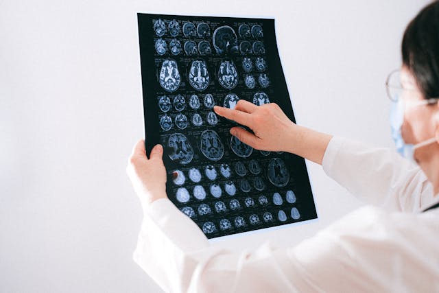
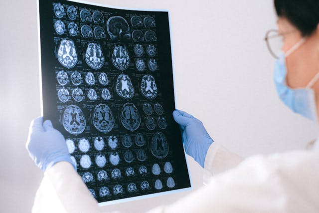
RSNA 2025 is putting real energy behind risk-adjusted screening and the evolving roles of contrast-enhanced mammography (CEM) and breast MRI. For breast programs, the takeaway is practical: risk tools are moving from the research poster to the reading room, and CEM/MRI decisions are becoming operational levers you can plan around—especially for dense-breast pathways and overflow routing to subspecialists.

What’s new at RSNA: risk from the image itself

RSNA’s breast-imaging preview highlights sessions on image-only, 5-year breast cancer risk models, external validation work, and how MRI adds value in multi-modal AI. It also calls out global screening updates and a deeper look at background parenchymal enhancement (BPE) on MRI. RSNA

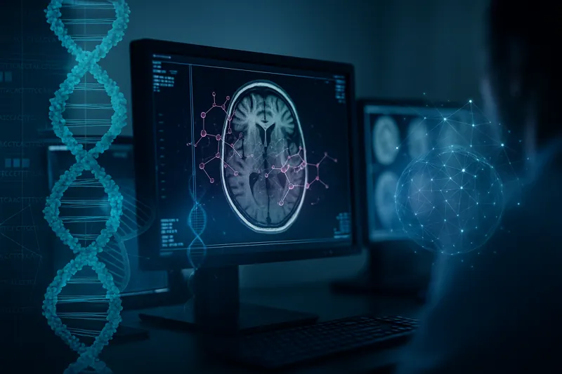
In parallel, the FDA granted De Novo authorization to the first image-only AI risk platform that predicts 5-year risk directly from a screening mammogram—an inflection point that makes risk-adjusted pathways far more scalable. Coverage from Radiology Business and BCRF explains the authorization and clinical intent. Radiology Business

Why it matters: average-risk guidance in the U.S. now begins screening at age 40 (USPSTF, 2024). Programs can layer image-based risk on top of that baseline to triage who needs annual vs. short-interval follow-up and who merits supplemental imaging. USPSTF

CEM is earning a seat next to MRI

Expect exhibits and sessions positioning CEM as a cost-effective, accessible adjunct—particularly for dense-breast populations and diagnostic workups. RSNA News recently framed CEM as a practical alternative to MRI in some screening/diagnostic scenarios, and new peer-review literature is refining technique (e.g., lower volume/higher-iodine contrast while preserving diagnostic performance). RSNA

On outcomes, the RACER trial in The Lancet Regional Health – Europe reported that using CEM as primary imaging for recalled women improved the accuracy and efficiency of the work-up compared with conventional imaging—evidence that will influence protocols beyond the show floor. The Lancet

MRI still leads for sensitivity—BPE is your underused signal

Breast MRI remains the sensitivity champion for high-risk patients and for problem solving. This year’s RSNA content spotlights BPE—how the level of background enhancement relates to tumor biology and outcomes. Recent reviews (2024–2025) synthesize BPE’s predictive/prognostic value, including associations with pathologic complete response after neoadjuvant therapy and survival in certain subtypes. SpringerLink

Practical move: standardize how you document BPE and incorporate it into structured reports and risk conferences; it’s becoming more than a descriptive footnote.

What to ask vendors at RSNA

  1. Risk engine proof: “Show external validation and calibration plots by density and race; how does your image-only model integrate into our mammography worklist and letters?”
  2. CEM logistics: “Demonstrate CEM acquisition workflows, contrast protocols, and how your viewer handles subtraction/kinetics alongside priors.”
  3. MRI + BPE analytics: “Can we standardize BPE capture in structured reports and trend it across treatment?”

As risk-first screening, CEM, and MRI gain real traction, the winners will be the programs that operationalize them quickly and consistently. If you’re planning your 2026 breast-imaging playbook, stop by Vesta at RSNA to see how our subspecialists, standardized templates, and overflow routing make risk-adjusted pathways usable on day one.

Imaging the Individual — In the Trenches: AI, Personalization & Equity at RSNA 2025

RSNA’s 2025 theme, Imaging the Individual, isn’t just about futuristic science—it’s about doing the basics better for each patient, every day. The official Trending Topics preview highlights three threads cutting across subspecialties: AI you can deploy, personalized care you can operationalize, and equity you can measure. This guide translates those themes into practical checkpoints hospitals and imaging centers can use right now. RSNA

1) AI that graduates from pilot to practice

This year’s agenda emphasizes real outcomes over proofs of concept: reader-in-the-loop tools, bias monitoring, and governance. In breast imaging alone, RSNA previews spotlight external validation for image-only risk models and integration of MRI signals into multimodal AI—clear signals that “personalization” is landing in routine workflows. Bring vendor questions that force specifics: external validation cohorts, drift detection, and how metrics (TAT, recalls, rework) appear in your dashboard. RSNA

What to set up before RSNA: define 3–5 outcome metrics and insist every demo shows pre/post performance tied to those measures. Use QIBA concepts to push for standardized inputs/outputs so results are reproducible across scanners and sites. QIBA Wiki

2) Personalization that reaches the reading room

Personalization isn’t only radiogenomics. RSNA’s preview points to risk-stratified pathways you can actually run: e.g., image-only 5-year breast cancer risk at the point of screening to route patients into annual vs. short-interval follow-up or supplemental imaging (CEM/MRI). That pairs well with updated U.S. recommendations: screening beginning at age 40 for average-risk women, then adjusting based on risk and local policy. Build routing rules, templates, and letters now, so RSNA demos can plug into your plan.

Operational checklist:

  • Map risk thresholds → next steps (annual vs. short-interval, CEM/MRI).
  • Standardize templates so risk outputs appear consistently in reports and patient letters.
  • Decide who reviews outlier risk flags and how quickly (SLA).

3) Equity you can instrument—not just endorse

RSNA is foregrounding health equity, with sessions on encoding equity in AI and addressing access gaps for underserved communities. Equity becomes real when you can see it in your data: turnaround times by language, missed-appointment patterns by zip code, recall rates by screening site, and AI performance by subgroup. Build those slices into your analytics now; then ask vendors to show subgroup performance in their dashboards.

Practical moves:

  • Add demographic and language filters to your TAT and recall reports.
  • Require AI vendors to show calibration and error analysis by subgroup.
  • Stand up multilingual patient letter templates to support new screening starts at 40. USPSTF

4) CEM/MRI momentum: choose the lever that fits your service line

RSNA coverage calls out CEM as an increasingly practical adjunct—especially useful for dense-breast populations and diagnostic workups where capacity or cost limits MRI. The RACER trial reported higher accuracy and efficiency for CEM as the primary exam for recalled women vs. conventional imaging—evidence that can justify protocol changes and equipment planning. Meanwhile, MRI retains the sensitivity crown, with renewed attention on background parenchymal enhancement (BPE) as a signal worth documenting consistently.


Action items:

  • Decide where CEM fits: diagnostic recall pathway, dense-breast supplemental strategy, or both.
  • Add BPE level to structured MRI reports and trend it during therapy response clinics.

5) Governance, not guesswork

If personalization is the “what,” governance is the “how.” Use QIBA ideas—claim definitions, acquisition standards, and profile adherence—to control variability across devices and shifts. Tie RSNA learnings to a written governance plan with three parts: 1) protocol book (who owns it, update cadence), 2) quality book (metrics, subgroup views), and 3) AI book (approval process, monitoring, rollback).

6) Where teleradiology extends your capacity

Personalization increases complexity at peaks (recalls, dense-breast seasons, MR backlogs). A teleradiology partner helps you keep individualized pathways moving: standardized templates, subspecialty over-reads, and after-hours coverage that adheres to your risk rules and equity metrics—so “Imaging the Individual” doesn’t stop at 5 p.m.

Headed to RSNA?


Visit Vesta at Booth 1346 (South Hall) to see how we make “Imaging the Individual” work in real clinics—then enter to win a 1-year Medality CME subscription. Don’t wait: email “RSNA CME Entry” to info@vestarad.com now for a reserved entry, and show your confirmation at the booth for a bonus entry.

Precision Imaging at RSNA 2025: Radiomics, Biomarkers, and the Era of Multi-Omics Integration

As radiology moves deeper into the era of precision medicine, quantitative imaging is transforming from a promising research tool to a clinical driver of individualized care. The convergence of radiomics, imaging biomarkers, and multi-omics integration represents one of the most exciting frontiers showcased under RSNA 2025’s theme, “Imaging the Individual.”

Radiomics — the extraction of high-dimensional quantitative features from medical images — allows the characterization of tissue heterogeneity beyond what can be perceived visually. These features, derived from modalities such as CT, MRI, or PET, have been linked to tumor phenotype, gene expression, and therapeutic response across oncology, neurology, and cardiology studies (Springer, 2024).

Imaging Biomarkers in Practice

Validated imaging biomarkers are redefining how clinicians stratify patients, monitor disease, and predict outcomes. Quantitative features from radiomics pipelines can act as noninvasive surrogates for histopathologic or molecular data, guiding therapy selection and prognosis assessment. For instance, radiomic signatures have shown potential in predicting response to immunotherapy and correlating with tumor-infiltrating lymphocytes in non-small cell lung cancer (ScienceDirect, 2020).

In cardiovascular and neuroimaging applications, biomarkers derived from texture and perfusion patterns are being explored to detect subclinical disease, assess ischemic risk, and evaluate treatment efficacy. The promise lies in moving from population averages toward individualized predictions based on each patient’s unique imaging phenotype.

Radiogenomics and Multi-Omics Integration

The next step in precision imaging is radiogenomics — linking imaging phenotypes with genomic and proteomic data to uncover biologically meaningful correlations. Integrating imaging with multi-omics datasets enables the creation of comprehensive disease models that reflect both spatial and molecular dimensions.

Recent reviews highlight the potential of AI-driven multi-omics integration to refine cancer subtyping, prognostication, and therapeutic decision-making (British Journal of Radiology, 2025) and (ScienceDirect, 2025). Federated approaches and multi-modal AI models are emerging to harmonize these heterogeneous datasets while preserving privacy and reproducibility.

Projects such as NAVIGATOR, a regional imaging biobank integrating multimodal imaging with molecular and clinical data, illustrate how research infrastructure is catching up to these ambitions (European Journal of Radiology, 2025).

From Quantitative Imaging to Clinical Translation

Despite the promise, clinical translation remains the critical frontier. Feature reproducibility, acquisition standardization, and regulatory validation continue to challenge adoption (Insights into Imaging, 2020). However, the increasing presence of quantitative imaging biomarkers in prospective trials, along with support from the Quantitative Imaging Biomarkers Alliance (QIBA) and FDA’s digital health framework, signals that this research is crossing the threshold into practice.

At RSNA 2025, expect sessions emphasizing standardization of radiomics workflows, reproducibility metrics, and AI-assisted integration of multi-omics data. Discussions will likely center on how to validate imaging biomarkers in multi-institutional settings and what infrastructure is required for clinical scalability.

The Role of Teleradiology in Precision Imaging

For teleradiology providers like Vesta, these developments offer both opportunity and responsibility. The same digital infrastructure that enables subspecialty coverage across time zones can support quantitative image analysis, data harmonization, and longitudinal tracking — essential foundations for radiomic and biomarker validation.

By aligning with quantitative imaging standards and collaborating with research institutions, teleradiology networks can help bring precision imaging insights into real-world practice — from oncology to cardiovascular disease management.

Precision imaging is not a distant future — it’s the next evolution of radiology happening now.


At RSNA 2025, Vesta will be on site to explore how radiomics, biomarkers, and AI-driven data integration are redefining what it means to truly “image the individual.”


Vizamyl’s New PET Label: Quantify & Monitor Amyloid—What It Means for Imaging Teams


What changed—and why it matters

The FDA has expanded the label for flutemetamol F 18 (Vizamyl), enabling quantification of amyloid plaque burden and long-term therapy monitoring in Alzheimer’s disease. This shift moves amyloid PET beyond a qualitative “positive/negative” decision toward objective, longitudinal assessment that can inform treatment choice, dose intervals, and discontinuation decisions. Business Wire

Professional groups report the update aligns amyloid PET with the clinical era of disease-modifying anti-amyloid therapies (e.g., lecanemab, donanemab), clarifying roles for baseline confirmation, on-treatment monitoring, and response tracking in routine care. Notably, SNMMI stated the FDA granted supplemental indications—including quantitative measurement and use for therapy monitoring—to three amyloid PET agents (flutemetamol F-18/Vizamyl, florbetapir F-18, and florbetaben F-18). SNMMI

Operational updates for radiology leaders

  • Protocols & quant pipelines: Build or validate a quant workflow (SUVr or comparable metrics) that’s scanner-calibrated and reproducible across sites. If you operate multi-vendor fleets, document harmonization steps in your SOPs.
  • Structured reports: Add fields for quantified burden at baseline, change from baseline, and interpretive guidance tied to therapeutic decisions (initiation, continuation, or discontinuation).
  • Scheduling & throughput: Expect rising referral volume from neurology and geriatrics as therapy monitoring enters routine practice; protect access with extended hours or overflow capacity.
  • Quality & governance: Define thresholds for biologically meaningful change, reader training for quant review, and reconciliation rules when quant and visual impressions diverge.

For additional context, trade coverage underscores that the updated label formally removes previous limitations around therapy monitoring and permits quant analysis in routine reporting. Empr

How Vesta Teleradiology helps

Vesta’s subspecialty neuro and nuclear medicine radiologists provide:

  • Amyloid PET expertise: Visual+quant reads with structured templates aligned to your therapy pathway.
  • Coverage when you need it: After-hours, weekends, or daytime overflow—without sacrificing turnaround time.
  • Interoperability: Seamless delivery to your PACS/RIS and EMR; clear flags for therapy decisions and recall intervals.
  • QA you can see: Peer review, consistency checks across readers, and optional double-reads during program ramp-up.

If you’re standing up or scaling amyloid PET services, we can supply immediate subspecialty coverage and templates tuned to your neurologists’ needs.


The Future of AI + Human Collaboration in Radiology

Artificial intelligence (AI) is playing an increasingly important role in radiology and diagnostic imaging. From workflow optimization to automated image analysis, AI tools are now assisting radiologists in more imaging departments than ever before. Right now, AI tools are assisting with tasks like automatically prioritizing critical cases, generating draft reports, and flagging potential abnormalities in studies such as chest X-rays, mammograms, and CT scans.”

At the same time, it’s clear that AI’s role is best seen as complementary to human expertise, not a replacement. In fact, a 2023 study published in JAMA Network Open found that radiologists using AI frequently sometimes experienced higher burnout rates—especially when workflows were not well integrated or added new demands.

This highlights an important lesson: for AI to truly benefit radiology, it must be thoughtfully implemented, supporting radiologists rather than complicating their work.

Why Human Expertise Remains Essential

While AI offers exciting capabilities—such as triaging cases, flagging abnormalities, or standardizing reports—there is no substitute for the experience and clinical judgment of a radiologist.

Subspecialty areas like:

  •         Neuroradiology
  •         Musculoskeletal imaging
  •         Cardiac imaging
  •         Pediatric radiology

…require nuanced interpretation that today’s AI tools simply cannot match.

Vesta Teleradiology supports healthcare facilities by ensuring that every read is performed by a board-certified U.S.-based radiologist—with subspecialty expertise available across all major modalities.


Balancing AI + Workflow: A Smarter Approach

Many imaging departments today are navigating how to integrate AI without adding unnecessary complexity.


At Vesta, we work with partner facilities to provide flexible teleradiology services that complement their existing workflows—whether or not they are using AI tools internally.


Our approach emphasizes:

✅ Efficient, reliable human reads

✅ Subspecialty expertise when needed

✅ Consistent communication with referring providers

✅ Flexibility to support 24/7 coverage and manage fluctuations in volume


By helping facilities maintain high-quality interpretations with efficient turnaround, Vesta supports radiology teams as they adopt new technologies and respond to growing imaging demand.


Looking Ahead: The Collaborative Future of Radiology

AI’s role in radiology will continue to evolve. The most effective imaging departments will combine:


  •         Advanced AI tools where they add value
  •         Skilled radiologists providing expert interpretation
  •         Clear, integrated workflows that reduce friction
  •         Strategic partnerships to ensure coverage and subspecialty access


At Vesta Teleradiology, we believe that human expertise will remain the foundation of diagnostic imaging—and that thoughtful integration of AI can enhance, not replace, that expertise.


We’re committed to working with healthcare facilities to build balanced solutions that support radiologists, improve patient care, and keep pace with the demands of modern imaging.


If your team is looking for flexible, expert support—whether for subspecialty reads, after-hours coverage, or help managing increased imaging demand—Vesta Teleradiology is here to help.


Contact us to learn more.


RSNA 2024 Highlights: AI, Imaging Advancements, and Industry Recognition

The Radiological Society of North America’s (RSNA) 2024 annual meeting showcased significant advancements in medical imaging, including artificial intelligence (AI), innovations in computed tomography (CT) and magnetic resonance imaging (MRI), and strategies to address the ongoing radiology staffing shortage. The event also honored leaders in the field for their exceptional contributions.

Advancements in Radiology Technology

AI Integration

Artificial intelligence was a major focus, with over 200 exhibitors showcasing AI-related innovations. The FDA has approved nearly 1,000 clinical AI algorithms, 80% of which pertain to medical imaging. AI’s expanding role includes rapid stroke detection, workflow orchestration, and FFR-CT assessments, now included in national guidelines. However, discussions emphasized the need for rigorous monitoring to prevent bias and performance degradation in these algorithms.

CT and MRI Innovations

The conference highlighted advancements in imaging technologies, unveiling three new AI-enhanced CT systems aimed at improving diagnostics and patient comfort. These innovations promise faster, more accurate imaging, enhancing both clinical outcomes and workflow efficiency.

Addressing the Staffing Shortage

The radiology workforce shortage remains a critical challenge. Proposed solutions include expanding residency programs, adopting AI to reduce workloads, and ensuring fair compensation. While AI shows promise in easing demands, experts caution it is not a standalone solution to the staffing crisis. Teleradiology companies like Vesta can play a vital role in addressing these shortages by providing access to qualified radiologists, ensuring timely interpretations, and supporting healthcare facilities in maintaining efficient workflows.

Industry Recognition

RSNA 2024 also celebrated the accomplishments of industry leaders:

  • Gold Medal Awards: Recognizing excellence in leadership and innovation, the awards were presented to James P. Borgstede, MD, Elizabeth S. Burnside, MD, MPH, and Beverly G. Coleman, MD, for their groundbreaking contributions to radiology and healthcare.
  • Alexander R. Margulis Award for Scientific Excellence: This award honored the authors of a 20-year study on low-dose CT screening for lung cancer, emphasizing its life-saving potential through early detection.
  • Outstanding Researcher Award: Jeffrey G. Jarvik, MD, MPH, was recognized for his impactful work in spine imaging and back pain research.

Conclusion

RSNA 2024 reinforced radiology’s pivotal role in healthcare, spotlighting transformative technologies, addressing workforce challenges, and honoring outstanding achievements. The integration of AI, combined with ongoing innovation in imaging and efforts to bolster the radiology workforce, ensures the field continues to thrive and evolve.


Sources:

Radiologbusiness.com
rsna.org
openai.com


Top 5 Trends Shaping Radiology in 2025

Radiology is constantly evolving, with advancements and challenges shaping how providers deliver care. As we step into 2025, exciting developments in technology, workforce dynamics, patient engagement, and regulatory compliance are transforming the landscape. In this blog, we’ll dive into the top five trends to watch in radiology this year and explore how they’re influencing the future of the field.


  1. Artificial Intelligence (AI): Revolutionizing Radiology in 2025

AI continues to make waves in radiology, offering improved diagnostic accuracy and efficiency. In 2025, AI tools are more refined than ever, assisting radiologists with cancer detection, anomaly identification, and image interpretation. Advanced algorithms can now process vast amounts of imaging data faster than ever, reducing turnaround times and enhancing patient outcomes.

radiology trendsHowever, challenges remain, including concerns about transparency in AI decision-making and biases in data sets. These hurdles are gradually being addressed with stricter regulations and improved algorithm training. AI isn’t just a tool; it’s becoming a trusted collaborator in radiology practices worldwide.

Read more about AI advancements in radiology here.


  1. Shifts in Diagnostic Imaging: The Rise of Independent Facilities

The trend of moving diagnostic imaging services away from hospitals and into Independent Diagnostic Testing Facilities (IDTFs) continues to grow in 2025. Patients and providers increasingly favor IDTFs for their cost-effectiveness and accessibility.


These facilities are adopting cutting-edge imaging technology, enabling faster and more accurate diagnoses. For healthcare providers, this trend presents an opportunity to collaborate with IDTFs or expand their own outpatient imaging services to meet the rising demand.

Learn more about the rise of IDTFs here.


  1. Addressing Workforce Shortages in Radiology

Workforce challenges remain a key issue in 2025. The demand for radiologists continues to outpace supply, especially as imaging volumes grow due to an aging population and the increased use of advanced diagnostic techniques. These shortages are felt acutely during peak times like the holiday season or in underserved areas.

To mitigate these challenges, healthcare organizations are relying on teleradiology to bridge gaps, ensuring 24/7 coverage without overburdening onsite staff. In addition, many practices are adopting flexible work schedules and prioritizing workplace wellness to attract and retain talent in this competitive market.

Explore workforce challenges and solutions here.


  1. Patient-Centered Care Takes Center Stage

Patient engagement continues to be a major focus in radiology in 2025. Programs like the FDA’s Patient and Caregiver Connection are pushing for more transparency and collaboration in radiology services. These initiatives encourage providers to involve patients in their care by offering clear, timely explanations of imaging results and personalized care recommendations.

Additionally, new tools, such as mobile apps that allow patients to access their imaging records and reports, are empowering individuals to take control of their health. Radiology practices that adopt these technologies are seeing improved patient satisfaction and stronger provider-patient relationships.

Learn more about patient-centered care here.


  1. New Breast Density Legislation in Effect

2025 marks the implementation of new breast density notification laws in many states. These laws require radiologists to inform patients if they have dense breast tissue, which can make it more difficult to detect cancer during mammograms. Dense tissue can also increase the risk of breast cancer, making this information critical for patients and their healthcare providers.

mammogramRadiology practices are adapting to these regulations by enhancing their reporting systems and educating patients about the implications of breast density. This legislation empowers patients to make informed decisions about supplemental screening options, improving early detection and outcomes.

Read more about breast density legislation here.


Looking Forward: Radiology’s Bright Future in 2025

Radiology is more integral to healthcare than ever before, and 2025 promises to be a transformative year. From leveraging AI to addressing workforce shortages, radiology providers are finding innovative ways to enhance care delivery. As patient engagement grows and new regulations take effect, the field is evolving to meet the demands of modern medicine.


For healthcare facilities looking to stay ahead of these trends, partnering with a trusted teleradiology provider can make all the difference. At Vesta Teleradiology, we specialize in sourcing skilled radiologists for both remote and onsite roles. Whether you’re navigating staff shortages, expanding diagnostic capabilities, or seeking flexible coverage, our experienced team can help. Let us be your partner in delivering exceptional care in 2025 and beyond.


Explore how we can support your radiology needs today.

Sources:

apnews.com
stout.com
fda.gov
theimagingwire.com
wikipedia.org
Openai.com

Errors in Radiology Interpretations in CT and MR imaging

In a recent study of 10,090 body CT scans, reviewed by attending physicians in the abdominal division, no significant difference in error rates was found between day and night studies. The study identified 44 errors in 2,164 day studies and 226 errors in 7,446 night studies. The research highlights that body CT studies often have higher error rates when interpreted by non-specialists due to the complexity of evaluating multiple organs. Specialists in body imaging, who reviewed the scans, often detected additional observations that generalists missed. To reduce errors, recommendations include using checklists, taking notes during reviews, making all observations before dictating, and thoroughly reviewing reports before finalizing them.


Additional Study

Research published in the Journal of the American College of Radiology highlights that up to 44% of diagnostic errors stem from improperly ordered laboratory tests and radiology exams. This issue is particularly significant for CT and MR imaging. A study led by Dr. Ariadne DeSimone at Brigham and Women’s Hospital found that implementing tailored order options and educational interventions significantly reduced these errors by 83%, from 16% to 3%. The study emphasizes the importance of improving the imaging order process to minimize diagnostic errors and enhance radiologists’ workflow by reducing time spent on correcting incorrect orders.


ChatGPT—Can it Reduce Errors?

chatgpt in interpretations
ChatGPT


A retrospective study explored the potential of ChatGPT-4 in assisting radiologists with error detection in radiology reports. The study involved 200 reports, with 150 common errors intentionally introduced. ChatGPT-4 identified 82.7% of errors, closely matching the performance of senior radiologists (89.3%) and surpassing assistants and residents (80.0%). Notably, ChatGPT-4 was significantly faster, taking just 3.5 seconds per report compared to 25.1 seconds for radiologists, and was more cost-effective. While promising, the study suggests that further prospective validation and specialized training are needed before widespread adoption of AI-assisted re-reading in radiology.


How Vesta can Help


Vesta is a leading teleradiology company offering comprehensive radiology support—partial, full, onsite or remotely. Our team of U.S. Board Certified radiologists encompasses various subspecialties, ensuring that interpretations are accurate and reliable, regardless of the complexity of the case. By leveraging our expertise and advanced technology, we provide seamless and precise radiology services, helping healthcare facilities enhance diagnostic accuracy and patient outcomes.


Sources:

Auntminnie.com
dotmed.com
Medscape.com
openai.com


An Update to the Physician Shortage Problem

The AAMC (Association of American Medical Colleges) has released new projections indicating a physician shortage of up to 86,000 physicians in the United States by 2036. This underscores the critical need for sustained and increased investments in training new physicians to address the country’s healthcare needs. The report, conducted by GlobalData Plc, includes various scenarios based on trends in healthcare delivery and the workforce. While the projected shortfall is smaller than previous estimates, it still highlights the necessity for additional investments in graduate medical education (GME). Demographics, particularly population growth and aging, are driving the increasing demand for physicians. The report also notes a significant portion of the physician workforce nearing retirement age, which will further decrease the physician supply. Addressing underserved communities could require approximately 202,800 more physicians than current estimates. Lifting the federal cap on Medicare support for GME and bipartisan legislation like the Resident Physician Shortage Reduction Act aim to alleviate the shortage, but further efforts are needed to meet future healthcare demands.


Rural Americans’ Healthcare Challenges

Rural Americans face significant healthcare challenges, with fewer available doctors compared to urban areas, exacerbating existing health issues. Dr. Bruce A. Scott, President of the American Medical Association, emphasizes the urgent need for policymakers to address these disparities. Rural communities experience higher rates of various illnesses, exacerbated by economic pressures and limited access to healthy living conditions. The shortage of specialists and the closure of rural hospitals further compound the problem. Insufficient access to primary care physicians is a pressing issue, with inadequate residency spots and decreasing applications from rural areas. The AMA advocates for changes to the Medicare physician payment system, which has seen a decline in rates over the years. Administrative burdens, such as prior authorizations, are also contributing to physician burnout and compromising patient care. To combat the doctor shortage and rural health challenges, the AMA advocates for healthcare reforms, including overhauling the Medicare payment system, expanding telehealth, increasing residency positions, incentivizing rural practice, and addressing workforce stresses.

hospitals in rural America

Radiology Is Being Hit, Too

Radiology departments are grappling with worsening staffing shortages alongside declining reimbursements. During the RSNA 2023 meeting, Ashish Sant from Merge by Merative discussed key trends and challenges. Staffing and cost management remain top concerns due to burnout and insufficient replacements for retiring radiologists. To address these issues, there’s a push towards cloud-based solutions, with a modular approach easing concerns about data security and patient information management. The pandemic has accelerated the shift towards cloud adoption, highlighting benefits such as accessibility and cost reduction. Integrating AI into radiology workflows is another focus, though challenges persist in seamlessly embedding AI solutions. Merge’s partnership with Microsoft Azure aims to provide customers with cloud solutions tailored to their needs.


Radiology Support for the US

Addressing radiology staffing shortages is crucial for ensuring efficient and effective healthcare delivery. Whether you’re a hospital, outpatient center, or part of the Indian Health Service (IHS), Vesta is here to help. Our team can provide on-site radiologists or teleradiologists to meet the specific needs of your facility. By partnering with us, you can ensure timely and accurate radiology services, ultimately improving patient care and outcomes. Don’t let staffing shortages hinder your operations – reach out today to learn how we can support your radiology department.


Sources:

Aamc.org
dtnpf.com
Healthimaging.com
openai.com


The Latest in Brain Imaging News

In recent years, awareness surrounding brain injuries has steadily risen, prompting significant strides in diagnostic technologies and treatment modalities. As we delve into the latest developments in this critical area of healthcare, it becomes increasingly apparent that advancements in medical imaging, particularly in the realm of neurological disorders, are poised to revolutionize the landscape of brain injury diagnosis and management.


AI-based Quantitative Brain Imaging System

Philips and Synthetic MR have joined forces to advance the diagnosis of neurological disorders through cutting-edge quantitative brain imaging tools. Their collaboration introduces the Smart Quant Neuro 3D MRI software suite, combining Philips’ SmartSpeed image-reconstruction technology, the 3D SyntAc clinical application, and SyntheticMR’s SyMRI NEURO 3D software. This innovation employs AI to analyze brain tissues, enhancing the detection and analysis of conditions like multiple sclerosis, traumatic brain injuries, and dementia.

The rise of AI in diagnostic imaging, projected to reach $1.2bn by 2027, signifies a transformative shift in improving accuracy and patient outcomes. With the diagnostic imaging market expected to grow to $9.1bn by 2030, fueled by demand for early disease diagnosis and personalized medicine, this partnership underscores the crucial role of AI in enhancing medical imaging.

Read the press release here.


A New Way of Diagnosing Mild TBIs

Researchers have developed a novel brain imaging method to diagnose mild traumatic brain injuries (mTBIs), which are often missed by standard techniques like MRI. This method involves loading gadolinium, a common MRI contrast agent, into micropatches attached to immune cells called macrophages. These cells migrate to areas of brain inflammation caused by mTBIs, enabling MRI detection. The technique, called M-GLAMs, was successfully tested in mice and pigs, showing promise for accurately diagnosing mTBIs. It also allows imaging at lower gadolinium doses, potentially benefiting patients with kidney issues. While unable to pinpoint injury locations, M-GLAMs could aid in identifying and treating brain inflammation. The researchers aim to bring this technology to clinical trials, with support from grants and intellectual property protection.

Read the study here.

tbi

New Imaging Tech that Captures Neuronal Activity Across the Brain During Recovery

Researchers at Tufts University School of Medicine have developed a novel imaging technology to monitor neuronal activity throughout the entire brain during the initial weeks of recovery from traumatic brain injury (TBI). Their study, published in Cerebral Cortex, reveals that TBI can induce changes in brain function beyond the injury site. Using a combination of fluorescent sensors and electrodes, they observed altered connectivity patterns in mice post-injury, even in regions distant from the impact. Despite the mice’s ability to perform physical tasks normally, their brain activity during both exercise and rest differed significantly from healthy brains. This impaired ability to switch between states suggests underlying brain state dysfunction post-injury. The findings highlight the brain’s plasticity in response to injury and have potential clinical implications for understanding TBI impacts and tailoring treatments. The researchers aim to further investigate long-term neural activity changes post-recovery and explore the technology’s potential in predicting specific dysfunctions or long-term outcomes of TBI. 

Read the study here.


Sources:

Medicaldevice-network.com
Otd.harvard.edu
Scitechdaily.com
Openai.com